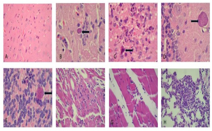
Figure 8.
Histological cuts from brain, cardiac muscle and lung in the acute-infected group. (A) Brain cortex illustrating neuronal hypoxia (400×), (B) gliosis with a Toxoplasma gondii bradyzoite cyst (400×), (C) site exhibiting glionecrosis with a neuron in apoptosis (1000×), (D,E) Toxoplasma cyst placed in the cerebellar molecular and the granular layers (1000×), (F) moderate myocarditis (400×), and (G) moderate myositis with a fragmented muscle fiber (400×), (H) focal pneumonitis with acute and chronic components (400×). The arrows indicate Toxoplasma cysts except in figure C wich indicates an apoptotic cell.

