Abstract
Background
Peptide transporter 1 (PepT1, alias Slc15a1) mediates the uptake of dietary di/tripeptides in all vertebrates. However, in teleost fish, more than one PepT1-type transporter might function, due to specific whole genome duplication event(s) that occurred during their evolution leading to a more complex paralogue gene repertoire than in higher vertebrates (tetrapods).
Results
Here, we describe a novel di/tripeptide transporter in the zebrafish (Danio rerio), i.e., the zebrafish peptide transporter 1a (PepT1a; also known as Solute carrier family 15 member a1, Slc15a1a), which is a paralogue (78% similarity, 62% identity at the amino acid level) of the previously described zebrafish peptide transporter 1b (PepT1b, alias PepT1; also known as Solute carrier family 15 member 1b, Slc15a1b). Also, we report a basic analysis of the pept1a (slc15a1a) mRNA expression levels in zebrafish adult tissues/organs and embryonic/early larval developmental stages. As assessed by expression in Xenopus laevis oocytes and two-electrode voltage clamp measurements, zebrafish PepT1a, as PepT1b, is electrogenic, Na+-independent, and pH-dependent and functions as a low-affinity system, with K0.5 values for Gly-Gln at − 60 mV of 6.92 mmol/L at pH 7.6 and 0.24 mmol/L at pH 6.5 and at − 120 mV of 3.61 mmol/L at pH 7.6 and 0.45 mmol/L at pH 6.5. Zebrafish pept1a mRNA is highly expressed in the intestine and ovary of the adult fish, while its expression in early development undergoes a complex trend over time, with pept1a mRNA being detected 1 and 2 days post-fertilization (dpf), possibly due to its occurrence in the RNA maternal pool, decreasing at 3 dpf (~ 0.5-fold) and increasing above the 1–2 dpf levels at 4 to 7 dpf, with a peak (~ 7-fold) at 6 dpf.
Conclusions
We show that the zebrafish PepT1a-type transporter is functional and co-expressed with pept1b (slc15a1b) in the adult fish intestine. Its expression is also confirmed during the early phases of development when the yolk syncytial layer is present and yolk protein resorption processes are active. While completing the missing information on PepT1-type transporters function in the zebrafish, these results open to future investigations on the similar/differential role(s) of PepT1a/PepT1b in zebrafish and teleost fish physiology.
Keywords: Di/tripeptide transport(ers), Dietary protein, Electrogenic transport, Heterologous expression, Peptide absorption, pH-dependence, Teleost fish, Whole genome duplication, Xenopus laevis oocytes
Background
The intestinal peptide transporter 1 (PepT1) plays a major role in protein nutrition as it mediates the luminal-to-cellular uptake of dietary amino acids in a di- and tripeptide (di/tripeptide) form at the brush-border membrane of the enterocytes [1, 2]. In this way, PepT1 allows absorption of large fractions of exogenous ingested proteins of animal, plant, and microorganism origin and/or endogenous luminal resident proteins of epithelial and microorganism origin, as they are terminally degraded by digestive and/or microbial enzymes [3–5]. PepT1 is also responsible for the absorption of orally active peptidomimetics, including β-lactam antibiotics and selected pro-drugs [2, 6, 7].
PepT1 belongs to the peptide transporter family [8], which members are found from bacteria to vertebrates [8–11]. In humans, it is referred to as the Solute Carrier 15 (SLC15) family member A1 (SLC15A1) [1, 2]. In higher vertebrates, PepT1 is a Na+-independent, H+-dependent electrogenic symporter, and by coupling substrate uptake to H+-movement down an inwardly directed electrochemical H+-gradient, it allows transport of peptides across the plasma membrane even against a substrate concentration gradient. The transport responds to membrane potential and extracellular pH, with extracellular pH-optima varying between 4.5 and 6.5 depending on the net charge of the transported substrate [1, 2, 10]. PepT1 function has also been described in detail in teleost fish [12, 13]. Zebrafish (Danio rerio) PepT1, the first teleost PepT1-type transporter cloned and functionally characterized [14], exhibited a unique pH dependence, with neutral-to-alkaline extracellular pH increasing its maximal transport rate (for information on human vs. zebrafish PepT1-type transporters and of their major features in the larger context of the human and zebrafish SLC15 transporters see Table 1). However, studies on European sea bass (Dicentrarchus labrax) [25], Atlantic salmon (Salmo salar) [26], and Antarctic icefish (Chionodraco hamatus) [27] PepT1 transporters revealed a more standard behavior with respect to the pH-optimum, with maximal transport rates independent of the extracellular pH in the alkaline to neutral-to-slightly-acidic range [25–28]. With respect to substrate specificity, as in higher vertebrates, all teleost PepT1 transporters also mediated the uptake of neutral and charged di/tripeptides [26, 28, 29].
Table 1.
The Solute Carrier 15 (proton oligopeptide cotransporter) family members in human (Homo sapiens) and zebrafish (Danio rerio)
| Human | Zebrafish | ||||||||||
|---|---|---|---|---|---|---|---|---|---|---|---|
| From http://www.bioparadigms.org | From http://www.guidetopharmacology.org | From http://www.ncbi.nlm.nih.gov/gene | https://www.ncbi.nlm.nih.gov/unigene/ | From http://zfin.org | |||||||
| SLC name | Protein name | Aliases | Transport type | Substrates | Tissue and cellular expression | Substrates | slc name | EST profile | Tissue and cellular expression | Stage range | References |
| SLC15A1 | PEPT1 | Oligopeptide transporter 1, H+-peptide transporter 1 | C/H+ | Di- and tripeptides, protons, β-lactam antibiotics | Small intestine, kidney, pancreas, bile duct, liver |
Endogenous substrates: 5-aminolevulinic acid, dipeptides, protons, tripeptides. Other substrates: fMet-Leu-Phe, muramyl dipeptide, D-Ala-Lys-AMCA, β-Ala-Lys-AMCA, His-Leu-lopinavir, alafosfalin. |
slc15a1a (Chr 9) | – | Details in this study | Details in this study | This study |
| slc15a1b (Chr 6) |
Developmental stage|larval > adult Adult|intestine |
Digestive system, gut, intestinal bulb, intestinal bulb enterocyte, intestinal epithelium, liver, muscle, squamous epithelial cell, whole organism | Prim-5 to adult | [14–22] | |||||||
| SLC15A2 | PEPT2 | Oligopeptide transporter 2, H+-peptide transporter 2 | C/H+ | Di- and tripeptides, protons, β-lactam antibiotics | Apical surface of epithelial cells from kidney and choroid plexus; neurons, astrocytes (neonates), lung, mammary gland, spleen, enteric nervous system |
Endogenous substrates: 5-aminolevulinic acid, dipeptides, protons, tripeptides. Other substrates: muramyl dipeptide, D-Ala-Lys-AMCA, β-Ala-Lys-AMCA, alafosfalin, γ-iE-DAP. |
slc15a2 (Chr 9) |
Developmental stage|adult Adult|intestine >> kidney > reproductive system |
Brain, eye, gill, gut, kidney, musculature system, otic vesicle, semicircular canal, ventricular system, whole organism | 26+ somites to day 6; days 30–44; adult | [21, 23] |
| SLC15A3 | PHT2 | Peptide/histidine transporter 2, PTR3 | C/H+ | Di- and tripeptides, protons, β-lactam antibiotics | Lung, spleen, thymus, intestine (faintly in brain, liver, adrenal gland, heart) |
Endogenous substrates: L-histidine, dipeptides, protons, tripeptides. Other substrates: muramyl dipeptide, MDP-rhodamine, Tri-DAP |
n.p. | n.p. | n.p. | n.p. | n.p. |
| SLC15A4 | PHT1 | Peptide/histidine transporter 1, PTR4 | C/H+ | Di- and tripeptides, protons, β-lactam antibiotics | Brain, eye, intestine (faintly in lung and spleen) |
Endogenous substrates: L-histidine, carnosine Other substrates: valacyclovir, muramyl dipeptide, His-Leu-lopinavir, glycyl-sarcosine, MDP-rhodamine, Tri-DAP, C12-iE-DAP |
slc15a4 (Chr 8) |
Developmental stage|adult Adult|brain ≈ reproductive system ≥ fin |
Epidermis, eye, immature eye, midbrain, periderm, ventricular zone, yolk syncytial layer | 50%-epiboly to Long-pec | [24] |
Abbreviations for transport type: C cotransporter, n.p. not present in the zebrafish genome
With several teleost genomes available in databanks, it was progressively clear that teleost PepT1-type proteins are the result of a gen(om)e duplication event, and, after an initial description of partial nucleotide sequences [30], the idea that in teleost genomes a peptide transporter 1a (pept1a; also known as solute carrier family 15 member 1a, slc15a1a) gene occurs beside a peptide transporter 1b (pept1b, alias pept1; also known as solute carrier family 15 member 1b, slc15a1b) gene fully emerged. Also, it was clear that all the functional data available in teleosts literature referred to PepT1b-type transporters only [12, 13].
The question whether or not teleost PepT1a-type transporters are functional has been answered recently, with first cloning, analysis of sequence, tissue expression of Atlantic salmon pept1a (slc15a1a), and its functional characterization in terms of transport kinetics and substrate specificity [31]. In this study, we report information on the functional expression of zebrafish pept1a (slc15a1a) and compare it to its species paralog pept1b (slc15a1b). These findings extend the data on the Atlantic salmon pept1a (slc15a1a) and indicate that this gene expresses a di/tripeptide transporter transporting peptide substrates across the membranes along the intestinal tract epithelial layer in feeding fish. Possibly, it also operates in an extra-embryonic tissue such as the yolk syncytial layer during the pre-feeding stages. Notably, our data fill the missing information and define the functional picture of the whole set of PepT-type (i.e., the PepT1- and PepT2-type; see Table 1) transporters in an “alternative model in nutrigenomics” such as the zebrafish.
Results
Sequence analysis
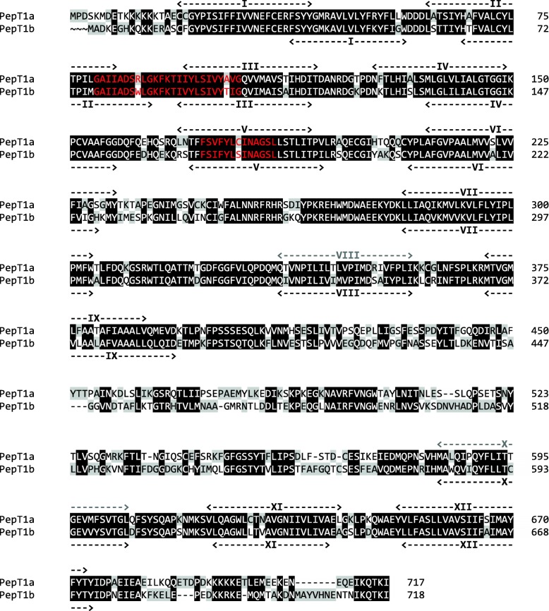
Zebrafish pept1a (slc15a1a) cDNA was 2478 bp long, with a coding sequence (CDS) of 2154 bp encoding a putative protein of 717 amino acids (Additional file 1: Figure S1). Zebrafish PepT1a (Slc15a1a) and PepT1b (Slc15a1b) amino acid sequences shared 78% similarity and 62% identity (Fig. 1). Hydropathy analysis predicted 12 potential transmembrane domains with a large extracellular loop between transmembrane domains IX and X (Fig. 1). Structural motifs such as the PTR2 family proton/oligopeptide symporter signatures could be found in zebrafish PepT1a (Slc15a1a) sequence (amino acid residues 80–104 for signature 1, PROSITE pattern: PS0102; amino acid residues 173–185 for signature 2, PROSITE pattern: PS01023) (Fig. 1). Three putative extracellular N-glycosylation sites, one intracellular consensus region containing protein kinase C motif, and three intracellular cAMP-dependent protein kinase sequences were also identified (Additional file 1: Figure S1).
Fig. 1.
Pairwise alignment between zebrafish PepT1a (Slc15a1a) and PepT1b (Slc15a1b) amino acid sequences obtained by using Clustal Omega and edited using GeneDoc 2.7 software. The predicted conserved PTR2 family proton/oligopeptide symporter signatures (in zebrafish PepT1a, motif 1—PROSITE pattern PS01022—amino acid residues 80–104; and motif 2—PROSITE pattern PS01023—amino acid residues 173–185) are colored in red. In the amino acid sequence, putative transmembrane domains are named I to XII. Weak predicted transmembrane domains (in zebrafish PepT1a, transmembrane domains VIII and X) are colored in gray
Basic function
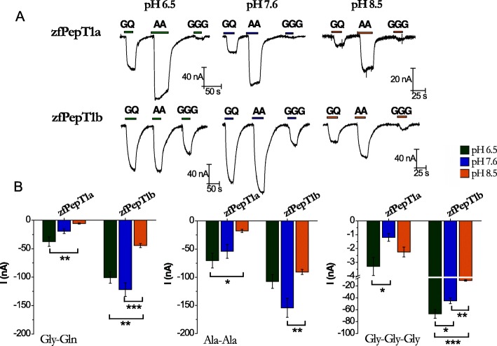
Figure 2 summarizes the first functional data about zebrafish PepT1a. Oocytes expressing the transporter were tested at the holding potential of − 60 mV in external control solution at pH 6.5, 7.6, or 8.5, and the substrate-induced currents (substrates: Gly-Gln, Ala-Ala, and Gly-Gly-Gly; concentration 1 mmol/L) were recorded. Representative traces for zebrafish PepT1a are in the upper part of Fig. 2a. The presence of inward currents of tens of nanoamperes amplitude clearly demonstrated that zebrafish PepT1a is electrogenic, like zebrafish PepT1b (see the lower part of Fig. 2a) and the other PepT1-type transporters so far characterized. As for the other PepT1-type transporters, the transport of zebrafish PepT1a was Na+-independent, regardless of the testing voltage (Additional file 1: Figure S2). In these experiments, the mean transport-associated currents of zebrafish PepT1a and PepT1b showed different profiles (Fig. 2b). In PepT1a, the amplitude of the currents (I) associated to the transport of the dipeptides decreased with the increase of pH from 6.5 to 8.5 (I6.5 > I7.6 > I8.5), with differences in the amplitude between the two tested pH extremes (P < 0.01 for Gly-Gln and P < 0.05 for Ala-Ala). PepT1b showed higher currents at pH 7.6 (I7.6 > I6.5 > I8.5), and the current amplitudes were different between pH 7.6 and pH 8.5 (P < 0.001 for Gly-Gln and P < 0.01 for Ala-Ala) and between pH 6.5 and pH 8.5 (P < 0.01 for Gly-Gln). Notably, although both transporters worked well with neutral dipeptides, PepT1a showed larger currents in the presence of Ala-Ala, while PepT1b exhibited similar current amplitudes for both substrates at the three pH tested. For the neutral tripeptide Gly-Gly-Gly, a current of ~ − 50 nA was recorded in PepT1b at pH 6.5 and pH 7.6 (with I6.5 > I7.6; P < 0.05), and the amplitude of current was reduced at pH 8.5 (P < 0.001 for pH 6.5 vs. pH 8.5, and P < 0.01 for pH 7.6 vs. pH 8.5). Conversely, in PepT1a, Gly-Gly-Gly elicited only very small currents regardless of the pH conditions (P < 0.05 for pH 6.5 vs. pH 7.6 only).
Fig. 2.
Transport activity and pH dependence of zebrafish PepT1a (Slc15a1a) and PepT1b (Slc15a1b). a Representative traces of transport currents in zebrafish PepT1a (zfPepT1a, top) and zebrafish PepT1b (zfPepT1b, bottom) heterologously expressed in Xenopus laevis oocytes. The currents in the presence of the substrates (1 mmol/L), indicated by bars, were recorded at the holding potential of − 60 mV and at pH 6.5 (left), 7.6 (middle), and pH 8.5 (right). b Transport-associated currents elicited by 1 mmol/L Gly-Gln (GQ) (left), Ala-Ala (AA) (middle), and Gly-Gly-Gly (GGG) (right) at − 60 mV at pH 6.5 (green), 7.6 (blue), and 8.5 (orange). Current values, shown in the histograms as the differences of the current recorded in the presence of the substrate and that in its absence, are reported as means ± SEM from 5 oocytes from 1 batch (one-way ANOVA test; *P < 0.05, **P < 0.01, and ***P < 0.001)
Kinetic parameters
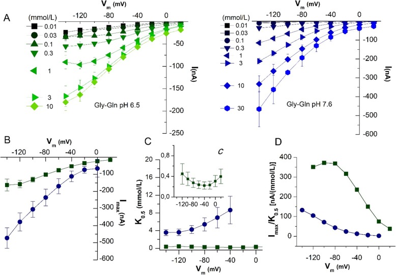
To characterize zebrafish PepT1a, the currents were recorded in the presence of increasing concentrations of Gly-Gln (from 0.01 to 10 mmol/L for pH 6.5, and to 30 mmol/L for pH 7.6) and in a range of voltage from − 140 to + 20 mV (Fig. 3a). The data of transport-associated current at pH 6.5 showed that Gly-Gln 3 mmol/L is the saturating value regardless of the voltage tested. Conversely, at pH 7.6, the transport-associated current constantly increased with the substrate concentration and it did not reach the maximal value even in the presence of Gly-Gln 30 mmol/L (Fig. 3a). The I/V relationships of PepT1a at the two pH values were used to calculate the kinetic parameters, i.e., the maximal transport current (Imax) and the apparent substrate affinity (i.e., the apparent concentration of peptide that yields one-half of Imax; K0.5). These (Fig. 3b, c) clearly showed the changes induced in substrate interaction by increasing the pH. For example, at pH 6.5, the Imax at − 140 mV was ~ − 160 nA (Imax = − 162.79 ± 35.35 nA), but it increased close to − 500 nA (Imax = − 473.10 ± 59.89 nA) at pH 7.6. As expected, the K0.5 values differed at the two pH tested, i.e., while at pH 6.5 PepT1a showed a high affinity that did not change with the voltage (K0.5 = 0.36 ± 0.24 mmol/L at − 140 mV and K0.5 = 0.22 ± 0.07 at − 40 mV), at pH 7.6 PepT1a affinity decreased and the K0.5 values became voltage-dependent passing from ~ 3.5 mmol/L (K0.5 = 3.55 ± 0.67 mmol/L) at − 140 mV to ~ 8.5 mmol/L (K0.5 = 8.63 ± 3.12 mmol/L) at − 40 mV. Accordingly, the transport efficiency, evaluated as Imax/K0.5 ratio, decreased with the increase of the pH. Also, if reported as a function of the voltage, Imax/K0.5 ratios showed a completely different pattern at the two pH values (Fig. 3d), i.e., a rather complete bell-shape at pH 6.5 with a maximum at ~ − 90 mV, and a left-shift of the curve at pH 7.6 suggesting that the maximal efficiency might be reached at potentials more negative than − 140 mV. Data about Imax, K0.5, and their ratio in the presence of Gly-Gln at pH 6.5 and 7.6 at the two reference membrane potentials of − 60 and − 120 mV for both PepT1a and PepT1b are summarized in Table 2.
Fig. 3.
Dose-response analysis. K0.5, Imax, and transport efficiency of zebrafish PepT1a (Slc15a1a) evaluated in the presence of Gly-Gln. a I/V relationships were obtained by subtracting the current traces in the absence to that in the presence of the indicated amounts of Gly-Gln, at pH 6.5 (green) and 7.6 (blue). The current values were fitted with the logistic equation to obtain K0.5, i.e., the substrate concentration that yields one-half of the maximal current (Imax), at each indicated voltage and at pH 6.5 (green square) and 7.6 (blue circle). b Imax at each voltage and pH. c K0.5 at each voltage and pH; the insert (c) is an enlargement of K0.5 at pH 6.5. d Transport efficiency, evaluated as the ratio of Imax/K0.5, and plotted vs. membrane potential for the two pH conditions. Imax, relative maximal current; K0.5, apparent substrate affinity; Imax/K0.5, transport efficiency
Table 2.
Kinetic parameters of the inwardly directed transport of Gly-Gln via the zebrafish PepT1a (Slc15a1a) and zebrafish PepT1b (Slc15a1b) measured in two-electrode voltage clamp experiments
| − 60 mV | − 120 mV | |||||||
|---|---|---|---|---|---|---|---|---|
| pH | Neutral form (%) | K0.5 (mmol/L) | Imax (nA) | Imax/K0.5 (nA/mmol/L) | K0.5 (mmol/L) | Imax (nA) | Imax/K0.5 (nA/mmol/L) | Oocytes/batches (n/N) |
| PepT1a | ||||||||
| 6.5 | 98.4 | 0.24 ± 0.07 | − 75.76 ± 6.04 | 316.37 | 0.45 ± 0.19 | − 157.39 ± 21.48 | 350.81 | 9/3 |
| 7.6 | 83.0 | 6.92 ± 2.34 | − 169.57 ± 43.75 | 24.51 | 3.61 ± 0.73 | − 378.82 ± 53.08 | 105.02 | 14/3 |
| PepT1b | ||||||||
| 6.5 | 98.4 | 0.13 ± 0.02 | − 195.73 ± 8.89 | 1535.32 | 0.13 ± 0.02 | − 396.24 ± 22.21 | 3032.16 | 7/1 |
| 7.6 | 83.0 | 2.22 ± 1.04 | 566.16 ± 212.44 | 254.54 | 1.01 ± 0.35 | 1142.30 ± 285.34 | 1129.67 | 7/1 |
Kinetic parameters (K0.5, Imax, and Imax/K0.5) were calculated on Xenopus laevis oocytes voltage clamped at − 60 mV and at − 120 mV and perfused with Gly-Gln in sodium chloride buffer solutions at pH 6.5 and 7.6. Values are expressed as means ± SEM of n oocytes (each oocyte represents an independent observation). Kinetic parameters were calculated by least-square fit to the logistic equation (Fig. 2). Imax/K0.5, transport efficiency
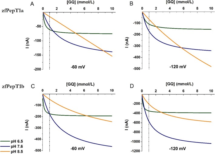
Considering the effect of the pH on the kinetic parameters and the results on the transport currents summarized in Fig. 2, the dose-response curves of Gly-Gln generated by oocytes expressing zebrafish PepT1a or PepT1b at pH 6.5, 7.6, and 8.5 were compared, and the transport-associated currents elicited by increasing concentrations of substrate from 0.01 to 10 mmol/L were plotted as current/concentration relationships and fitted with a logistic equation at − 60 and − 120 mV (see Fig. 4a, b for PepT1a and Fig. 4c, d for PepT1b). The data show that the acidic pH similarly affected the function of PepT1a and PepT1b. Due to the high affinity for Gly-Gln of both transporters at pH 6.5, the transport-associated currents reached the Imax value (see Fig. 3c for PepT1a and [14] for PepT1b) in the presence of Gly-Gln concentration lower than 10 mmol/L at the two potentials tested. At pH 7.6, the current generated by the concentration of 1 mmol/L (see Fig. 4, dashed line) is far from the maximal current in both transporters, regardless of the voltage considered. At this pH, the proteins bind the substrate with lower affinity and the current increases with increasing the concentration without showing saturation in the range of concentrations tested. At pH 8.5, the fitted curves suggest a further increase of K0.5 values, particularly for PepT1a. Notably, the transport currents elicited by concentrations of Gly-Gln up to 1 mmol/L differ of few nanoamperes only between pH 6.5 and 7.6 for both transporters at the membrane potential of − 60 mV. When the voltage is set at − 120 mV, the currents are similar at the two pH with substrate concentration up to 0.3 mmol/L, and when the substrate is 1 mmol/L at pH 7.6, the currents are larger than those at pH 6.5 for both transporters. As already reported for PepT1b [14], the curves at pH 8.5 for PepT1a suggest that the proton decrease in the external solution markedly affects the kinetic parameters, emphasizing the effects already seen at pH 7.6.
Fig. 4.
Fitting of the Gly-Gln (GQ) transport-associated currents as a function of substrate concentration (from 0.01 to 10 mmol/L) at different pH (pH 6.5 in green, pH 7.6 in blue, and pH 8.5 in orange) for two different membrane potentials: − 60 mV (left) and − 120 mV (right). a, b Zebrafish PepT1a (zfPepT1a). c, d Zebrafish PepT1b (zfPepT1b). The dashed line indicates 1 mmol/L Gly-Gln concentration
Pre-steady-state currents
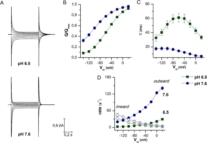
In Fig. 5, the characteristics of the transport cycle were analyzed by investigating the behavior of the transient currents at pH 6.5 and 7.6. Pre-steady-state currents were isolated as reported in [25, 32]. From these, it is possible to calculate the total amount of charge moved in the membrane electric field, the time of relaxation decay, and the rate of outward and inward constants [28]. The representative recordings reported in Fig. 5a clearly underline the accelerating effect of reducing the number of extracellular protons. PepT1a shows the complete sigmoidal curve for the normalized Q/V relationship at pH 6.5 (Fig. 5b), a consistent reduction of the decay time constant (τ), and a complete bell-shaped curve for τ/V relationship (Fig. 5c). These curves are left-shifted by increasing the pH at 7.6. Consequently, the τ maximal value and the V0.5 move to more negative voltage. These data are very similar to those recorded in rabbit PepT1, and different to those of the zebrafish PepT1b in which τ/V and Q/V curves are left-shifted even at pH 6.5 and when the pH increases only a slight shift toward negative potentials is predicted by fitting with the Boltzmann equation. The parameters for zebrafish PepT1a, zebrafish PepT1b, and rabbit PepT1 are summarized in Table 3.
Fig. 5.
Biophysical parameters of PepT1a. a Representative trace of current elicited by voltage pulses in the range − 140 to + 20 mV (20 mV steps from Vh = − 60 mV) in the absence of substrate at pH 6.5 and pH 7.6 as indicated. b–d Analysis of pre-steady-state currents at pH 6.5 (green square) and 7.6 (blue circle) obtained from the slow component of a double exponential fitting of the corresponding traces in the absence of the substrate. b Charge/voltage (Q/V) curves obtained by integration of the pre-steady-state isolated at the two pH values. c Time constant/voltage (τ/V) relation; the values were estimated from the on transients, except at − 60 mV (Vh), which was estimated from the off transients. d Unidirectional rate constants, inward (open symbols) and outward (solid symbols), of the intramembrane charge movement in function of different tested voltage conditions, derived from the τ/V relationship and the Q/V relationship at two pH conditions. Data are mean ± SEM from 10 oocytes of 3 different batches. Vh, holding potential
Table 3.
Boltzmann equation parameters of zebrafish PepT1a (Slc15a1a), compared to zebrafish PepT1b (Slc15a1b) and rabbit PepT1 (Slc15a1)
| Zebrafish PepT1a | Zebrafish PepT1b | Rabbit PepT1 | ||||
|---|---|---|---|---|---|---|
| pH | 6.5 | 7.6 | 6.5 | 7.5 | 6.5 | 7.5 |
| Qmax (nC) | 41 ± 0.7 | 53 ± 0.6 | 11 ± 0.3 | 9.9 ± 1.1 | 33.2 ± 1.9 | 31.5 ± 1.2 |
| V0.5 (mV) | − 57.6 ± 0.6 | − 110 ± 0.7 | − 108 ± 1.6 | − 119 ± 7.2 | − 41.4 ± 2.5 | − 100 ± 2.3 |
| σ (mV) | 33.6 ± 0.8 | 39.3 ± 0.4 | 31.1 ± 0.9 | 33.5 ± 3.3 | 42.9 ± 3.1 | 39.5 ± 1.7 |
Boltzmann equation parameters were calculated at two pH conditions: 6.5 and 7.6 for zebrafish PepT1a (data from Fig. 5) and 6.5 and 7.5 for zebrafish PepT1b and rabbit PepT1 (data from [28]). Qmax, the maximal moveable charge; V0.5, the voltage at which half of the charge is moved; σ, slope factor of sigmoidal curve
The Q/V curve represents the steady-state distribution of the transporter proteins between two conformations with the charge (intrinsic or extrinsic) in two opposite locations of the membrane electrical field. The reaction describes the movement of the charge between the two positions. The outward and inward rates are the unidirectional rate constants, and Qout and Qin are the amount of charge respectively at the outer and inner position of the membrane electric field. Determining these rates according to [28] allows to better appreciate the effect of the pH (Fig. 5d). These data clearly show that alkalization accelerates the outward rate constant, i.e., when the pH increases to pH 7.6, the transporter completes the cycle faster, reducing the time needed for substrate translocation [33, 34].
Another piece of information coming from pre-steady-state currents records is the amount of maximal charge moved in the membrane electric field consequent to voltage steps (Qmax). Zebrafish PepT1a, in the range of potentials tested, has the highest values among the tested transporters and, differently from the other PepT1 proteins, its parameters are affected by external pH (Table 3). Moreover, the slope of the Q/V curves (σ) suggests that charge movement in zebrafish PepT1a might occur over a smaller fraction of the electrical membrane field than in zebrafish PepT1b. For both transporters, when the external pH is set at 7.6, the fraction of electrical membrane field is reduced, and this effect is more evident in PepT1a.
Tissue distribution of zebrafish pept1a (slc15a1a) in adult fish
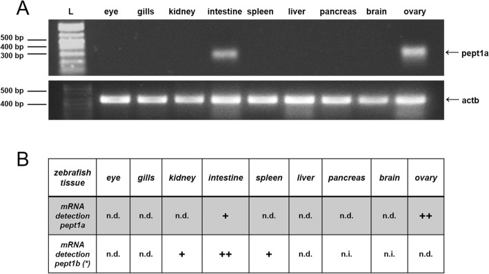
Using zebrafish pept1a-specific primers, a 350-bp RT-PCR product was amplified from total RNA isolated from the intestine of adult zebrafish, as well as from the ovary, while no signal was detected in the eye, gills, kidney, spleen, pancreas, and brain (Fig. 6a). As internal control to assess RNA quality, detection of β-actin (actb) mRNA was performed using zebrafish actb-specific primers, showing comparable 442-bp amplification products for all tested tissues (Fig. 6a). When investigated in the separated intestinal (rostral) bulb, mid and posterior intestine, pept1a (slc15a1a) mRNA amplification was obtained in each of the three consecutive tracts (Additional file 1: Figure S3). Notably, pept1a (slc15a1a) and pept1b (slc15a1b) were found to share the same “intestinal” localization (Fig. 6b).
Fig. 6.
Expression analysis by RT-PCR on pept1a (slc15a1a) mRNA in adult zebrafish tissues. a RT-PCR assay on cDNA templates from total RNA extracted from various tissues; a PCR product of ~ 350 bp related to pept1a (slc15a1a) mRNA is present in samples from the intestine and ovary, while it is absent in the eye, gills, kidney, spleen, liver, pancreas, and brain; using the same cDNA templates, a PCR product of ~ 440 bp related to the actb mRNA is present in all tissue samples; L: 1 Kb Plus DNA ladder (Thermo Fisher Scientific). b Comparative table of pept1a (slc15a1a) vs. pept1b (slc15a1b) mRNA presence in the different zebrafish tissues analyzed. pept1b (slc15a1b) tissue expression data are from [14]. +, positive detection; n.d., not detected; n.i., not investigated
Expression of zebrafish pept1a (slc15a1a) during larval development
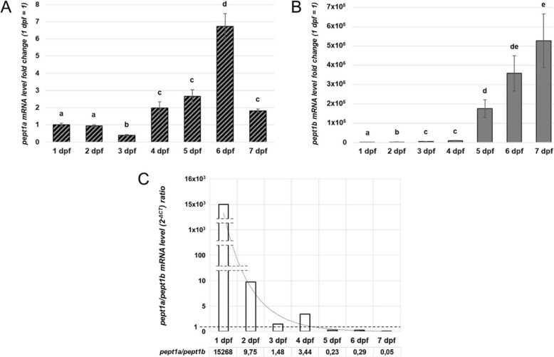
Zebrafish pept1a (slc15a1a) mRNA expression profile was quantitatively evaluated by qPCR during embryonic/early larval developmental stages. A polyphasic trend of mRNA expression was detected over time, passing from 1 to 7 days post-fertilization (dpf) (Fig. 7a). In particular, a negative fold-change (− 0.61) of pept1a (slc15a1a) mRNA levels was registered at 3 dpf with respect to 1 dpf (fold-change = 1), while a time-dependent increase of the signal was observed at the next stages, i.e., 4, 5, and 6 dpf (+ 2.00, + 2.66, and + 6.73 fold-change, respectively). At 7 dpf, pept1a (slc15a1a) mRNA levels were significantly reduced with respect to 6 dpf, but still slightly higher than at 1 dpf (+ 1.81). The analysis of pept1b (slc15a1b) mRNA levels in the same developmental stages revealed a time-dependent trend with a strong increase from 1 up to 7 dpf (Fig. 7b). In detail, a very faint signal for zebrafish pept1b-specific mRNA was detected at 1 dpf (fold-change = 1), which increased ~ + 1.5 × 10³-fold already at 2 dpf, reaching ~ + 8.8 × 10³- at 4 dpf and + 5.3 × 10⁵-fold increase at 7 dpf. A relative comparison of the expression level data (2-ΔCT values) was inferred by calculating the pept1a-to-pept1b ratio at each developmental stage. The ratio between the expression levels was > 1 from 1 to 4 dpf and < 1 from 5 to 7 dpf (Fig. 7c).
Fig. 7.
Quantitative expression analysis of zebrafish pept1a (slc15a1a) and pept1b (slc15a1b) mRNAs during early development. a mRNA expression analysis by qPCR in zebrafish embryos/larvae from 1 to 7 days post-fertilization (dpf). The levels of pept1a (slc15a1a) mRNA were calculated as 2-ΔCT mean values obtained from two rounds of qPCR assays for each of three independent biological replicates (pools of 10–15 embryos/larvae), and then they were expressed as fold-change (y-axis) with respect to the 1 dpf stage taken as control value (1 dpf = 1). b mRNA expression analysis by qPCR of the pept1b (slc15a1b) gene in zebrafish embryos and larvae from 1 to 7 dpf. Statistical analysis of variance of the means was assessed by one-way ANOVA and Tukey's post hoc test. In histograms, different letters indicate statistically different values (n = 3 independent biological replicates; P < 0.05). c Representation of the trend of the pept1a/pept1b mRNA level ratio at 1 to 7 dpf, based on the 2-ΔCT mean values obtained from the output data deriving from qPCR assays performed, with the same primer efficiency values, for both the pept1a- and pept1b-specific primer pairs
Discussion
Like many other animal models, zebrafish is a highly tractable organism and despite being a vertebrate it offers an extremely high potential for genetic analysis and cellular observation. Also, it is readily available and easy to breed, and its transparent embryo develops quickly. The zebrafish “model system” now comprises a sequenced genome, thousands of mutants, transgenic tools, staging series, know-how for imaging, embryological manipulation, drug discovery, and a lot more (among many other papers see, e.g., [24, 35]; among many other excellent reviews see, e.g., [36–38]). However, to join the rank of top model organism for biomedical research, the suite of tools and resources already available needs to be implemented with robust phenotyping and functional analyses at the same performance levels of investigation and with the same advanced experimental approaches and methods as those considered standard for mammalian models (e.g., rodents) and humans. Searching in detail for (a) species-specific molecular phenotype(s) offers the possibility to define how many orthologous/paralogous proteins from same/different species operate similarly or differently, from one another, thus opening to the comprehension of the multiplicity of structural-functional solutions at the molecular level in the various animal bio-systems analyzed. This is particularly true for epithelial physiology, and in transport and transporters functional analyses, in which the complexity of the substrate specificities largely meets the complexity of the model species. Thus, in the context of this discussion, which evaluates the zebrafish as a suitable model organism in nutrigenomics and nutrition research, emphasis should be given to the concept that a very careful evaluation of the impact of a study on (a) membrane transporter(s) in a non-human/non-rodent model system is always required (see, e.g., [39]).
In this “case” study, we report the functional characterization of the zebrafish PepT1a-type transporter, which completes the functional picture of the triad of PepT-type transporters, i.e., PepT1a (Slc15a1a), PepT1b (Slc15a1b), and PepT2 (Slc15a2), expressed by the zebrafish (for details, see Table 1). This study confirms and extends results recently obtained in the Atlantic salmon [31] and leads to the general assumption that PepT1a-type transporters physiologically operate in teleost fish. In particular, zebrafish pept1a (slc15a1a), which is expressed in the intestinal tract and in the ovary of adult fish, generates a protein product, i.e., PepT1a (Slc15a1a), that is able to mediate the transport of neutral di/tripeptides, such as Gly-Gln, Ala-Ala, and Gly-Gly-Gly. However, PepT1a (Slc15a1a) differs from the already well-characterized zebrafish PepT1b (Slc15a1b) in terms of transport kinetics, substrate specificity, and transport efficiency. Notably, pept1a (slc15a1a) mRNA expression profile during the first 7 dpf also differs from the pept1b (slc15a1b) [14], highlighting a possible role of pept1a (slc15a1a) mRNA in the first 3 days of embryonic development and as a component of the maternal mRNA pool. Whether or not pept1a (slc15a1a) plays a physiological role in extra-embryonic tissue(s), such as the yolk syncytial layer, remains a relevant, yet unanswered, question.
Function
The basic transport currents recorded (and reported in Fig. 2 and Additional file 1: Figure S2) confirm that zebrafish PepT1a (Slc15a1a), like the recently characterized Atlantic salmon PepT1a (Slc15a1a) [31], is electrogenic and capable of transporting di/tripeptides in a H+-dependent manner and independently of the presence of sodium ions. However, while eliciting transport currents, the 1 mmol/L substrate condition is not the most adequate to test the pH dependence of the transport because of the considerable effects of pH on substrate affinity. Nevertheless, the representative traces in Fig. 2a and the analysis of the transport-associated currents in Fig. 2b clearly indicate that the differences in amino acid sequences between the two transporters may have functional implication(s) in both pH dependence and substrate preferences. In fact, if compared to the well-characterized zebrafish PepT1b (Slc15a1b), PepT1a (Slc15a1a) (i) prefers Ala-Ala to Gly-Gln, (ii) works well at acidic pH, and (iii) gives rise to relatively smaller currents in all conditions tested. Moreover, Gly-Gly-Gly appears to be a poor substrate for this transporter. When the transport-associated currents are recorded in the presence of increasing concentrations of Gly-Gln by using a standard step protocol, PepT1a (Slc15a1a) shows some common features with PepT1b (Slc15a1b), but also its own. In particular, the pH has a large effect on the affinity of the PepT1a transporter, which is evident by observing the behavior of the I/V curves reported in Fig. 3 and comparing the K0.5 and Imax values of the PepT1a and PepT1b proteins (Table 2). At pH 6.5, both proteins work similarly, as it results when considering the data reported for − 120 and − 60 mV in Fig. 4 and Table 2, but when the pH increases to 7.6, the two proteins work differently, and PepT1a affinity for Gly-Gln is largely influenced by pH. For instance, when the external pH is set at 7.6, the amount of substrate to reach one-half of the maximal currents (K0.5) increases in PepT1a [e.g., at − 60 mV, K0.5 is ~ 6.92 mmol/L at pH 7.6 and ~ 0.24 mmol/L at pH 6.5 (ratio 28.83)] more than it occurs in PepT1b [e.g., at − 60 mV, K0.5 ~ 2.22 mmol/L at pH 7.6 and ~ 0.13 mmol/L at pH 6.5 (ratio 17.07)]. This suggests that PepT1a has very strong pH dependence, which is confirmed by data reported in Fig. 4 where the fitting dose-response of the two transporters is compared.
The pre-steady-state currents are elicited by voltage steps and are due to the charges moved inside the membrane electric field. In many solute carriers, they are recordable and related to the first steps of the transport cycle. These currents are due to one or more intrinsic (internal, that is one or more protein residues, i.e., charged amino acids) or extrinsic (external, that is ion or proton) charges, present or entered in the membrane electric field that are moved by voltage changes. As suggested by the analysis of the pre-steady-state currents, the main effects of protons are on the turnover rate of the transporter (1/τ), i.e., the two proteins cycle differently and are differently affected by the pH. In response to voltage steps, the amount of charges moved by PepT1a is higher, while the time decay of pre-steady-state currents and the unidirectional rate constants are lower (slower) if compared to PepT1b [28]. At pH 6.5, the τ/V relationship for PepT1a shows a complete bell-shape with the slower value at − 60 mV, like the rabbit PepT1 reported in [28]. This curve is reduced and left-shifted at pH 7.6. In PepT1b, the curves are left-shifted and faster even at pH 6.5 and changing the pH decreases τ values and only slightly moves the curve to a more negative potential. In this transporter, the slower value for τ is recorded at pH 6.5 at − 140 mV. For PepT1a, the Q/V relationship is a complete sigmoidal curve in the range of voltage tested, confirming the symmetrical behavior of the transient current at this pH for PepT1a. As for other PepT1 transporters, also for zebrafish PepT1a with the increase of the pH both Q/V and τ/V curves shift to more negative voltage, increasing the transport rate (1/τ). The effect of protons is evident on the unidirectional rate constants, with a main effect on the outward rate, which greatly increases its value by changing the pH (from ~ 30 s−1 at pH 6.5 to ~ 140 s−1 at pH 7.6, at + 20 mV).
Summarizing the data from the kinetic parameters and the pre-steady-state currents, we suggest that zebrafish PepT1a and PepT1b residues involved in protons and substrates binding/interaction are differently located in the membrane electric field and that the two transport proteins are differently affected by changes in external proton concentration. Consequently, the pH might alter in a different way the steps of transport cycle differently influencing the transport rate of the two proteins. Interestingly, PepT1a shows a “mammalian” behavior [28, 34]. In addition, the data collected on the biophysical parameters make it more similar to rabbit PepT1 than to the other fish transporters. To date, we have not been able to identify any obvious amino acid residues along the primary sequences and/or in the three-dimensional structures deposited in databanks that can possibly be associated to protons and/or substrates binding/interaction. However, a systematic analysis, mainly based on site-directed mutagenesis of amino acid residues selected by means of sequence and structure coevolution computational criteria (see, e.g., [40]) and subsequent electrophysiological analysis in oocytes, will answer this question.
Expression
Similar to recent findings in Atlantic salmon [31], the analysis of the expression profile clearly shows that zebrafish pept1a (slc15a1a) mRNA is present in the intestine of the adult fish. In particular, zebrafish pept1a (slc15a1a) mRNAs are detected in the intestinal bulb, mid and posterior intestinal segments (see Additional file 1: Figure S3). In this respect, zebrafish pept1a (slc15a1a) partially overlaps zebrafish pept1b (slc15a1b) since the latter is very strongly expressed in the proximal intestine of this teleost fish (see, e.g., [12–14]). However, unlike the Atlantic salmon [26, 31], further and ad hoc studies are still needed to precisely define the relative mRNA amounts of pept1a (slc15a1a) alone and vs. pept1b (slc15a1b) in the various segments of the alimentary canal of the adult zebrafish.
During early development (1–7 dpf), pept1a (slc15a1a) mRNA expression seems to undergo a multi-phasic trend over time. The mRNA levels already present (in the ovary of the adult fish and thus in the unfertilized eggs and) at 1 and 2 dpf, possibly due to the occurrence of pept1a (slc15a1a) mRNA in the maternal RNA pool, significantly decrease at 3 dpf. Interestingly, recovery and significant increase of mRNA levels are measured from 4 to 6 dpf, which is in line with pept1a (slc15a1a) baseline expression as extrapolated from a recent transcriptional profiling (high-resolution mRNA expression time course) of zebrafish embryonic developmental stages [35]. It is worth noting that this phenomenon parallels maturation of the gut and achievement of the full digestive/absorptive function (see, e.g., [41, 42]), during which pept1a (slc15a1a) mRNA new-synthesis seems to occur. Remarkably, a third phase of expression is hinted by the reduced levels of pept1a (slc15a1a) at 7 dpf with respect to 6 dpf, which suggests a time frame-specific functional expression of pept1a (slc15a1a) that needs to be further addressed. This pept1a (slc15a1a) expression trend during development appears even more interesting when compared to the qPCR expression data for the pept1b (slc15a1b) mRNA levels which, as expected (see, e.g., [12–14]), are found to increase day by day strongly and progressively, starting from the very faint signal at 1 dpf and then increasing to more than 5 × 10⁵-fold at 7 dpf. Assuming that the “quantitative” comparative evaluation of the pept1a (slc15a1a) vs. pept1b (slc15a1b) expression levels goes beyond the aim of this paper and the analyses in question, we cannot but noticing that the “raw” calculation (based on 2-ΔCT values) of the pept1a (slc15a1a)-to-pept1b (slc15a1b) expression ratio seems to indicate that pept1a (slc15a1a) expression prevails on pept1b (slc15a1b) during the 1-to-4 dpf period, while the expression level ratio becomes lower than 1 from 5 dpf on, hinting that pept1a (slc15a1a) may be the predominant pept1-type mRNA species at the immediacy over time. Whether or not this expression trend is general or zebrafish-specific remains an open question. In fact, at least to our knowledge, the only study available in the literature that specifically compares pept1a (scl15a1a) and pept1b (slc15a1b) in pre-feeding stages larvae refers to the Mozambique tilapia (Oreochromis mossambicus) and is limited to the intestinal organ only. In this case, pept1a (slc15a1a) and pept1b (slc15a1b) temporal trend of expression in the intestine goes parallel from 3 to 14 dpf (for details, see Table 4, and literature therein [49]).
Table 4.
Organ/tissue distribution of pept1a (slc15a1a) and pept1b (slc15a1b) mRNA in teleost fish species for which the expression of the two genes has contemporarily been studied. Whenever co-analyzed pept2 (slc15a2) mRNA expression has also been considered
| Species [order] | Developmental stage | Description | GenBank Acc. No. | Organ/tissue distribution (observed in the study) | Distribution along the post-gastric alimentary canal (observed in the study) | Notes | References |
|---|---|---|---|---|---|---|---|
| Mummichog (Fundulus heteroclitus) [Cyprinodontiformes] | Adults (~ 9 g) | pept1a (slc15a1a) | JN615008.1 | Intestine | Anterior intestine ≈ posterior intestine | Environmental (freshwater acclimation vs. seawater acclimation) and nutritional (fasting vs. re-feeding) regulation of pept1a (slc15a1a) and pept1b (slc15a1b) | [43] |
| pept1b (slc15a1b) | JN615007.1 | Anterior intestine ≈ posterior intestine | |||||
| Nile Tilapia (Oreochromis niloticus) [Cichliformes] | Juveniles (~ 12 g) | pept1a (slc15a1a) | XM_005452882 | Intestine >>> stomach > brain > gill > liver | Proximal intestine >> mid intestine >>> distal intestine | Environmental (waterborne copper exposure) and/or nutritional (fasting vs. re-feeding) regulation of pept1a (slc15a1a), pept1b (slc15a1b) and pept2 (slc15a2) | [44] |
| pept1b (slc15a1b) | XM_005465251 | Intestine >>> brain ≈ stomach | Mid intestine > proximal intestine >>> distal intestine | ||||
| pept2 (slc15a2) | XM_005475385 | Intestine >> stomach > kidney > liver ≥ gill ≈ brain > spleen > muscle | Mid intestine >>> distal intestine > proximal intestine | ||||
| Adults (~ 62 g) | pept1a (slc15a1a) | XM_013267250.1 | Intestine | Anterior intestine > middle intestine >>>> posterior intestine | Environmental (high-salinity acclimation) regulation of pept1b (slc15a1b) | [45] | |
| pept1b (slc15a1b) | XM_005452882.2 | Anterior intestine ≈ middle intestine >>>> posterior intestine | |||||
| pept2 (slc15a2) | XM_005475385 | Posterior intestine > middle intestine >> anterior intestine | |||||
| Adults (~ 125 g) | pept1a (slc15a1a) | XM_003459630 | Intestine | Anterior intestine > middle intestine >>> posterior intestine | Nutritional (dietary salt supplementation) regulation of pept1a (slc15a1a), pept1b (slc15a1b) and pept2 (slc15a2) | [46] | |
| pept1b (slc15a1b) | XM_003447363 | Middle intestine > anterior intestine >>> posterior intestine | |||||
| pept2 (slc15a2) | XM_003454878 | Posterior intestine ≥ middle intestine >> anterior intestine | |||||
| Mozambique tilapia (Oreochromis mossambicus) [Cichliformes] | Adults (~ 97 g) | pept1a (slc15a1a) | XM_003459630 | Intestine | Anterior and middle intestine | Environmental (salinity-dependent) nutritional regulation of pept1a (slc15a1a), pept1b (slc15a1b) and pept2 (slc15a2) | [47] |
| pept1b (slc15a1b) | XM_003447363 | Anterior and middle intestine | |||||
| pept2 (slc15a2) | XM_003454878 | Middle and posterior intestine | |||||
| Adults (100–250 g) | pept1a (slc15a1a) | LC197343 | Intestine | Hepatic loop > proximal major coil >>> gastric loop ≈ distal major coil ≈ terminal segment | Nutritional (fasting vs. re-feeding) regulation of pept1a (slc15a1a) | [48] | |
| Adults (~ 24 g) | pept1a (slc15a1a) | XM_013267250.1 | Intestine | Anterior intestine > middle intestine >>>> posterior intestine | Environmental (high-salinity acclimation) regulation of pept1a (slc15a1a) and pept2 (slc15a2) | [45] | |
| pept1b (slc15a1b) | XM_005452882.2 | Anterior intestine ≈ middle intestine >>>> posterior intestine | |||||
| pept2 (slc15a2) | XM_005475385 | Posterior intestine > middle intestine >> anterior intestine | |||||
| Adults (~ 54 g) | pept1a (slc15a1a) | KX034112.1 | Intestine >>>> pituitary ≈ skin ≈ muscle ≈ kidney ≥ heart ≈ brain ≈ gills ≥ liver ≈ fat ≥ stomach ≈ esophagus ≈ spleen | Anterior intestine >>> middle intestine >> posterior intestine | – | [49] | |
| pept1b (slc15a1b) | KX034110.1 | Intestine >>>> brain > pituitary > muscle > skin ≈ gills ≈ heart ≈ liver ≈ fat ≈ spleen ≈ kidney ≈ esophagus ≈ stomach | Anterior intestine >>>> middle intestine > posterior intestine | ||||
| pept2 (slc15a2) | KX034111.1 | Intestine ≥ kidney >> muscle ≥ liver > brain ≈ pituitary ≈ skin ≈ stomach > heart ≈ spleen ≈ heart > gills | Middle intestine >>>> posterior intestine >> anterior intestine | ||||
| Pre-feeding larvae (3–14 dpf) | pept1a (slc15a1a) | KX034112.1 | Intestine | Whole intestine | Temporal (time course from the pre-hatching to completion of yolk sac resorption stage) regulation of pept1a (slc15a1a), pept1b (slc15a1b) and pept2 (slc15a2) | ||
| pept1b (slc15a1b) | KX034110.1 | ||||||
| pept2 (slc15a2) | KX034111.1 | ||||||
| European seabass (Dicentrarchus labrax) [Perciformes] | Juveniles (~ 1.2 g) | pept1a (slc15a1a) | – | Intestine | Whole intestine | Environmental (short- and long-term low-salinity acclimation) regulation of pept1a (slc15a1a), pept1b (slc15a1b) and pept2 (slc15a2) | [50] |
| pept1b (slc15a1b) | – | ||||||
| pept2 (slc15a2) | – |
Assayed by quantitative real-time PCR, dpf days post-fertilization
Understanding the functional importance, and thus the physiological implications, of having two similar transporters that work in the same biological district(s) with dissimilar kinetics and possibly dissimilar expression levels is a major topic of peptide transport research in teleost fish, with promising implications in higher vertebrate and human physiology. In the case of our PepT1-type transporters, the expression data bring our attention to both the intestine and the yolk syncytial layer.
The presence of PepT1a and PepT1b in the intestine has been related to the possible variability of the natural environment where the fish live, to the nutritional input and to the peculiarities of the digestive system of the various fish species and to a variety of other challenges (reviewed in [12, 13]). In particular, in a large number of teleost fish species, zebrafish and other cyprinids included, the spatio-temporal expression of PepT1b intestinal mRNA largely varies during ontogeny, in response to nutritional states (e.g., food deprivation/re-feeding), dietary challenges, and/or environmental conditions (e.g., in freshwater/seawater adaptation), as well as under certain disease states (e.g., gut inflammation) [14–22, 26, 30, 51–63]. But, in the light of the most recent findings, the new view that PepT1a and PepT1b may both be expressed and operate in teleost fish models and similarly or differently respond to the various internal and external solicitations should always be taken into account (for details, see Table 4, and literature cited therein [43–48, 50]).
Moreover, the findings during zebrafish early development open to novel interesting scenarios in applied nutrition and nutrigenomics. In fact, in a perspective, the expression of specific genes involved in nutrients utilization, such as pept1a (slc15a1a), if located at the level of the yolk sac structures might become functional to the systematic comprehension of the uptake processes of all the nutritional resources in it contained. In particular, the presence of selected transporters that operate with their kinetic properties on selected and rather homogenously represented yolk protein degradation products (e.g., those from vitellogenin, phosvitin, and lipovitellin; see, e.g., [64, 65]) might be highly informative to fully understand the rules and the dynamics of the proteolysis process(es) as a whole. In addition, it could help to address specifically the fate of a variety of highly relevant nutritional, immunological, and/or differently bioactive peptides there generated.
Conclusions
Molecular cloning and functional expression in a heterologous system has allowed the characterization of a second PepT1-type transporter, after PepT1b alias PepT1, in the zebrafish. The zebrafish represents the second PepT1a-type transporter, after the Atlantic salmon, for which thorough functional characterization by two-electrode voltage clamp (TEVC) has been achieved. Therefore, the concept that PepT1a does functionally act in teleost fish model systems can be fully asserted. In this context, re-evaluation of the di/tripeptide absorptive model along the alimentary canal of teleost fish (for review see, e.g., [12–14]) should be considered in the light of the fact that two PepT1-type transporters—and not one like in higher vertebrates such as mammals and birds—operate at the intestinal level. Whether or not PepT1a and PepT1b transporters share physiological roles, cellular localization in the intestinal epithelium, sub-cellular localization in the intestinal epithelial cells, type of regulation, etc. are questions to be addressed, but in this respect, the zebrafish model and its toolbox represent the most suitable teleost fish experimental system to answer these questions. All together, the molecular and functional data obtained for zebrafish (and Atlantic salmon) PepT1a, together with the molecular and functional data already available and extended from zebrafish (and Atlantic salmon) PepT1b, allow combinatorial analysis of kinetics properties vs. primary amino acid sequences, which might help in identifying specific amino acids along the primary sequences relevant for substrate specificity, pH dependence, transport efficiency, turnover rate, etc. Comparison studies with higher vertebrate orthologs, such as human PEPT1 (SLC15A1) and murine PepT1 (Slc15a1), might be translational to human physiology and pharmacology, and in the context of this discussion in nutrigenomics, dietetics, and nutrition research, and in developing new model(s) of substrate-transporter interaction(s), pharmacophore(s), etc. In this respect, this set of PepT1a- plus PepT1b-type transporters from teleost fish may represent an original tool to support structure-function studies at the molecular level. Moreover, there are several pieces of evidence suggesting that PepT1-type proteins operate in the membrane in oligomeric (tetrameric) state (for review, see, e.g., [13]). Were they co-expressed in the same cell type, PepT1a- and PepT1b-type transporters could form hetero-tetramers and possibly interact cooperatively for optimal di/tripeptide transport function. Another functional consideration regards the question of whether or not teleost fish PepT1-type transporters are linked to any Na+/H+ exchanger(s) at the apical membrane of the enterocyte, like it occurs in the mammalian systems where the antiporter plays a major role in building the inwardly directed H+-gradient that supports H+-dependent peptide uptake (see, e.g., [66, 67]). The comparison between agastric, such as the zebrafish, and gastric, such as the Atlantic salmon, teleost fish models might help going more systematically into the details of such a physiological question departing from the singularity of the zebrafish model (see, e.g., [12–14]). Last but not least, due to its expression during the early embryonic development, it has to be fully considered the hypothesis that zebrafish pept1a (slc15a1a) (like other solute carriers involved in sugar, lipid, amino acid, anion, and metal ion uptake [68–74]) is part of the maternal machinery that supports early developmental stages and/or it is expressed in an extra-embryonic tissue such as the yolk syncytial layer. If so, it could operate in specifically mediating the uptake of the di/tripeptides that derive from the yolk protein degradation processes, thus strategically contributing to provide the bulk of protein nitrogen for early embryo development and growth. If so, the kinetic properties of pept1a (slc15a1a) would well support yolk protein uptake in the embryonic and larval zebrafish, making pept1a (slc15a1a) a specific marker of the yolk protein degradation process.
Methods
Animals
Zebrafish (wild-type AB) were maintained and bred at the High Technology Centre (HIB), Department of Biological Sciences, University of Bergen, according to standard protocols as described elsewhere [75]. Zebrafish embryos were obtained from natural mating. The developing embryos/larvae were incubated at 28.5 °C until use. Developmental stages of zebrafish embryos/larvae were expressed as dpf at 28.5 °C [76].
Adult fish were anesthetized by immersion in 0.2 g/l MS-222 and then killed by decapitation prior to organ removal and dissection. Developing embryos/larvae were euthanized by anesthetic overdose before sampling into RNALater (Qiagen, Hilden, Germany).
Molecular cloning
Zebrafish pept1a (slc15a1a) gene sequence was retrieved from the Genome Data Viewer (GDV) tool at the NIH U.S. National Library of Medicine (NCBI) from the zebrafish GRCz11 Genome Assembly (RefSeq Acc. No. GCF_000002035.6; GenBank Acc. No. GCA_000002035.4; submitter: Genome Reference Consortium; annotation release 106; release date 26 June 2017), where it is located on chromosome (Chr) 9: 1,136,369-1,163,151 (GenBank Acc. No. NC_007120.7). To amplify pept1a (slc15a1a), specific primers were designed on the genomic sequence (GenBank Acc. No. NC_007120.7), in the untranslated regions (UTR) flanking the CDS upstream exon 1 (5′ UTR) and downstream exon 24 (3′ UTR) (Additional file 1: Table S1). Total RNA was isolated from zebrafish intestine as described below. cDNA was synthesized from 5 μg of total RNA using SuperScript III First-Strand Synthesis system for RT-PCR kit (Thermo Fisher Scientific, Monza, Italy) with Oligo (dT) primers according to the manufacturer’s protocol. pept1a (slc15a1a) cDNA was amplified using specific primers and Platinum® Taq DNA Polymerase High Fidelity (Thermo Fisher Scientific) according to the manufacturer’s protocol, with a T100™ Thermal Cycler (Bio-Rad). PCR products were checked on 1% (w/v) agarose gel, purified using QIAquick Gel Extraction Kit (Qiagen), and cloned into a StrataClone blunt PCR cloning vector pSC-B (Agilent Technologies, La Jolla, CA, USA) following the manufacturer’s protocol. Sequencing was performed at the University of Insubria (Varese, Italy), and sequence identity was confirmed by tBLASTx analysis against the GenBank database.
Sequence analysis
Pairwise alignment of zebrafish PepT1a and PepT1b protein sequences was performed using Clustal Omega (https://www.ebi.ac.uk/Tools/msa/clustalo/) [77] with default parameters (Gonnet series matrix, Gap opening penalty 10, Gap extension 0.2). Alignment was displayed in GeneDoc 2.7 software [78], and the percentage of sequence identity and similarity between the paralogue proteins calculated. The putative transmembrane domains were predicted using TMHMM v. 2.0 program as implemented in SMART (http://smart.embl-heidelberg.de/) [79, 80]. Potential N-glycosylation sites, cAMP/cGMP-dependent protein kinase phosphorylation sites, and protein kinase C phosphorylation sites were predicted using the ScanProsite tool (https://prosite.expasy.org/scanprosite/) [81].
Expression in Xenopus laevis oocytes and electrophysiology
The full length of cDNA encoding zebrafish PepT1a was subcloned in pSPORT1 for Xenopus laevis oocyte expression. The construct was verified by sequencing.
The recombinant plasmids (pSPORT1-zfPepT1a) were linearized with NotI and purified with Wizard SV Gel and PCR clean-up system (Promega Italia, Milan, Italy), in vitro capped and transcribed using T7 RNA polymerase. The purified cRNA was quantified by NanoDrop™ 2000 Spectrophotomer (Thermo Fisher Scientific). All enzymes used were supplied by Promega Italia.
The oocytes were obtained by laparotomy from adult female Xenopus laevis (Envigo, San Pietro al Natisone, Italy). The frogs were anesthetized by immersion in MS222 0.10% w/v solution in tap water adjusted at final pH 7.5 with bicarbonate, and after the treatment with an antiseptic agent (povidone-iodine 10%), the frog abdomen was incised and the portions of the ovary removed. The oocytes were treated with 1 mg/mL collagenase (Sigma Collagenase from Clostridium histolyticum) in calcium-free ND96 (NaCl 96 mmol/L, KCl 2 mmol/L, CaCl2 1.8 mmol/L, MgCl2 1 mmol/L, HEPES 5 mmol/L, pH 7.6) for at least 1 h at 18 °C. The healthy and full-grown oocytes were selected and separated manually in NDE solution (ND96 plus 2.5 mmol/L pyruvate and 0.05 mg/mL gentamycin sulphate). After 24 h at 18 °C, the oocytes were injected with 25 ng (in 50 nL of water) of in vitro synthesized zebrafish PepT1a cRNA using a manual microinjection system (Drummond Scientific Company, Broomall, PA, USA). Before electrophysiological studies, the oocytes were incubated at 18 °C for 3–4 days in NDE [82].
The membrane currents under voltage clamp conditions controlled by Clampex 10.2 (Molecular Devices, Sunnyvale, CA, USA) were recorded by TEVC (Oocyte Clamp OC-725C, Warner Instruments, Hamden, CT, USA). The electrodes, with a tip resistance of 0.5–4 MΩ, were filled with 3 mol/L KCl. Bath electrodes were connected to the experimental oocyte chamber via agar bridges (3% agar in 3 mol/L KCl). The holding potential was kept at − 60 mV; the voltage pulse protocol consisted of 10 square pulses from − 140 to + 20 mV (20 mV increment) of 700 ms each. Signals were filtered at 0.1 kHz and sampled at 200 Hz or 0.5 kHz and at 1 kHz. Transport-associated currents were calculated by subtracting the traces in the absence of substrate from those in its presence. Data was analyzed using Clampfit 10.7 (Molecular Devices). Transient currents were analyzed using double exponential methods in order to separate the endogenous capacitive component of the oocytes. The equilibrium distribution of the charge moved during the pre-steady-state currents was fitted with the Boltzmann equation:
where Qmax is the maximal moveable charge, V0.5 is the voltage at which half of the charge is moved (that is, the midpoint of the sigmoidal), and σ = kT/qδ represents a slope factor, in which q is the elementary electronic charge, k is the Boltzmann constant, T is the absolute temperature, and δ is the fraction of electrical field over which the charge movement occurs [28]. All figures were prepared with Origin 8.0 (OriginLab, Northampton, MA, USA). The external control solution had the following composition: NaCl (or TMA) 98 mmol/L, MgCl2 1 mmol/L, and CaCl2 1.8 mmol/L. For pH 6.5, the buffer solution Pipes 5 mmol/L was used; Hepes 5 mmol/L was used to obtain a pH 7.6 and pH 8.5. The final pH values were adjusted with HCl or NaOH. The substrates tested were Gly-Gln, Ala-Ala, and Gly-Gly-Gly (Sigma-Aldrich). Every oligopeptide was added at the indicated concentrations (from 0.1 to 30 mmol/L) in the NaCl or TMA buffer solutions with appropriate pH.
RNA extraction
RNA was extracted from adult tissues, embryos, and larvae by using the RNeasy® Plus mini kit (Qiagen) protocol, according to the manufacturer’s instructions, and implemented with the on-column PureLink DNase (Qiagen) treatment to eliminate possible genomic DNA contamination. Briefly, after removal of RNALater excess, tissues were lysed in the kit lysis buffer, until complete homogenization. At the end of the extraction protocol, RNA aliquots were stored at − 80 °C until use. RNA concentrations were calculated by spectrophotometry, and the λ260/λ280 ratios were calculated to evaluate possible protein contamination. The RNA was evaluated, qualitatively and quantitatively, in an agarose gel.
Reverse transcription, RT-PCR and real-time PCR (qPCR)
For each total RNA extraction, two reverse transcriptions were performed on 500 ng RNA each, using the Bio-Rad iScriptTM Select cDNA Synthesis kit (Bio-Rad, Segrate, MI, Italy) and random primers according to the manufacturer’s instructions.
RT-PCR amplification assays were performed using Platinum® Taq DNA Polymerase (Thermo Fisher Scientific) according to the manufacturer’s protocol [10× PCR Buffer Minus Mg 5 μl; 10 mmol/L dNTP mixture 1 μl; 50 mmol/L MgCl2 1.5 μl; Primer mix (10 μM each) 1 μl; Template cDNA ≥ 1 μl (as required); Platinum® Taq DNA Polymerase 0.2 μl; in a final volume of 50 μl]. A CFX96 Touch™ Real-Time PCR device (Bio-Rad) was used.
qPCR was performed using the IQ SYBR GREEN SUPERMIX protocol (Bio-Rad) on a CFX96 Touch™ Real-Time PCR device (Bio-Rad). Primer efficiencies in qPCR protocols for the expression of pept1b (slc15a1b), pept1a (slc15a1a), and the housekeeping gene 28S were calculated according to the efficiency parameters proposed by [83]. Briefly, tenfold serial dilutions (1:1, 1:10, 1:100) of cDNA template were used in the presence of primers for the gene of interest and the 28S rRNA. Threshold cycle (CT) output values (y-axis) were plotted vs. log of cDNA dilution (x-axis) to determine the slope of the line. qPCR efficiencies were then calculated by the equation m = −(1/logE), where m is the slope of the line and E is the efficiency. In the qPCR analysis, mRNA relative quantification was calculated analyzing the output CT values by the comparative CT method (also referred to as the 2-ΔCT or 2-ΔΔCT method [83, 84]); the qPCR data are shown as 2-ΔCT values, which are taken as proportional to the amount of the target mRNA. ΔCT values (ΔCT = target gene CT − housekeeping gene CT) were obtained from two different rounds of qPCR (starting from two different retro-transcribed cDNA templates, each consisting of n = 3 biological replicates) for both the target and the 28S internal control. According to [83], statistical analyses (see paragraph below) were performed after the 2-ΔCT transformation.
Sequences and details on the specific primers used for PCR assays are reported in Additional file 1: Table S1.
Statistical analysis
For functional analysis, descriptive statistic and logistic fit were applied; numbers of samples and of batch were reported in each figure. The analysis of the statistical significance between transport-associated currents under different experimental conditions was done using one-way ANOVA followed by Bonferroni’s post hoc multiple comparison test (differences were considered significant with at least P < 0.05). For embryos/larval stages, mRNA distribution analysis of the statistical significance among sample mRNA levels was done using one-way ANOVA followed by Tukey’s post hoc multiple comparison test (differences were considered significant with at least P < 0.05). All statistical analyses were conducted in R 3.5.1 [85].
Supplementary information
Additional file 1: Table S1. List of the specific primers used for cloning and qPCR analysis. Sequence accession numbers, primer sequences and amplicon sizes are shown. Figure S1. Nucleotide and predicted amino acid sequence of zebrafish pept1a (slc15a1a) obtained using ORFfinder (https://www.ncbi.nlm.nih.gov/orffinder/). Numbers on the left refer to the nucleotide (upper row) and amino acid (lower row) positions. Nucleotides are numbered, starting from the first ATG initiation codon. * indicates the stop codon. The specific primers used for cloning and PCR analyses (Additional file 1: Table S1) are indicated in red and green, respectively. In the amino acid sequence, putative transmembrane domains, obtained using the TMHMM v. 2.0 program as implemented in SMART, are indicated and named I to XII. Potential extracellular N-glycosylation sites (white boxes), potential cAMP/cGMP-dependent protein kinase phosphorylation sites at the cytoplasmic surface (light gray boxes) and potential protein kinase C phosphorylation sites at the cytoplasmic surface (dark gray boxes) were obtained using the ScanProsite tool. Figure S2. Current-voltage relationships of transport-associated currents in zebrafish PepT1a, in the presence of 3 mmol/L Gly-Gln in sodium (NaCl) saline buffer (black square) and tetramethylammonium (TMACl) saline buffer (empty circle) at pH 7.6 (see Methods for details). Values are means ± SEM from 5 oocytes from one batch each group. The transport-associated current values reported were obtained by subtracting the current recorded in the absence of the substrate to that recorded in its presence. Figure S3. Expression analysis by RT-PCR on pept1a (slc15a1a) mRNA in different sections of adult zebrafish intestine. a RT-PCR assay on cDNA templates from total RNA extracted from whole gut (gut), intestinal bulb (I. bulb), mid intestine (mid) and posterior intestine (posterior); a PCR product of ~ 350 bp related to pept1a (slc15a1a) mRNA is present in all intestinal samples; L: 1 Kb Plus DNA ladder (Thermo Fisher Scientific). b A graphic representation of the adult zebrafish gut anatomy with its major adjacent tracts.
Acknowledgements
We thank Dr. Antonio Danieli for his excellent technical assistance and Dr. Roberta Schiavone, Dr. Vincenzo Zonno, and Prof. Sebastiano Vilella for the critical reading of the manuscript.
Abbreviations
- CDS
Coding sequence
- Chr
Chromosome
- GDV
Genome Data Viewer
- I/V
Current/voltage
- Imax
Maximal transport current
- K0.5
Apparent substrate affinity (i.e., apparent concentration of peptide that yields one-half of Imax)
- MS222
Tricaine methanesulfonate
- PepT1
Peptide transporter 1 protein
- pept1a
Peptide transporter 1a gene
- PepT1a
PEPTIDE transporter 1a protein
- pept1b or pept1
Peptide transporter 1b gene
- PepT1b
Peptide transporter 1b protein
- PepT2
Peptide transporter 2 protein
- qPCR
Quantitative real-time PCR
- Slc15a1 or SLC15A1
Solute carrier family 15 member 1 protein
- slc15a1a
Solute carrier family 15 member 1a gene
- Slc15a1a
Solute carrier family 15 member 1a protein
- slc15a1b
Solute carrier family 15 member 1b gene
- Slc15a1b
Solute carrier family 15 member 1b protein
- Slc15a2
Solute carrier family 15 member 2 protein
- TEVC
Two-electrode voltage clamp
- UTR
Untranslated regions
- WGD
Whole genome duplication
Authors’ contributions
FV, AB, ASG, EB, IR, and TV designed the research; FV, AB, ASG, AM, BP, RC, GDV, AR, and EB conducted the research; AR, IR, EB, and TV provided essential reagents, essential materials, and/or essential contributions to protocols/setups; FV, AB, ASG, EB, and TV analyzed the data and/or performed the statistical analysis; FV, AB, ASG, IR, EB, and TV wrote the paper; IR, EB, and TV had primary responsibility for the final content. All authors read and approved the final manuscript.
Funding
Supported by Apulia Region (Italy) (JUMP UP 2, Grant No. 78M4CM5) (TV), University of Salento (Internal Funds, Grant No. Fondi ex-60%) (TV), University of Insubria (Fondo di Ateneo per la Ricerca, Grant No. FAR2017) (EB), Research Council of Norway (RCN) (GUTASTE, Grant No. 262096) (ASG), and RCN and Norwegian Centre for International Cooperation in Education (ExcelAQUA, Grant No. 261753) (IR).
Availability of data and materials
Zebrafish pept1a (slc15a1a) nucleotide sequence has been submitted to GenBank (https://www.ncbi.nlm.nih.gov/nuccore/) and is available with the following accession number: GenBank Acc. No. [to be assigned]; GenBank Submission No. 2285160, via BankIt; release date June 21, 2020.
Ethics approval and consent to participate
Zebrafish were maintained, bred, and reared in compliance with the Norwegian Animal Welfare Act guidelines. Sampling was conducted according to the EU Directive 2010/63/EU on the protection of animals used for scientific purposes and Norwegian Food Safety Authority permit nos. 16/137750 (larval stages) and 17/205656 (adults).
The research involving the Xenopus laevis was conducted using an experimental protocol approved locally by the Committee of the ‘Organismo Preposto al Benessere degli Animali’ of the University of Insubria (OPBA-permit no. 02_15) and by the Italian Ministry of Health (permit no. 1011/2015).
Consent for publication
Not applicable.
Competing interests
The authors declare that they have no competing interests.
Footnotes
Publisher’s Note
Springer Nature remains neutral with regard to jurisdictional claims in published maps and institutional affiliations.
Francesca Vacca and Amilcare Barca contributed equally to this work.
Contributor Information
Ivar Rønnestad, Email: Ivar.Ronnestad@uib.no.
Elena Bossi, Email: Elena.Bossi@uninsubria.it.
Tiziano Verri, Email: tiziano.verri@unisalento.it.
Supplementary information
Supplementary information accompanies this paper at 10.1186/s12263-019-0657-3.
References
- 1.Daniel H. Molecular and integrative physiology of intestinal peptide transport. Annu Rev Physiol. 2004;66:361–384. doi: 10.1146/annurev.physiol.66.032102.144149. [DOI] [PubMed] [Google Scholar]
- 2.Smith DE, Clémençon B, Hediger MA. Proton-coupled oligopeptide transporter family SLC15: physiological, pharmacological and pathological implications. Mol Asp Med. 2013;34:323–336. doi: 10.1016/j.mam.2012.11.003. [DOI] [PMC free article] [PubMed] [Google Scholar]
- 3.Conceição LEC, Aragão C, Rønnestad I. Proteins. In: Holt J, editor. Larval fish nutrition. 1. London: Wiley; 2011. pp. 83–116. [Google Scholar]
- 4.Conceição LE, Aragão C, Dias J, Costas B, Terova G, Martins C, Tort L. Dietary nitrogen and fish welfare. Fish Physiol Biochem. 2012;38:119–141. doi: 10.1007/s10695-011-9592-y. [DOI] [PubMed] [Google Scholar]
- 5.Hou Y, Yin Y, Wu G. Dietary essentiality of “nutritionally non-essential amino acids” for animals and humans. Exp Biol Med (Maywood) 2015;240:997–1007. doi: 10.1177/1535370215587913. [DOI] [PMC free article] [PubMed] [Google Scholar]
- 6.Brandsch M. Drug transport via the intestinal peptide transporter PepT1. Curr Opin Pharmacol. 2013;13:881–887. doi: 10.1016/j.coph.2013.08.004. [DOI] [PubMed] [Google Scholar]
- 7.Nakanishi T, Tamai I. Interaction of drug or food with drug transporters in intestine and liver. Curr Drug Metab. 2015;16:753–764. doi: 10.2174/138920021609151201113537. [DOI] [PubMed] [Google Scholar]
- 8.Steiner HY, Naider F, Becker JM. The PTR family: a new group of peptide transporters. Mol Microbiol. 1995;16:825–834. doi: 10.1111/j.1365-2958.1995.tb02310.x. [DOI] [PubMed] [Google Scholar]
- 9.Daniel H, Spanier B, Kottra G, Weitz D. From bacteria to man: archaic proton-dependent peptide transporters at work. Physiology (Bethesda) 2006;21:93–102. doi: 10.1152/physiol.00054.2005. [DOI] [PubMed] [Google Scholar]
- 10.Gilbert ER, Wong EA, Webb KE., Jr Board-invited review: peptide absorption and utilization: implications for animal nutrition and health. J Anim Sci. 2008;86:2135–2155. doi: 10.2527/jas.2007-0826. [DOI] [PubMed] [Google Scholar]
- 11.Léran S, Varala K, Boyer JC, Chiurazzi M, Crawford N, Daniel-Vedele F, David L, Dickstein R, Fernandez E, Forde B, Gassmann W, Geiger D, Gojon A, Gong JM, Halkier BA, Harris JM, Hedrich R, Limami AM, Rentsch D, Seo M, Tsay YF, Zhang M, Coruzzi G, Lacombe B. A unified nomenclature of nitrate transporter 1/peptide transporter family members in plants. Trends Plant Sci. 2014;19:5–9. doi: 10.1016/j.tplants.2013.08.008. [DOI] [PubMed] [Google Scholar]
- 12.Romano A, Barca A, Storelli C, Verri T. Teleost fish models in membrane transport research: the PEPT1(SLC15A1) H+-oligopeptide transporter as a case study. J Physiol. 2014;592:881–897. doi: 10.1113/jphysiol.2013.259622. [DOI] [PMC free article] [PubMed] [Google Scholar]
- 13.Verri T, Barca A, Pisani P, Piccinni B, Storelli C, Romano A. Di- and tripeptide transport in vertebrates: the contribution of teleost fish models. J Comp Physiol B. 2017;187:395–462. doi: 10.1007/s00360-016-1044-7. [DOI] [PubMed] [Google Scholar]
- 14.Verri T, Kottra G, Romano A, Tiso N, Peric M, Maffia M, Boll M, Argenton F, Daniel H, Storelli C. Molecular and functional characterisation of the zebrafish (Danio rerio) PEPT1-type peptide transporter. FEBS Lett. 2003;549:115–122. doi: 10.1016/S0014-5793(03)00759-2. [DOI] [PubMed] [Google Scholar]
- 15.Zecchin E, Filippi A, Biemar F, Tiso N, Pauls S, Ellertsdottir E, Gnügge L, Bortolussi M, Driever W, Argenton F. Distinct delta and jagged genes control sequential segregation of pancreatic cell types from precursor pools in zebrafish. Dev Biol. 2007;301:192–204. doi: 10.1016/j.ydbio.2006.09.041. [DOI] [PubMed] [Google Scholar]
- 16.Flores MV, Hall CJ, Davidson AJ, Singh PP, Mahagaonkar AA, Zon LI, Crosier KE, Crosier PS. Intestinal differentiation in zebrafish requires Cdx1b, a functional equivalent of mammalian Cdx2. Gastroenterology. 2008;135:1665–1675. doi: 10.1053/j.gastro.2008.07.024. [DOI] [PubMed] [Google Scholar]
- 17.Chen YH, Lu YF, Ko TY, Tsai MY, Lin CY, Lin CC, Hwang SP. Zebrafish cdx1b regulates differentiation of various intestinal cell lineages. Dev Dyn. 2009;238:1021–1032. doi: 10.1002/dvdy.21908. [DOI] [PubMed] [Google Scholar]
- 18.Li IC, Chan CT, Lu YF, Wu YT, Chen YC, Li GB, Lin CY, Hwang SP. Zebrafish krüppel-like factor 4a represses intestinal cell proliferation and promotes differentiation of intestinal cell lineages. PLoS One. 2011;6:e20974. doi: 10.1371/journal.pone.0020974. [DOI] [PMC free article] [PubMed] [Google Scholar]
- 19.Koven W, Schulte P. The effect of fasting and refeeding on mRNA expression of PepT1 and gastrointestinal hormones regulating digestion and food intake in zebrafish (Danio rerio) Fish Physiol Biochem. 2012;38:1565–1575. doi: 10.1007/s10695-012-9649-6. [DOI] [PubMed] [Google Scholar]
- 20.Hu B, Chen H, Liu X, Zhang C, Cole GJ, Lee JA, Chen X. Transgenic overexpression of cdx1b induces metaplastic changes of gene expression in zebrafish esophageal squamous epithelium. Zebrafish. 2013;10:218–227. doi: 10.1089/zeb.2012.0784. [DOI] [PMC free article] [PubMed] [Google Scholar]
- 21.Tian J, He G, Mai K, Liu C. Effects of postprandial starvation on mRNA expression of endocrine-, amino acid and peptide transporter-, and metabolic enzyme-related genes in zebrafish (Danio rerio) Fish Physiol Biochem. 2015;41:773–787. doi: 10.1007/s10695-015-0045-x. [DOI] [PubMed] [Google Scholar]
- 22.Li JM, Li LY, Qin X, Ning LJ, Lu DL, Li DL, Zhang ML, Wang X, Du ZY. Systemic regulation of L-carnitine in nutritional metabolism in zebrafish, Danio rerio. Sci Rep. 2017;7:40815. doi: 10.1038/srep40815. [DOI] [PMC free article] [PubMed] [Google Scholar]
- 23.Romano A, Kottra G, Barca A, Tiso N, Maffia M, Argenton F, Daniel H, Storelli C, Verri T. High-affinity peptide transporter PEPT2 (SLC15A2) of the zebrafish Danio rerio: functional properties, genomic organization, and expression analysis. Physiol Genomics. 2006;24:207–217. doi: 10.1152/physiolgenomics.00227.2005. [DOI] [PubMed] [Google Scholar]
- 24.Howe DG, Bradford YM, Conlin T, Eagle AE, Fashena D, Frazer K, Knight J, Mani P, Martin R, Moxon SA, Paddock H, Pich C, Ramachandran S, Ruef BJ, Ruzicka L, Schaper K, Shao X, Singer A, Sprunger B, Van Slyke CE, Westerfield M. ZFIN, the Zebrafish Model Organism Database: increased support for mutants and transgenics. Nucleic Acids Res. 2013;41:D854–D860. doi: 10.1093/nar/gks938. [DOI] [PMC free article] [PubMed] [Google Scholar]
- 25.Sangaletti R, Terova G, Peres A, Bossi E, Cora S, Saroglia M. Functional expression of the oligopeptide transporter PepT1 from the sea bass (Dicentrarchus labrax) Pflugers Arch. 2009;459:47–54. doi: 10.1007/s00424-009-0700-0. [DOI] [PubMed] [Google Scholar]
- 26.Rønnestad I, Murashita K, Kottra G, Jordal AE, Narawane S, Jolly C, Daniel H, Verri T. Molecular cloning and functional expression of Atlantic salmon peptide transporter 1 in Xenopus oocytes reveals efficient intestinal uptake of lysine-containing and other bioactive di- and tripeptides in teleost fish. J Nutr. 2010;140:893–900. doi: 10.3945/jn.109.118240. [DOI] [PubMed] [Google Scholar]
- 27.Rizzello A, Romano A, Kottra G, Acierno R, Storelli C, Verri T, Daniel H, Maffia M. Protein cold adaptation strategy via a unique seven-amino acid domain in the icefish (Chionodraco hamatus) PEPT1 transporter. Proc Natl Acad Sci U S A. 2013;110:7068–7073. doi: 10.1073/pnas.1220417110. [DOI] [PMC free article] [PubMed] [Google Scholar]
- 28.Renna MD, Sangaletti R, Bossi E, Cherubino F, Kottra G, Peres A. Unified modeling of the mammalian and fish proton-dependent oligopeptide transporter PepT1. Channels (Austin) 2011;5:89–99. doi: 10.4161/chan.5.1.13505. [DOI] [PubMed] [Google Scholar]
- 29.Margheritis E, Terova G, Oyadeyi AS, Renna MD, Cinquetti R, Peres A, Bossi E. Characterization of the transport of lysine-containing dipeptides by PepT1 orthologs expressed in Xenopus laevis oocytes. Comp Biochem Physiol A Mol Integr Physiol. 2013;164:520–528. doi: 10.1016/j.cbpa.2012.12.016. [DOI] [PubMed] [Google Scholar]
- 30.Gonçalves AF, Castro LF, Pereira-Wilson C, Coimbra J, Wilson JM. Is there a compromise between nutrient uptake and gas exchange in the gut of Misgurnus anguillicaudatus, an intestinal air-breathing fish? Comp Biochem Physiol Part D Genomics Proteomics. 2007;2:345–355. doi: 10.1016/j.cbd.2007.08.002. [DOI] [PubMed] [Google Scholar]
- 31.Gomes AS, Vacca F, Cinquetti R, Murashita K, Barca A, Bossi E, Rønnestad I, Verri T. Functional expression of Atlantic salmon peptide transporter 1a discloses efficient uptake of intestinal di/tripeptides via a second PepT1-type transport route. Am J Physiol Cell Physiol. 2019. 10.1152/ajpcell.00360.2019 [Epub ahead of print].
- 32.Mertl M, Daniel H, Kottra G. Substrate-induced changes in the density of peptide transporter PEPT1 expressed in Xenopus oocytes. Am J Physiol Cell Physiol. 2008;295:C1332–C1343. doi: 10.1152/ajpcell.00241.2008. [DOI] [PubMed] [Google Scholar]
- 33.Peres A, Giovannardi S, Bossi E, Fesce R. Electrophysiological insights into the mechanism of ion-coupled cotransporters. News Physiol Sci. 2004;19:80–84. doi: 10.1152/nips.01504.2003. [DOI] [PubMed] [Google Scholar]
- 34.Bossi E, Cherubino F, Margheritis E, Oyadeyi AS, Vollero A, Peres A. Temperature effects on the kinetic properties of the rabbit intestinal oligopeptide cotransporter PepT1. Pflugers Arch. 2012;464:183–191. doi: 10.1007/s00424-012-1125-8. [DOI] [PubMed] [Google Scholar]
- 35.White RJ, Collins JE, Sealy IM, Wali N, Dooley CM, Digby Z, Stemple DL, Murphy DN, Billis K, Hourlier T, Füllgrabe A, Davis MP, Enright AJ, Busch-Nentwich EM. A high-resolution mRNA expression time course of embryonic development in zebrafish. Elife. 2017;6:e30860. doi: 10.7554/eLife.30860. [DOI] [PMC free article] [PubMed] [Google Scholar]
- 36.Braasch I, Peterson SM, Desvignes T, McCluskey BM, Batzel P, Postlethwait JH. A new model army: emerging fish models to study the genomics of vertebrate Evo-Devo. J Exp Zool B Mol Dev Evol. 2015;324:316–341. doi: 10.1002/jez.b.22589. [DOI] [PMC free article] [PubMed] [Google Scholar]
- 37.Parichy DM. Advancing biology through a deeper understanding of zebrafish ecology and evolution. Elife. 2015;4:e05635. 10.7554/eLife.05635. [DOI] [PMC free article] [PubMed]
- 38.Bambino K, Chu J. Zebrafish in toxicology and environmental health. Curr Top Dev Biol. 2017;124:331–367. doi: 10.1016/bs.ctdb.2016.10.007. [DOI] [PMC free article] [PubMed] [Google Scholar]
- 39.Kottra G, Spanier B, Verri T, Daniel H. Peptide transporter isoforms are discriminated by the fluorophore-conjugated dipeptides β-Ala- and d-Ala-Lys-N-7-amino-4-methylcoumarin-3-acetic acid. Physiol Rep. 2013;1:e00165. doi: 10.1002/phy2.165. [DOI] [PMC free article] [PubMed] [Google Scholar]
- 40.Nicoludis JM, Gaudet R. Applications of sequence coevolution in membrane protein biochemistry. Biochim Biophys Acta Biomembr. 2018;1860(4):895–908. doi: 10.1016/j.bbamem.2017.10.004. [DOI] [PMC free article] [PubMed] [Google Scholar]
- 41.Wallace KN, Pack M. Unique and conserved aspects of gut development in zebrafish. Dev Biol. 2003;255:12–29. doi: 10.1016/S0012-1606(02)00034-9. [DOI] [PubMed] [Google Scholar]
- 42.Wallace KN, Akhter S, Smith EM, Lorent K, Pack M. Intestinal growth and differentiation in zebrafish. Mech Dev. 2005;122:157–173. doi: 10.1016/j.mod.2004.10.009. [DOI] [PubMed] [Google Scholar]
- 43.Bucking C, Schulte PM. Environmental and nutritional regulation of expression and function of two peptide transporter (PepT1) isoforms in a euryhaline teleost. Comp Biochem Physiol A Mol Integr Physiol. 2012;161:379–387. doi: 10.1016/j.cbpa.2011.12.008. [DOI] [PubMed] [Google Scholar]
- 44.Huang Q, Vera Delgado JM, Seni Pinoargote OD, Llaguno RA. Molecular evolution of the Slc15 family and its response to waterborne copper and mercury exposure in tilapia. Aquat Toxicol. 2015;163:140–147. doi: 10.1016/j.aquatox.2015.04.011. [DOI] [PubMed] [Google Scholar]
- 45.Chourasia TK, D’Cotta H, Baroiller JF, Slosman T, Cnaani A. Effects of the acclimation to high salinity on intestinal ion and peptide transporters in two tilapia species that differ in their salinity tolerance. Comp Biochem Physiol A Mol Integr Physiol. 2018;218:16–23. doi: 10.1016/j.cbpa.2018.01.004. [DOI] [PubMed] [Google Scholar]
- 46.Hallali E, Kokou F, Chourasia TK, Nitzan T, Con P, Harpaz S, Mizrahi I, Cnaani A. Dietary salt levels affect digestibility, intestinal gene expression, and the microbiome, in Nile tilapia (Oreochromis niloticus) PLoS One. 2018;13:e0202351. doi: 10.1371/journal.pone.0202351. [DOI] [PMC free article] [PubMed] [Google Scholar]
- 47.Con P, Nitzan T, Cnaani A. Salinity-dependent shift in the localization of three peptide transporters along the intestine of the Mozambique tilapia (Oreochromis mossambicus) Front Physiol. 2017;8:8. doi: 10.3389/fphys.2017.00008. [DOI] [PMC free article] [PubMed] [Google Scholar]
- 48.Orozco ZG, Soma S, Kaneko T, Watanabe S. Effects of fasting and refeeding on gene expression of slc15a1a, a gene encoding an oligopeptide transporter (PepT1), in the intestine of Mozambique tilapia. Comp Biochem Physiol B Biochem Mol Biol. 2017;203:76–83. doi: 10.1016/j.cbpb.2016.09.006. [DOI] [PubMed] [Google Scholar]
- 49.Con P, Nitzan T, Slosman T, Harpaz S, Cnaani A. Peptide transporters in the primary gastrointestinal tract of pre-feeding Mozambique tilapia larva. Front Physiol. 2019;10:808. doi: 10.3389/fphys.2019.00808. [DOI] [PMC free article] [PubMed] [Google Scholar]
- 50.Kokou F, Con P, Barki A, Nitzan T, Slosman T, Mizrahi I, Cnaani A. Short- and long-term low-salinity acclimation effects on the branchial and intestinal gene expression in the European seabass (Dicentrarchus labrax) Comp Biochem Physiol A Mol Integr Physiol. 2019;231:11–18. doi: 10.1016/j.cbpa.2019.01.018. [DOI] [PubMed] [Google Scholar]
- 51.Amberg JJ, Myr C, Kamisaka Y, Jordal AE, Rust MB, Hardy RW, Koedijk R, Rønnestad I. Expression of the oligopeptide transporter, PepT1, in larval Atlantic cod (Gadus morhua) Comp Biochem Physiol B Biochem Mol Biol. 2008;150:177–182. doi: 10.1016/j.cbpb.2008.02.011. [DOI] [PubMed] [Google Scholar]
- 52.Ostaszewska T, Szatkowska I, Verri T, Dabrowski K, Romano A, Barca A, Muszynska M, Dybus A, Grochowski P, Kamaszewski M. Cloning two PepT1 cDNA fragments of common carp. Cyprynus carpio Acta Ichthyol Piscat. 2009;39:81–86. doi: 10.3750/AIP2009.39.2.01. [DOI] [Google Scholar]
- 53.Terova G, Corà S, Verri T, Rimoldi S, Bernardini G, Saroglia M. Impact of feed availability on PepT1 mRNA expression levels in sea bass (Dicentrarchus labrax) Aquaculture. 2009;294:288–299. doi: 10.1016/j.aquaculture.2009.06.014. [DOI] [Google Scholar]
- 54.Bakke S, Jordal AE, Gómez-Requeni P, Verri T, Kousoulaki K, Aksnes A, Rønnestad I. Dietary protein hydrolysates and free amino acids affect the spatial expression of peptide transporter PepT1 in the digestive tract of Atlantic cod (Gadus morhua) Comp Biochem Physiol B Biochem Mol Biol. 2010;156:48–55. doi: 10.1016/j.cbpb.2010.02.002. [DOI] [PubMed] [Google Scholar]
- 55.Ostaszewska T, Dabrowski K, Kamaszewski M, Grochowski P, Verri T, Rzepkowska M, Wolnicki J. The effect of plant protein-based diet supplemented with dipeptide or free amino acids on digestive tract morphology and PepT1 and PepT2 expressions in common carp (Cyprinus carpio L.) Comp Biochem Physiol A Mol Integr Physiol. 2010;157:158–169. doi: 10.1016/j.cbpa.2010.06.162. [DOI] [PubMed] [Google Scholar]
- 56.Ostaszewska T, Kamaszewski M, Grochowski P, Dabrowski K, Verri T, Aksakal E, Szatkowska I, Nowak Z, Dobosz S. The effect of peptide absorption on PepT1 gene expression and digestive system hormones in rainbow trout (Oncorhynchus mykiss) Comp Biochem Physiol A Mol Integr Physiol. 2010;155:107–114. doi: 10.1016/j.cbpa.2009.10.017. [DOI] [PubMed] [Google Scholar]
- 57.Kamalam BS, Panserat S, Aguirre P, Geurden I, Fontagné-Dicharry S, Médale F. Selection for high muscle fat in rainbow trout induces potentially higher chylomicron synthesis and PUFA biosynthesis in the intestine. Comp Biochem Physiol A Mol Integr Physiol. 2013;164:417–427. doi: 10.1016/j.cbpa.2012.11.020. [DOI] [PubMed] [Google Scholar]
- 58.Liu Z, Zhou Y, Feng J, Lu S, Zhao Q, Zhang J. Characterization of oligopeptide transporter (PepT1) in grass carp (Ctenopharyngodon idella) Comp Biochem Physiol B Biochem Mol Biol. 2013;164:194–200. doi: 10.1016/j.cbpb.2012.11.008. [DOI] [PubMed] [Google Scholar]
- 59.Terova G, Robaina L, Izquierdo M, Cattaneo A, Molinari S, Bernardini G, Saroglia M. PepT1 mRNA expression levels in sea bream (Sparus aurata) fed different plant protein sources. Springerplus. 2013;2:17. doi: 10.1186/2193-1801-2-17. [DOI] [PMC free article] [PubMed] [Google Scholar]
- 60.Wang P, Lu YQ, Wen Y, Yu DY, Ge L, Dong WR, Xiang LX, Shao JZ. IL-16 induces intestinal inflammation via PepT1 upregulation in a pufferfish model: new insights into the molecular mechanism of inflammatory bowel disease. J Immunol. 2013;191:1413–1427. doi: 10.4049/jimmunol.1202598. [DOI] [PubMed] [Google Scholar]
- 61.Liu Z, Zhou Y, Liu S, Zhao Q, Feng J, Lu S, Xiong G, Xie D, Zhang J, Liu Y. Characterization and dietary regulation of oligopeptide transporter (PepT1) in different ploidy fishes. Peptides. 2014;52:149–156. doi: 10.1016/j.peptides.2013.12.017. [DOI] [PubMed] [Google Scholar]
- 62.Lo Cascio P, Calabrò C, Bertuccio C, Paterniti I, Palombieri D, Calò M, Albergamo A, Salvo A, Gabriella DM. Effects of fasting and refeeding on the digestive tract of zebrafish (Danio rerio) fed with Spirulina (Arthrospira platensis), a high protein feed source. Nat Prod Res. 2017;31:1478–1485. doi: 10.1080/14786419.2016.1274893. [DOI] [PubMed] [Google Scholar]
- 63.Lo Cascio P, Calabrò C, Bertuccio C, Iaria C, Marino F, Denaro MG. Immunohistochemical characterization of PepT1 and ghrelin in gastrointestinal tract of zebrafish: effects of Spirulina vegetarian diet on the neuroendocrine system cells after alimentary stress. Front Physiol. 2018;9:614. doi: 10.3389/fphys.2018.00614. [DOI] [PMC free article] [PubMed] [Google Scholar]
- 64.Li H, Zhang S. Functions of Vitellogenin in eggs. Results Probl Cell Differ. 2017;63:389–401. doi: 10.1007/978-3-319-60855-6_17. [DOI] [PubMed] [Google Scholar]
- 65.Sun C, Zhang S. Immune-relevant and antioxidant activities of vitellogenin and yolk proteins in fish. Nutrients. 2015;7(10):8818–8829. doi: 10.3390/nu7105432. [DOI] [PMC free article] [PubMed] [Google Scholar]
- 66.Thwaites DT, Anderson CM. H+-coupled nutrient, micronutrient and drug transporters in the mammalian small intestine. Exp Physiol. 2007;92(4):603–619. doi: 10.1113/expphysiol.2005.029959. [DOI] [PMC free article] [PubMed] [Google Scholar]
- 67.Anderson CM, Thwaites DT. Hijacking solute carriers for proton-coupled drug transport. Physiology (Bethesda) 2010;25(6):364–377. doi: 10.1152/physiol.00027.2010. [DOI] [PubMed] [Google Scholar]
- 68.Sharma MK, Liu RZ, Thisse C, Thisse B, Denovan-Wright EM, Wright JM. Hierarchical subfunctionalization of fabp1a, fabp1b and fabp10 tissue-specific expression may account for retention of these duplicated genes in the zebrafish (Danio rerio) genome. FEBS J. 2006;273:3216–3229. doi: 10.1111/j.1742-4658.2006.05330.x. [DOI] [PubMed] [Google Scholar]
- 69.Castillo J, Crespo D, Capilla E, Díaz M, Chauvigné F, Cerdà J, Planas JV. Evolutionary structural and functional conservation of an ortholog of the GLUT2 glucose transporter gene (SLC2A2) in zebrafish. Am J Physiol Regul Integr Comp Physiol. 2009;297:R1570–R1581. doi: 10.1152/ajpregu.00430.2009. [DOI] [PubMed] [Google Scholar]
- 70.Chiarelli N, Ritelli M, Zoppi N, Benini A, Borsani G, Barlati S, Colombi M. Characterization and expression pattern analysis of the facilitative glucose transporter 10 gene (slc2a10) in Danio rerio. Int J Dev Biol. 2011;55:229–236. doi: 10.1387/ijdb.103179nc. [DOI] [PubMed] [Google Scholar]
- 71.Hong SK, Levin CS, Brown JL, Wan H, Sherman BT, Huang da W, Lempicki RA, Feldman B. Pre-gastrula expression of zebrafish extraembryonic genes. BMC Dev Biol. 2010;10:42. doi: 10.1186/1471-213X-10-42. [DOI] [PMC free article] [PubMed] [Google Scholar]
- 72.Xia Z, Wei J, Li Y, Wang J, Li W, Wang K, Hong X, Zhao L, Chen C, Min J, Wang F. Zebrafish slc30a10 deficiency revealed a novel compensatory mechanism of Atp2c1 in maintaining manganese homeostasis. PLoS Genet. 2017;13:e1006892. doi: 10.1371/journal.pgen.1006892. [DOI] [PMC free article] [PubMed] [Google Scholar]
- 73.Takesono A, Moger J, Farooq S, Cartwright E, Dawid IB, Wilson SW, Kudoh T. Solute carrier family 3 member 2 (Slc3a2) controls yolk syncytial layer (YSL) formation by regulating microtubule networks in the zebrafish embryo. Proc Natl Acad Sci U S A. 2012;109:3371–3376. doi: 10.1073/pnas.1200642109. [DOI] [PMC free article] [PubMed] [Google Scholar]
- 74.Xu C, Fan ZP, Müller P, Fogley R, DiBiase A, Trompouki E, Unternaehrer J, Xiong F, Torregroza I, Evans T, Megason SG, Daley GQ, Schier AF, Young RA, Zon LI. Nanog-like regulates endoderm formation through the Mxtx2-Nodal pathway. Dev Cell. 2012;22:625–638. doi: 10.1016/j.devcel.2012.01.003. [DOI] [PMC free article] [PubMed] [Google Scholar]
- 75.Westerfield M. The Zebrafish Book. A guide for the laboratory use of zebrafish (Danio rerio). 5th ed. Eugene: University of Oregon Press; 2007.
- 76.Kimmel CB, Ballard WW, Kimmel SR, Ullmann B, Schilling TF. Stages of embryonic development of the zebrafish. Dev Dyn. 1995;203(3):253–310. doi: 10.1002/aja.1002030302. [DOI] [PubMed] [Google Scholar]
- 77.Sievers F, Wilm A, Dineen D, Gibson TJ, Karplus K, Li W, Lopez R, McWilliam H, Remmert M, Söding J, Thompson JD, Higgins DG. Fast, scalable generation of high-quality protein multiple sequence alignments using Clustal Omega. Mol Syst Biol. 2011;7:539. doi: 10.1038/msb.2011.75. [DOI] [PMC free article] [PubMed] [Google Scholar]
- 78.Nicholas KB, Nicholas HB, Jr, Deerfield DW., II . GeneDoc. Analysis and visualization of genetic variation. Embnet.news. 1997. pp. 1–4. [Google Scholar]
- 79.Letunic I, Doerks T, Bork P. SMART: recent updates, new developments and status in 2015. Nucleic Acids Res. 2015;43:D257–D260. doi: 10.1093/nar/gku949. [DOI] [PMC free article] [PubMed] [Google Scholar]
- 80.Letunic I, Bork P. 20 years of the SMART protein domain annotation resource. Nucleic Acids Res. 2018;46:D493–D496. doi: 10.1093/nar/gkx922. [DOI] [PMC free article] [PubMed] [Google Scholar]
- 81.Hulo N, Bairoch A, Bulliard V, Cerutti L, De Castro E, Langendijk-Genevaux PS, Pagni M, Sigrist CJA. The PROSITE database. Nucleic Acids Res. 2006;34:D227–D230. doi: 10.1093/nar/gkj063. [DOI] [PMC free article] [PubMed] [Google Scholar]
- 82.Bossi E, Fabbrini MS, Ceriotti A. Exogenous protein expression in Xenopus oocytes: basic procedures. Methods Mol Biol. 2007;375:107–131. doi: 10.1007/978-1-59745-388-2_6. [DOI] [PubMed] [Google Scholar]
- 83.Schmittgen TD, Livak KJ. Analyzing real-time PCR data by the comparative C(T) method. Nat Protoc. 2008;3:1101–1108. doi: 10.1038/nprot.2008.73. [DOI] [PubMed] [Google Scholar]
- 84.Livak KJ, Schmittgen TD. Analysis of relative gene expression data using real-time quantitative PCR and the 2(−delta delta C(T)) method. Methods. 2001;25:402–408. doi: 10.1006/meth.2001.1262. [DOI] [PubMed] [Google Scholar]
- 85.R Core Team. R . A language and environment for statistical computing. Vienna: R Foundation for statistical Computing; 2018. [Google Scholar]
Associated Data
This section collects any data citations, data availability statements, or supplementary materials included in this article.
Supplementary Materials
Additional file 1: Table S1. List of the specific primers used for cloning and qPCR analysis. Sequence accession numbers, primer sequences and amplicon sizes are shown. Figure S1. Nucleotide and predicted amino acid sequence of zebrafish pept1a (slc15a1a) obtained using ORFfinder (https://www.ncbi.nlm.nih.gov/orffinder/). Numbers on the left refer to the nucleotide (upper row) and amino acid (lower row) positions. Nucleotides are numbered, starting from the first ATG initiation codon. * indicates the stop codon. The specific primers used for cloning and PCR analyses (Additional file 1: Table S1) are indicated in red and green, respectively. In the amino acid sequence, putative transmembrane domains, obtained using the TMHMM v. 2.0 program as implemented in SMART, are indicated and named I to XII. Potential extracellular N-glycosylation sites (white boxes), potential cAMP/cGMP-dependent protein kinase phosphorylation sites at the cytoplasmic surface (light gray boxes) and potential protein kinase C phosphorylation sites at the cytoplasmic surface (dark gray boxes) were obtained using the ScanProsite tool. Figure S2. Current-voltage relationships of transport-associated currents in zebrafish PepT1a, in the presence of 3 mmol/L Gly-Gln in sodium (NaCl) saline buffer (black square) and tetramethylammonium (TMACl) saline buffer (empty circle) at pH 7.6 (see Methods for details). Values are means ± SEM from 5 oocytes from one batch each group. The transport-associated current values reported were obtained by subtracting the current recorded in the absence of the substrate to that recorded in its presence. Figure S3. Expression analysis by RT-PCR on pept1a (slc15a1a) mRNA in different sections of adult zebrafish intestine. a RT-PCR assay on cDNA templates from total RNA extracted from whole gut (gut), intestinal bulb (I. bulb), mid intestine (mid) and posterior intestine (posterior); a PCR product of ~ 350 bp related to pept1a (slc15a1a) mRNA is present in all intestinal samples; L: 1 Kb Plus DNA ladder (Thermo Fisher Scientific). b A graphic representation of the adult zebrafish gut anatomy with its major adjacent tracts.
Data Availability Statement
Zebrafish pept1a (slc15a1a) nucleotide sequence has been submitted to GenBank (https://www.ncbi.nlm.nih.gov/nuccore/) and is available with the following accession number: GenBank Acc. No. [to be assigned]; GenBank Submission No. 2285160, via BankIt; release date June 21, 2020.


