

Abstract
Objective
Parafilm M® is a moisture-resistant thermoplastic commonly used to seal Nematode Growth Media (NGM) agar plates on which the nematode Caenorhabditis elegans is cultured. This practice reduces media dehydration and microbial contamination. However, the effects on C. elegans individuals of placing this barrier between the external environment and the interior of the NGM plate are currently unknown. Our research aims to determine if this common practice engenders developmental changes, such as growth, that could subsequently and unintentionally alter experimental data. We compared the larval growth over 48 h of animals cultured on Parafilm-wrapped and unwrapped control NGM plates.
Results
Wrapping culture plates with Parafilm significantly accelerated and increased larval growth, with a 0.87 μm/h increase in growth rate (~ 6%) and a 37.90 μm increase in the change in growth (Δgrowth; ~ 5%) over 48 h. Therefore, C. elegans investigators should be aware that wrapping their experimental cultures with Parafilm may result in statistically detectable changes in worm growth and possibly other developmental processes.
Keywords: Nematode Growth Media, NGM, C. elegans, Parafilm, Larval growth
Introduction
Caenorhabditis elegans (C. elegans), a microscopic and self-fertilizing hermaphroditic nematode, is an exceedingly well-established model organism [1]. It is used in diverse research areas ranging from environmental and natural history studies to biomedical and chemical research [1–5]. C. elegans individuals naturally colonize humid and nutrient-rich soil habitats such as compost piles, garden soil, and decomposing plant material. They coexist with and consume microorganisms including bacteria and microscopic eukaryotes [4, 6–10]. In the lab, they are typically cultured in Petri dishes on a nutrient rich agar media, Nematode Growth Media (NGM). The media is inoculated with an Escherichia coli (E. coli) food source, usually a uracil auxotroph mutant strain of E. coli (OP50; [11]). Unfortunately, these nutrient-rich laboratory cultures of C. elegans can easily succumb to microbial contamination despite careful use of sterile technique.
Caenorhabditis elegans researchers commonly wrap and seal their NGM agar culture plates with Parafilm M® (Parafilm), which is a moisture-resistant thermoplastic tape used in research laboratories (Fig. 1), to avoid contamination and media dehydration. Despite the prevalence of this practice, it is unknown if Parafilm sealing causes any secondary effects to worm experiments. Given that the design of Petri plates allows for moisture and air exchange between the external and internal environments, the Parafilm might alter gas exchange and/or increase humidity in the culture environment under which the worms are developing compared to cultures not wrapped with Parafilm. Under laboratory conditions, C. elegans individuals may be unable to avoid changes to environmental conditions within the culture that result from Parafilm wrapping, and it is possible that these altered conditions may affect worm development.
Fig. 1.
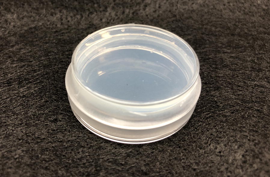
Parafilm M® is commonly used to seal NGM culture plates. Parafilm wrapped one time around the circumference of a NGM culture plate
In this study, we aimed to identify if sealing NGM culture plates with Parafilm altered larval growth. We found statistically significant, moderate increases in larval growth in worms cultured in plates wrapped with Parafilm compared to control worms. These results necessitate an awareness of C. elegans biologists to the unintended secondary effects this common lab practice could have on C. elegans biology and experimental datasets.
Main text
Materials and methods
Culture conditions
The Bristol wild-type C. elegans strain (N2) obtained from the Caenorhabditis Genetics Center (CGC) was grown in vented 35 mm plates (Tritech Research, Inc., #T3500) containing Nematode Growth Media with 2 mg/mL uracil (NGM). NGM was inoculated with the E. coli strain OP50 (CGC) grown in Miller’s Luria Broth (Fisher Scientific, #50-488-764). Worms were cultured at 20 °C ± 0.2 °C (Conviron I25L Growth Chamber), and standard culture conditions and methods were used [11, 12].
Larval growth
Larvae were synchronized at the L1 stage according to Brockett et al. [13]. L1 hermaphrodite worms were individually transferred at t = 0 h to NGM plates inoculated with 50 μL of the same overnight OP50 culture supplied ad libitum. Each plate was assigned to a treatment group using a random number table (n = 41 plates per group, each with one worm). Plates in the Parafilm treatment group were wrapped one time around the circumference of the plate with Parafilm™ M Wrapping Film (Fisher Scientific, #S37440), sealing the lid to the bottom of the Petri dish (Fig. 1). Control plates were not wrapped with Parafilm. There was no significant difference in median worm length between the Parafilm-treated and control worms at the time of transfer (Mann–Whitney U test, W = 755, p = 0.674). Cultures were incubated in vented plastic storage bins on the same shelf of the growth chamber. Temperature did not vary across the shelf of the growth chamber.
A still image of each worm was taken on a stereomicroscope at 0, 18, 24, 36, and 48 h after transfer. Images were taken without removing the culture plate lids since the lids were not removable in the Parafilm group. Worms and cultures were censored for sex, death, starvation, microbial contamination, and abnormalities in movement, behavior, and observable anatomical irregularities; 2 Parafilm-treated worms were removed from the dataset prior to analysis. Most worms produced embryos around 48 h, indicating the adult stage had been reached and thus terminating data collection. The length of each worm in the still images at each time point was measured down the midline from the tip of the lips to the end of the tail using the freehand tool in ImageJ [14]. All measurements were taken blind by one observer through randomized labeling of image files by a different individual.
We calculated the change in length (Δgrowth) from 0 to 48 h as well as the slope of the best-fit linear growth curve (growth rate) through the 5 time points for each worm. The data were not normally distributed for the two growth variables, so we used Mann–Whitney U tests to compare median Δgrowth and growth rate between the Parafilm and control groups in R version 3.5.2 with α = 0.05 [15]. We also calculated the standardized treatment effect size (Cohen’s d) for each growth variable [16, 17].
Results
Median larval length (Fig. 2) at each time point appeared similar with small differences between the Parafilm and control groups. However, over the 48 h period, worms in cultures wrapped with Parafilm showed significantly greater Δgrowth (Mann–Whitney U test, W = 472, p = 0.001) and a significantly faster growth rate (Mann–Whitney U test, W = 479, p = 0.002) than worms in the control group (Table 1). Furthermore, Cohen’s d indicated a moderate impact of Parafilm wrapping on the Δgrowth and growth rate of C. elegans over 48 h (Cohen’s d = 0.75 and d = 0.74 respectively).
Fig. 2.

Larval growth over time. Median length (μm) of worms in each treatment group at 0, 18, 24, 36, and 48 h after L1 transfer to plates. Data are displayed as boxplots overlaid with scatterplots of individual worm lengths (open circles) at each time point. Bold line within each box shows median length, and box shows Q3 (upper quartile) and Q1 (lower quartile). n = 39 Parafilm and n = 41 control
Table 1.
Significant differences in C. elegans larval growth over 48 h on Parafilm-wrapped (n = 39) and control (n = 41) plates
| Median (IQR) length (μm) | ||
|---|---|---|
| Parafilm | Control | |
| ΔGrowth | 764.25 (39.3) | 726.35 (97.3) |
| Growth rate | 16.43 (1.3) | 15.56 (1.7) |
Discussion
This study aimed to determine if C. elegans larval growth is affected by wrapping NGM culture plates with Parafilm. We found that larval Δgrowth and growth rate (Table 1) were significantly greater in worms on Parafilm-wrapped cultures than worms in control cultures. Our results suggest that wrapping culture plates with Parafilm alters conditions in the internal plate environment, causing statistically detectable differences in larval growth over time compared to control conditions.
The median values for the measured variables (Δgrowth and growth rate) were rather similar between the control and Parafilm groups (Table 1), and Cohen’s d indicated a moderate treatment effect. The moderate treatment effect size suggests that the measured differences may not reflect biologically significant differences for all experiments, depending upon the sensitivity to which the researcher needs to quantify and compare growth among treatment groups. For example, worm length can be used as a determinate of larval stage [18] along with developmental hallmarks [19, 20]. The median Δgrowth difference between our Parafilm and control groups over 48 h (37.90 μm; Table 1) would be unlikely to cause a mislabeling of larval stage since worms grow approximately 690 μm (20 °C) between the first molt to egg-laying onset [18], which are the stages we observed over the course of our measurements. Conversely, the small but significant differences in median growth rate (0.87 μm/h; Table 1) that we observed from wrapping cultures with Parafilm could unintentionally lead to misinterpretation of growth rates taken at finer and more precise time scales, as growth rates are linear within a larval stage but change between larval stages [21].
Wrapping culture dishes with Parafilm is a common practice with model organisms other than C. elegans. For example, researchers studying the plant model Arabidopsis thaliana (A. thaliana) wrap their Petri dish cultures with Parafilm for many of the same reasons as worm researchers. Similar to our study, recent research on A. thaliana sought to determine if wrapping plant cultures with Parafilm affected the culture environment and plant growth [22]. In both their and our studies, there were not only changes in organismal growth as a result of Parafilm-wrapping but also reduced variability in growth among Parafilm-wrapped replicates (Fig. 1F in [22]; IQR in Table 1 and Fig. 2 in this study), suggesting that Parafilm creates standardized conditions that reduce variability in organismal responses.
Environmental factors such as diet, population density, and temperature are known to affect worm body size [23–28]. In our study, it is unlikely that diet or population density contributed to differences between worms in each group because all worms were fed the same bacterial strain ad libitum and they all experienced the same population density effects by being individually cultured. Instead, we hypothesize that C. elegans grown in Parafilm-wrapped cultures showed moderately significant increases in growth compared to control worms because of differences in humidity, temperature, and/or gas levels (i.e. oxygen and/or carbon dioxide).
Caenorhabditis elegans is capable of sensing humidity [29–32], but the direct effects of humidity on growth have not been studied to date. However, Wang et al. postulated that the amount of water content in cultures might affect the temperature in or near the agar surface [29], and the Bristol N2 wild-type strain, which was used in our study, follows the temperature-size rule by growing larger at lower temperatures [33]. Since Parafilm prevents media desiccation, Parafilm-wrapped cultures likely have differences in relative humidity and, in turn, possibly temperature near and/or above the agar–air interface where worms live compared to cultures not wrapped with Parafilm. Humidity-mediated differences in temperature could have contributed to our observed differences in growth if we consider the temperature-size rule [33], and/or humidity could have directly affected growth by undetermined mechanisms. Worms were only exposed to Parafilm or control conditions for 48 h so minimal moisture would have been lost from the control cultures, but this short exposure time could explain the similar medians measured between the Parafilm and control groups.
Recent research on A. thaliana cultures demonstrated that gas exchange may be affected by Parafilm wrapping. Parafilm-wrapped cultures had significantly different carbon dioxide, but not oxygen, levels compared to control cultures over short (min) and long (days) time periods [22] even though the Parafilm manufacturer [34] and distributors [35] claim that Parafilm is permeable to gases. Interestingly, previous studies suggest that starved worms, which were used in our study, are more attracted to bacterial strains that produce higher levels of carbon dioxide during respiration and this affects life history traits [36]. Taken together, it is plausible that worm and bacterial respiration caused a higher accumulation of carbon dioxide in Parafilm-wrapped compared to control cultures, resulting in increased worm attraction to and consumption of the bacteria. This increase in feeding could explain increased worm growth in the Parafilm-treated worms as compared to the control worms, especially between 0 and 18 h where we see the greatest difference in median growth between the groups, at the time when worms are initiating growth from L1 starvation (Table 1; Fig. 2).
The increased growth we observed provides evidence that there is some effect on C. elegans from wrapping NGM cultures with Parafilm, and supports similar effects seen with A. thaliana [22]. Complementary studies that measure culture conditions (e.g. humidity, temperature, oxygen and carbon dioxide) and life-history traits under Parafilm-wrapped conditions should be done in an animal model such as C. elegans to more comprehensively explore the effects of Parafilm on laboratory cultures. Until then, researchers of all organisms should first test for the effects that this practice could have on their measured variables. This will eliminate potential secondary effects that could inadvertently influence dependent variables, especially those for which slight differences will affect data analysis and interpretation.
Limitations
Our study did not assess morphological or behavioral developmental hallmarks at each time point for developmental correlations to larval growth and development, nor did we evaluate development at a finer timescale. However, we observed no prominent differences in the timing of initial egg-laying between groups, suggesting worms in each group reached adulthood at approximately the same time. We also did not measure temperature, humidity, or carbon dioxide and oxygen levels inside the culture dishes to determine potential causes of differences in worm growth between Parafilm-wrapped and control cultures, and we suggest that this important issue be addressed by future studies.
Acknowledgements
We thank Thomas McCarthy and Daniel Kurtz for statistical analysis consultations, and Brandee Decker-Rockefeller and Adam Pack for manuscript consultation. Strains were provided by the CGC, which is funded by NIH Office of Research Infrastructure Programs (P40 OD010440).
Abbreviations
- NGM
Nematode Growth Media
- C.
Caenorhabditis
- E.
Escherichia
- CGC
Caenorhabditis Genetics Center
- Δgrowth
change in length
- IQR
interquartile range
- A.
Arabidopsis
Authors’ contributions
JST conceived and developed the research aims, supervised the research, and acquired necessary resources. JST, PS, and EK contributed to preliminary experimental designs. SS participated in evolving research goals and experimental design. PS performed preliminary experiments with assistance from MB, and HN contributed to data collection. HN performed experiments and collected data presented in this manuscript. SS maintained data and performed most statistical analyses, including validation. SS prepared figures with contribution from JST. JST wrote the manuscript with the exception of the Results and parts of the Materials and Methods, which were written by SS. SS contributed substantial feedback and editing. All authors read and provided feedback about the manuscript. All authors read and approved the final manuscript.
Funding
The Utica College Faculty Resources Committee and Office of Academic Affairs supported this work. They were not involved in the design of the study and collection, analysis, and interpretation of data and in writing the manuscript.
Availability of data and materials
The datasets used and/or analyzed during the current study are available from the corresponding author on reasonable request.
Ethics approval and consent to participate
Not applicable.
Consent for publication
Not applicable.
Competing interests
The authors declare that they have no competing interests.
Footnotes
Publisher's Note
Springer Nature remains neutral with regard to jurisdictional claims in published maps and institutional affiliations.
Contributor Information
Jessica H. Shinn-Thomas, Email: jhthomas@utica.edu
Sara E. Scanga, Email: sescanga@utica.edu
Patrick S. Spica, Email: paspica@utica.edu
Hardik K. Nariya, Email: hknariya@utica.edu
Emra Klempic, Email: Emra_Klempic@URMC.Rochester.edu.
Mary R. Brockett, Email: mary.brockett.ctr@usuhs.edu
References
- 1.Corsi AK, Wightman B, Chalfie M. A transparent window into biology: a primer on Caenorhabditis elegans. Genetics. 2015;200:387–407. doi: 10.1534/genetics.115.176099. [DOI] [PMC free article] [PubMed] [Google Scholar]
- 2.Ankeny RA. The natural history of Caenorhabditis elegans research. Nat Rev Genet. 2001;2:474–479. doi: 10.1038/35076538. [DOI] [PubMed] [Google Scholar]
- 3.Hulme SE, Whitesides GM. Chemistry and the worm: caenorhabditis elegans as a platform for integrating chemical and biological research. Angew Chem Int Ed. 2011;50:4774–4807. doi: 10.1002/anie.201005461. [DOI] [PubMed] [Google Scholar]
- 4.Frézal L, Félix MA. C. elegans outside the Petri dish. ELife. 2015;4:e05849. doi: 10.7554/eLife.05849. [DOI] [PMC free article] [PubMed] [Google Scholar]
- 5.Leung MCK, Williams PL, Benedetto A, Au C, Helmcke KJ, Aschner M, et al. Caenorhabditis elegans: an emerging model in biomedical and environmental toxicology. Toxicol Sci. 2008;106:5–28. doi: 10.1093/toxsci/kfn121. [DOI] [PMC free article] [PubMed] [Google Scholar]
- 6.Evans KS, Zhao Y, Brady SC, Long L, McGrath PT, Andersen EC. Correlations of genotype with climate parameters suggest Caenorhabditis elegans niche adaptations. G3 Genes Genomes Genetics. 2016;7:289–298. doi: 10.1534/g3.116.035162. [DOI] [PMC free article] [PubMed] [Google Scholar]
- 7.Félix M-A, Braendle C. The natural history of Caenorhabditis elegans. Curr Biol. 2010;20:R965–R969. doi: 10.1016/j.cub.2010.09.050. [DOI] [PubMed] [Google Scholar]
- 8.Félix M-A, Duveau F. Population dynamics and habitat sharing of natural populations of Caenorhabditis elegans and C. briggsae. BMC Biol. 2012;10:59. doi: 10.1186/1741-7007-10-59. [DOI] [PMC free article] [PubMed] [Google Scholar]
- 9.Petersen C, Dirksen P, Prahl S, Strathmann EA, Schulenburg H. The prevalence of Caenorhabditis elegans across 1.5 years in selected North German locations: the importance of substrate type, abiotic parameters, and Caenorhabditis competitors. BMC Ecol. 2014;14:4. doi: 10.1186/1472-6785-14-4. [DOI] [PMC free article] [PubMed] [Google Scholar]
- 10.Schulenburg H, Félix M-A. The natural biotic environment of Caenorhabditis elegans. Genetics. 2017;206:55–86. doi: 10.1534/genetics.116.195511. [DOI] [PMC free article] [PubMed] [Google Scholar]
- 11.Stiernagle T. Maintenance of C. elegans. WormBook. 2006. 10.1895/wormbook.1.101.1. [DOI] [PMC free article] [PubMed]
- 12.Brenner S. The genetics of Caenorhabditis elegans. Genetics. 1974;77:71–94. doi: 10.1093/genetics/77.1.71. [DOI] [PMC free article] [PubMed] [Google Scholar]
- 13.Brockett MR, Spica PS, Shinn-Thomas JH. C. elegans synchronization: Small- and large-scale protocols to isolate synchronized L1 larvae and beyond, vol. 20. The Worm Breeder’s Gazette. 2016. http://wbg.wormbook.org/2016/05/16/c-elegans-synchronization-small-and-large-scale-protocols-to-isolate-synchronized-l1-larvae-and-beyond/. Accessed 13 June 2016.
- 14.Schneider CA, Rasband WS, Eliceiri KW. NIH image to imagej: 25 years of image analysis. Nat Methods. 2012;9:671–675. doi: 10.1038/nmeth.2089. [DOI] [PMC free article] [PubMed] [Google Scholar]
- 15.R Core Team. R: A language and environment for statistical computing. R foundation for statistical computing. Vienna, Austria. 2018. https://www.R-project.org. Accessed 2 Dec 2019.
- 16.Cohen J. Statistical power analysis for the behavioral sciences. 2. New York: Routledge; 2013. [Google Scholar]
- 17.Lenhard W, Lenhard A. Calculation of effect sizes. 2016. Retrieved by: https://www.psychometrica.de/effect_size.html, 10.13140/rg.2.1.3478.4245. Accessed 22 June 2018.
- 18.Byerly L, Cassada RC, Russell RL. The life cycle of the nematode Caenorhabditis elegans. Dev Biol. 1976;51:23–33. doi: 10.1016/0012-1606(76)90119-6. [DOI] [PubMed] [Google Scholar]
- 19.Altun ZF, Herndon LA, Wolkow CA, Crocker C, Lints R, Hall DH (eds). WormAtlas. 2019. http://www.wormatlas.org. Accessed 23 June 2017.
- 20.Mok DZL, Sternberg PW, Inoue T. Morphologically defined sub-stages of C. elegans vulval development in the fourth larval stage. BMC Dev Biol. 2015;15(1):26. doi: 10.1186/s12861-015-0076-7. [DOI] [PMC free article] [PubMed] [Google Scholar]
- 21.Knight CG, Patel MN, Azevedo RBR, Leroi AM. A novel mode of ecdysozoan growth in Caenorhabditis elegans. Evol Dev. 2002;4:16–27. doi: 10.1046/j.1525-142x.2002.01058.x. [DOI] [PubMed] [Google Scholar]
- 22.Banerjee S, Siemianowski O, Liu M, Lind KR, Tian X, Nettleton D, et al. Stress response to CO2 deprivation by Arabidopsis thaliana in plant cultures. PLoS ONE. 2019;14:e0212462. doi: 10.1371/journal.pone.0212462. [DOI] [PMC free article] [PubMed] [Google Scholar]
- 23.Nagashima T, Ishiura S, Suo S. Regulation of body size in Caenorhabditis elegans: effects of environmental factors and the nervous system. Int J Dev Biol. 2017;61:367–374. doi: 10.1387/ijdb.160352ss. [DOI] [PubMed] [Google Scholar]
- 24.So S, Miyahara K, Ohshima Y. Control of body size in C.elegans dependent on food and insulin/IGF-1 signal. Genes Cells. 2011;16:639–651. doi: 10.1111/j.1365-2443.2011.01514.x. [DOI] [PubMed] [Google Scholar]
- 25.Mörck C, Pilon MC. elegans feeding defective mutants have shorter body lengths and increased autophagy. BMC Dev Biol. 2006;6:39. doi: 10.1186/1471-213X-6-39. [DOI] [PMC free article] [PubMed] [Google Scholar]
- 26.Lenaerts I, Walker GA, Van Hoorebeke L, Gems D, Vanfleteren JR. Dietary restriction of Caenorhabditis elegans by axenic culture reflects nutritional requirement for constituents provided by metabolically active microbes. J Gerontol A Biol Sci Med Sci. 2008;63:242–252. doi: 10.1093/gerona/63.3.242. [DOI] [PMC free article] [PubMed] [Google Scholar]
- 27.MacNeil L, Watson E, Arda HE, Zhu LJ, Walhout AJM. Diet-induced developmental acceleration independent of TOR and insulin in C. elegans. Cell. 2013;153(1):240–252. doi: 10.1016/j.cell.2013.02.049. [DOI] [PMC free article] [PubMed] [Google Scholar]
- 28.Rose JK, Sangha S, Rai S, Norman KR, Rankin CH. Decreased sensory stimulation reduces behavioral responding, retards development, and alters neuronal connectivity in Caenorhabditis elegans. J Neurosci. 2005;25:7159–7168. doi: 10.1523/JNEUROSCI.1833-05.2005. [DOI] [PMC free article] [PubMed] [Google Scholar]
- 29.Wang W, Qin L-W, Wu T-H, Ge C-L, Wu Y-Q, Zhang Q, et al. cGMP signalling mediates water sensation (hydrosensation) and hydrotaxis in Caenorhabditis elegans. Sci Rep. 2016;6:19779. doi: 10.1038/srep19779. [DOI] [PMC free article] [PubMed] [Google Scholar]
- 30.Russell J, Vidal-Gadea AG, Makay A, Lanam C, Pierce-Shimomura JT. Humidity sensation requires both mechanosensory and thermosensory pathways in Caenorhabditis elegans. Proc Natl Acad Sci USA. 2014;111:8269–8274. doi: 10.1073/pnas.1322512111. [DOI] [PMC free article] [PubMed] [Google Scholar]
- 31.Zhao B, Khare P, Feldman L, Dent JA. Reversal frequency in Caenorhabditis elegans represents an integrated response to the state of the animal and its environment. J Neurosci. 2003;23:5319–5328. doi: 10.1523/JNEUROSCI.23-12-05319.2003. [DOI] [PMC free article] [PubMed] [Google Scholar]
- 32.Filingeri D. Humidity sensation, cockroaches, worms, and humans: are common sensory mechanisms for hygrosensation shared across species? J Neurophysiol. 2015;114:763–767. doi: 10.1152/jn.00730.2014. [DOI] [PMC free article] [PubMed] [Google Scholar]
- 33.Kammenga JE, Doroszuk A, Riksen JAG, Hazendonk E, Spiridon L, Petrescu A-J, et al. A Caenorhabditis elegans wild type defies the temperature-size rule owing to a single nucleotide polymorphism in tra-3. PLoS Genet. 2007;3:e34. doi: 10.1371/journal.pgen.0030034. [DOI] [PMC free article] [PubMed] [Google Scholar]
- 34.Parafilm® M all-purpose laboratory film. http://www.bemis.com/na/products/parafilm-floratape/parafilm-lab. Accessed 31 Jul 2019.
- 35.Parafilm® product information sheet. https://www.sigmaaldrich.com/content/dam/sigma-aldrich/docs/Sigma/Product_Information_Sheet/1/p7543pis.pdf. Accessed 31 Jul 2019.
- 36.Yu L, Yan X, Ye C, Zhao H, Chen X, Hu F, et al. Bacterial respiration and growth rates affect the feeding preferences, brood size and lifespan of Caenorhabditis elegans. PLoS ONE. 2015;10:e0134401. doi: 10.1371/journal.pone.0134401. [DOI] [PMC free article] [PubMed] [Google Scholar]
Associated Data
This section collects any data citations, data availability statements, or supplementary materials included in this article.
Data Availability Statement
The datasets used and/or analyzed during the current study are available from the corresponding author on reasonable request.