ABSTRACT
In this supplement, we build on work previously published under the Human Connectome Project. Specifically, we show a comprehensive anatomic atlas of the human cerebrum demonstrating all 180 distinct regions comprising the cerebral cortex. The location, functional connectivity, and structural connectivity of these regions are outlined, and where possible a discussion is included of the functional significance of these areas. In part 5, we specifically address regions relevant to the insula and opercular cortex.
Keywords: Anatomy, Cerebrum, Connectivity, DTI, Functional connectivity, Human, Parcellations
ABBREVIATIONS
- AAIC
anterior agranular insular cortex
- AVI
anterior ventral insula
- FOP5
frontal operculum 5
- IFOF
inferior fronto-occipital fasciculus
- Ig
insula granular
- LBelt
lateral belt
- MBelt
medial belt
- MI
middle insula
- OP4
operculum 4
- PBelt
parabelt
- PFcm
parietal F, region cm
- PI
parainsular
- PoI1
posterior insula 1
- PSL
perisylvian language
- RI
retroinsular
- SLF
superior longitudinal fasciculus
- STGa
superior temporal gyrus region a
- STV
superior temporal visual
- TA
TA, temporal region A
- TPOJ
temporo-parieto-occipital junction
The notion that the insula is a complex anatomic area for surgery should require no additional justification to those familiar with the topic. The operculoinsular relations have been the object of study and struggle for some time.1,2 Thus, it is with some trepidation that we submit the following report which only makes the area more complex than it already is. We will note, though, that learning these areas pointed out details of insular anatomy that we had previously not considered, making the task of sorting out these areas quite useful. There is a surprising amount of cortex contained in the insular and opercular clefts. In contrast to how most of us think of this area in daily practice, every single crevice of this region has a unique area, including the underside of the opercula and the depths of the circular sulcus of the insula.
BASIC ORIENTATION AND INSULAR SUBDIVISIONS
We do not aim to repeat the work of previous authors on this subject, but we will point out a few well-known facts on the basic scheme of the insula which we will use to simplify the parcellation scheme. The shape of the insula is roughly a right triangle, with the right angle being located in the anterior superior corner, and the hypotenuse running along the posterior and inferior edges from one apex at the limen insula to a posterior superior apex roughly underlying the parietal operculum. The circular sulcus is the cleft delineating this triangle. The opercula begin at the circular sulcus and fold back over the insula to cover it.
Obviously, this is a bit of a simplification. The apices are not sharp angles, but are rounded. In addition, the sides of the triangle are not perfectly straight, and the anteroinferior apex folds somewhat onto the orbitofrontal surface to form the piriform regions. Despite this, we think this construct provides a useful method of splitting up these areas into 3 manageable subregions:
Anterior superior apex: Anterior and middle insula, FOP regions
Posterior parietal apex: Posterior insula, OP, retroinsular and supramarginal gyrus regions
Temporal hypotenuse: Opercular STG, inferior circular sulcus, and Heschl's gyrus regions
A Brief Word on Opercular Nomenclature
Sixteen different parcellated areas comprise the opercular regions proper. This can be quite confusing, and this was the major rationale for grouping the regions the way we have. A brief summary of the nomenclature is as follows:
FOP regions: There are 5 frontal opercular regions, FOP 1 to 5, which combined with area 43 make up the frontal operculum.
OP regions: The OP regions include OP1, OP2-3, and OP4. Combined with area PFcm and the supramarginal gyrus regions, PSL and STV, these make up the parietal operculum.
STG regions: These include areas A4, A5, STGa, and TA2, which make up the temporal operculum. The remainder of the temporal lobe is discussed in Chapter 6 of this series.
Anterior Apex Regions
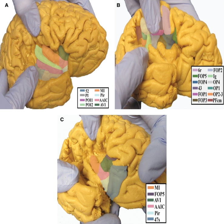
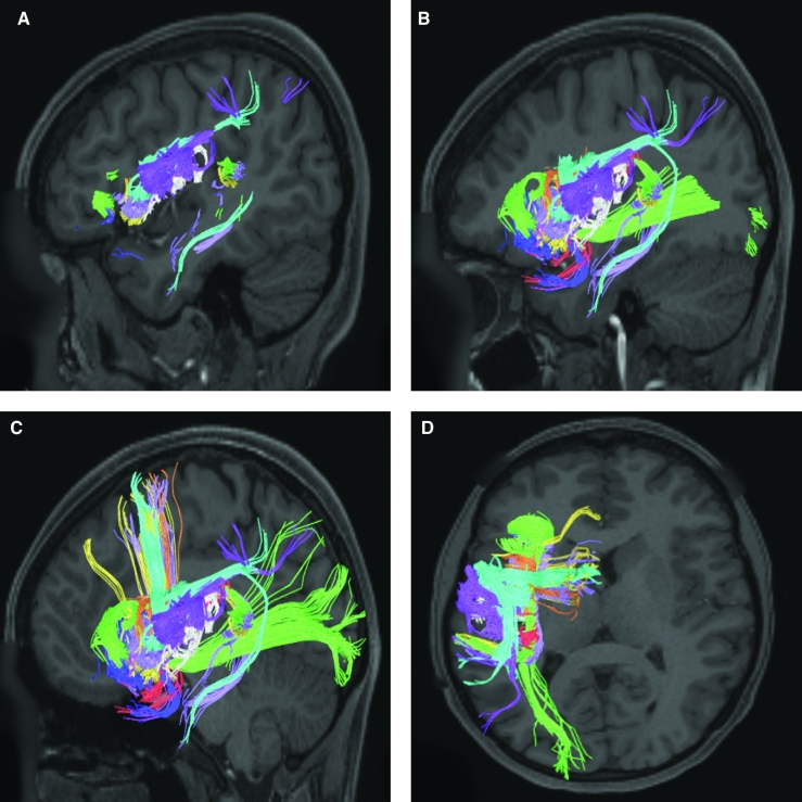
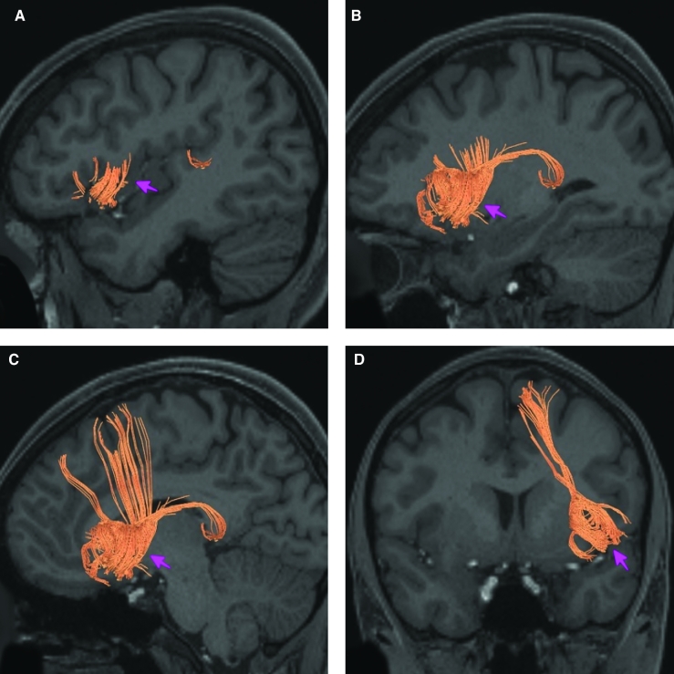
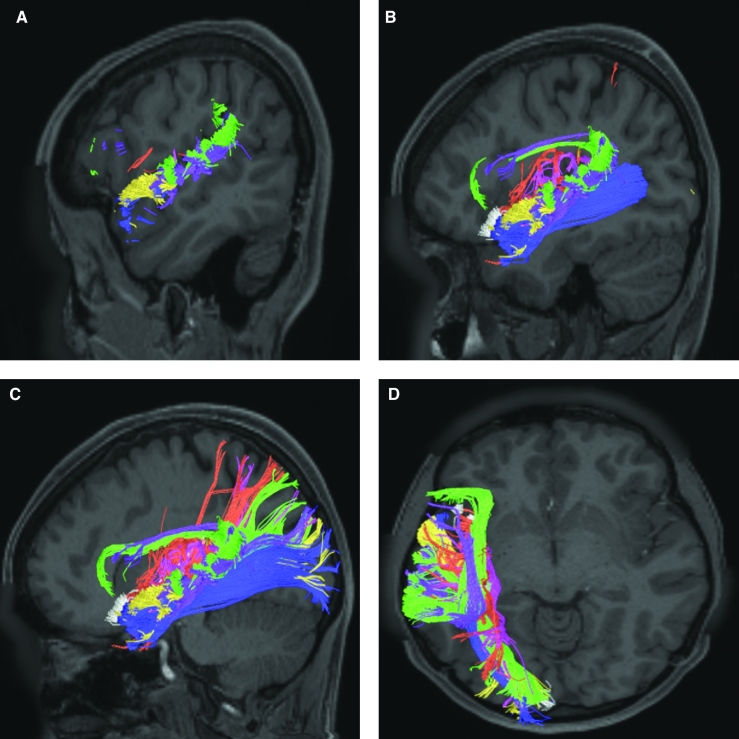
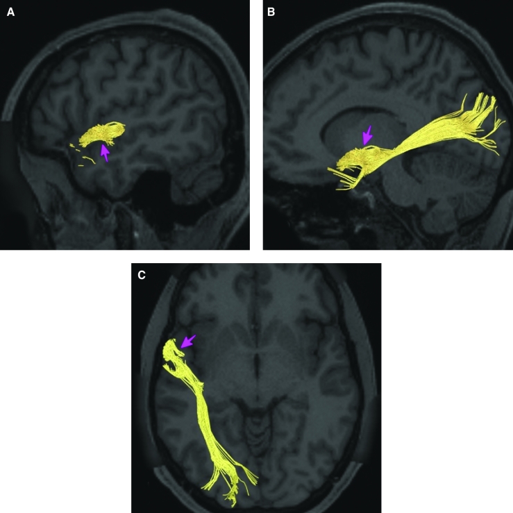
The parcellations comprised by the anterior apex region are Pir, AAIC, AVI, MI, FOP5, FOP4, FOP3, FOP2, FOP1, and 43. These areas roughly comprise the short and transverse insular gyri and their associated opercula. The anatomic location of these parcellations is shown on a cadaver brain in Figure 1. This region has consistent white matter connections with the arcuate/superior longitudinal fasciculus (SLF) and local insular parcellations. Area FOP5 is also connected with the inferior fronto-occipital fasciculus (IFOF). The combined tractography of these parcellations is shown in Figure 2.
FIGURE 1.
Anatomical location of the anterior apex parcellations of the frontal operculum and insula shown on the right hemisphere of a cadaver brain. A, Lateral view of the posterior insula visualized by widening the lateral sulcus. B, Inferior view of the right hemisphere demonstrating the parietal opercular regions visualized by widening the lateral sulcus. C, Oblique lateral view of the insular apex and frontal operculum visualized by widening the lateral sulcus.
FIGURE 2.
Combined structural connectivity of the anterior apex parcellations in the left hemisphere, shown on T1-weighted MR images. Sagittal views of A, lateral, B, medial, and C, far medial planes. D, Axial view.
Area Pir
Where is it?
Area Pir (piriform cortex) is located in the pyriform cortex, which itself is located just anterior to the anterior perforated substance, at the point where the limen insula folds onto the orbitofrontal surface.
What are its borders?
Area Pir borders AAIC and a small part of area 47s anteriorly, pOFC medially, and POI1 POI2, PI, and TA2 laterally. Its posterior border is with the anterior perforated substance.
What is its functional connectivity?
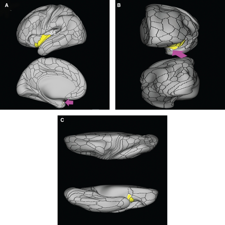
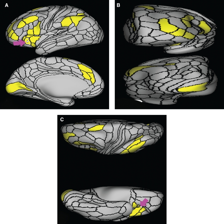
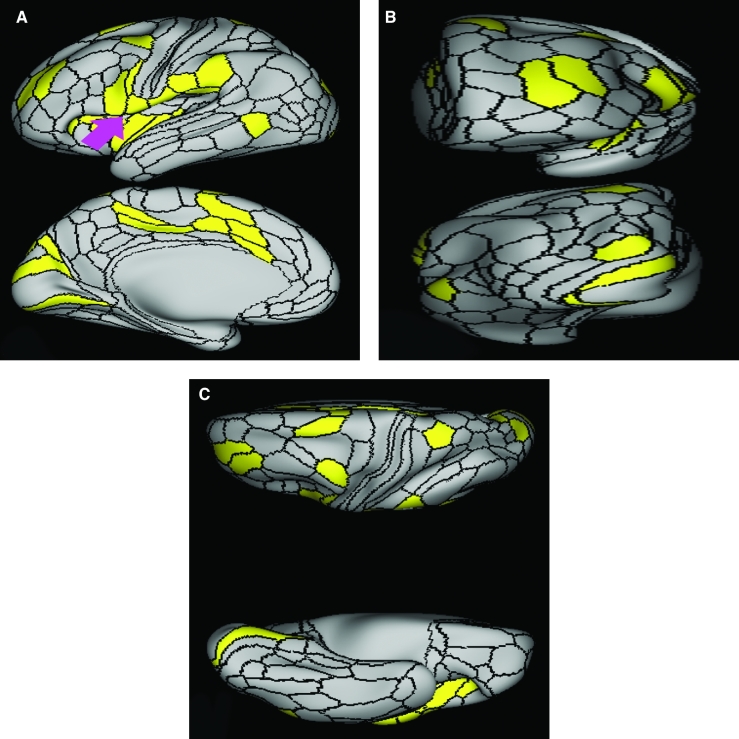
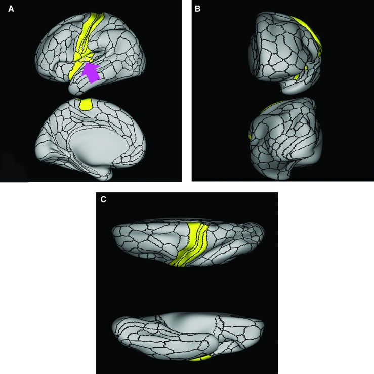
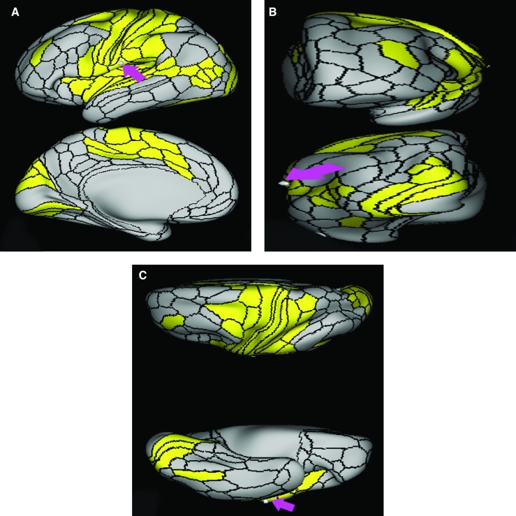
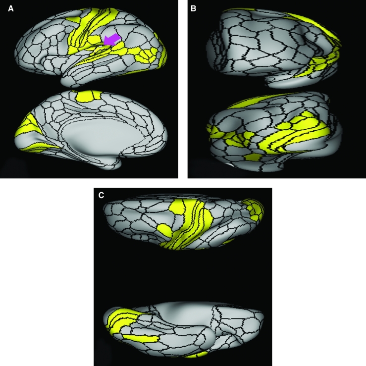
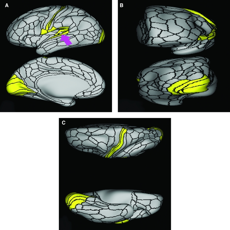
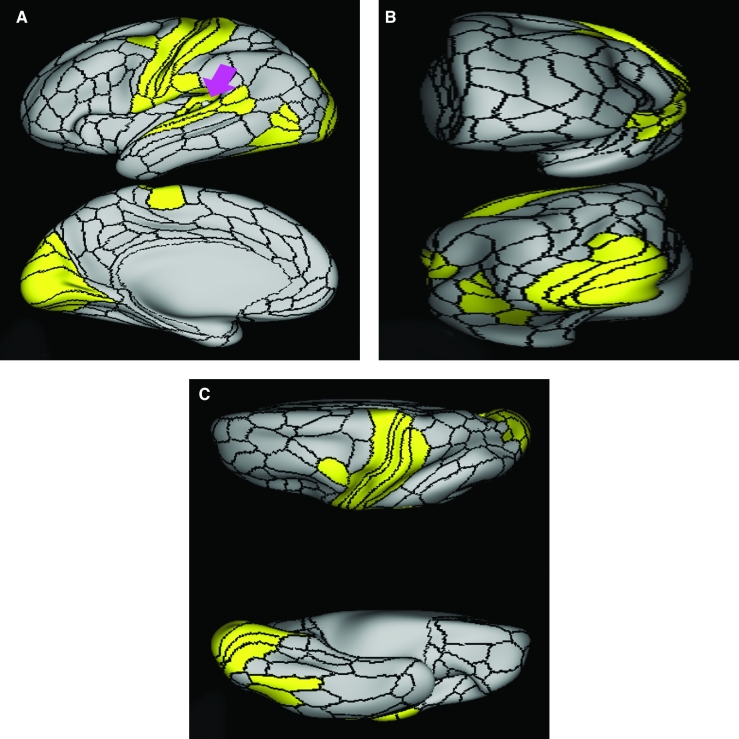
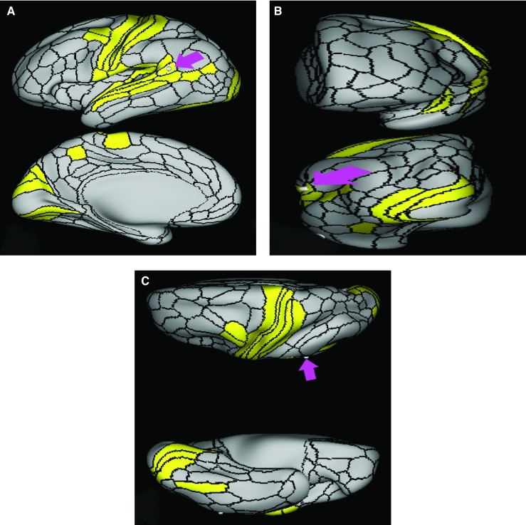
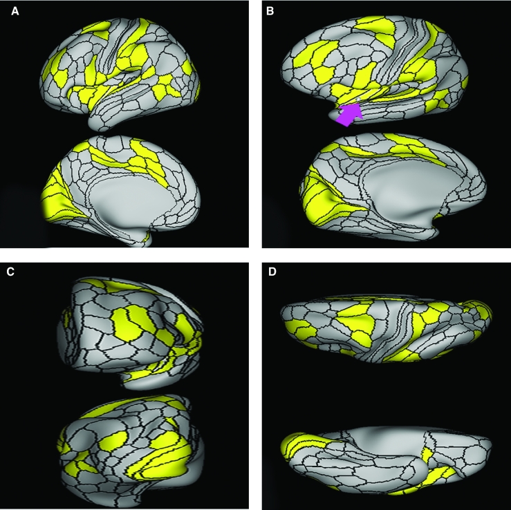
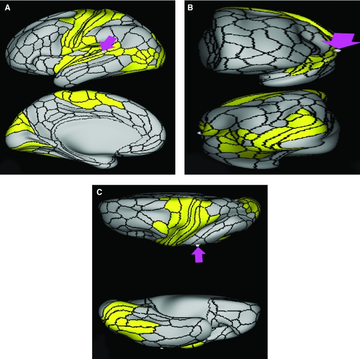
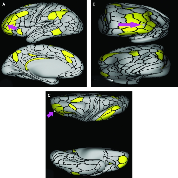
Area Pir demonstrates functional connectivity to areas AAIC, PoI1, and PoI2 in the insula (Figure 3).
FIGURE 3.
Functional connectivity of Pir demonstrated on an inflated left hemisphere. A, Lateral and medial views. B, Rostral and caudal views. C, Dorsal and ventral views. Parcellations with the strongest functional connectivity are shown in yellow. Pink arrows designate the parcellation of interest.
What are its white matter connections?
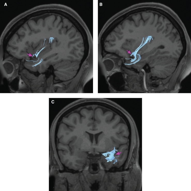
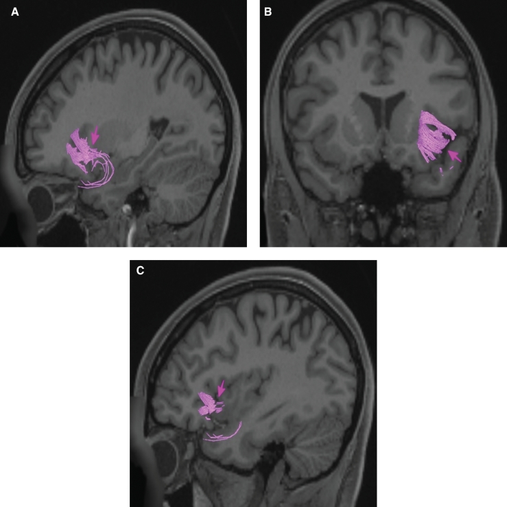
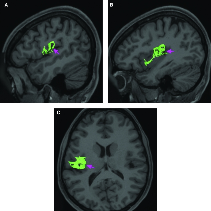
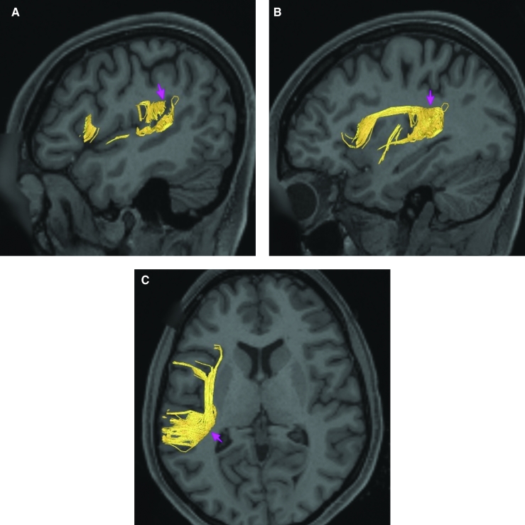
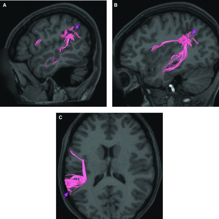
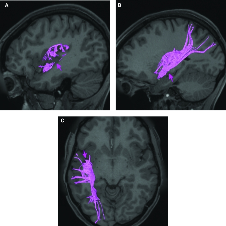
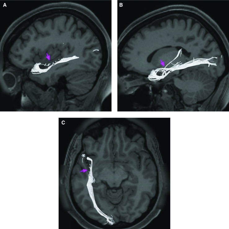
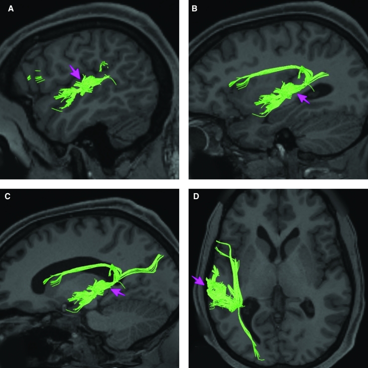
Area Pir is structurally connected to local parcellations and posterior insular regions. The white matter tracts of this parcellation are difficult to delineate due to the proximity of the area to underlying white matter tracts. Connections to posterior insular regions project from Pir to PoI2, Ig, and OP2-3. Local short association bundles connect with AAIC and TGd (Figure 4).
FIGURE 4.
Structural connectivity of Pir in the left hemisphere, shown on T1-weighted MR images. Sagittal views of A, lateral and B, medial planes. C, Coronal view. Light blue: white matter tracts of Pir demonstrating connections with the temporal pole and local parcellations. Pink arrows designate the parcellation of interest.
What is known about its function?
The piriform cortex is a region of the anterior apex that has been studied previously. Area Pir has been found to contain axons that distribute widely and extensively branch throughout other cortical regions with functional roles attributed to cognition, behavior, emotion, and memory.3 Area Pir is also the largest area of the brain to receive olfactory signals.3 Area Pir shows activation with all olfactory tasks and plays a pivotal role in the memory of olfaction, discrimination of odors, and distribution of this information to other regions of the brain.4
Area AAIC
Where is it?
Area AAIC (anterior agranular insular cortex) is located in the anteroinferior insula near the limen insula and in the transverse insular gyrus.
What are its borders?
Area AAIC is a small triangularly shaped area which borders areas 47s and AVI anteriorly, areas POI2 and Pir posteriorly and area 25 superomedially, and MI superiorly.
What is its functional connectivity?
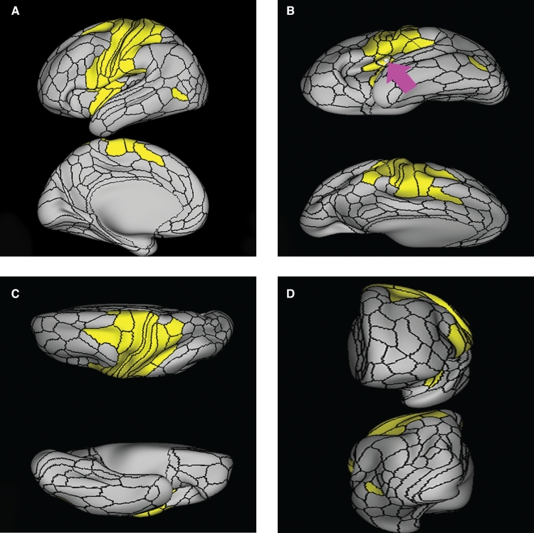
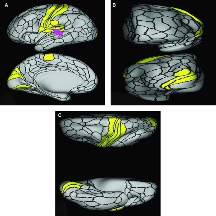
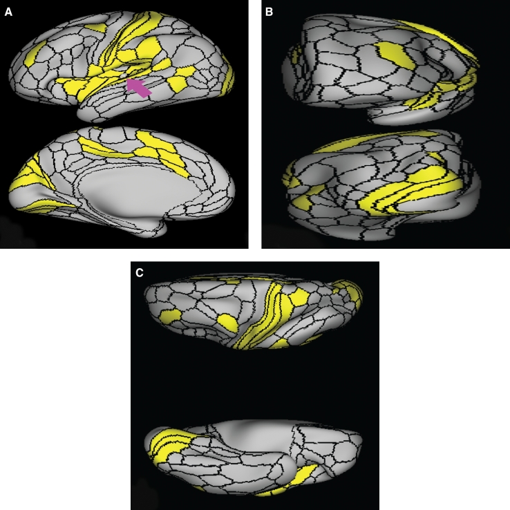
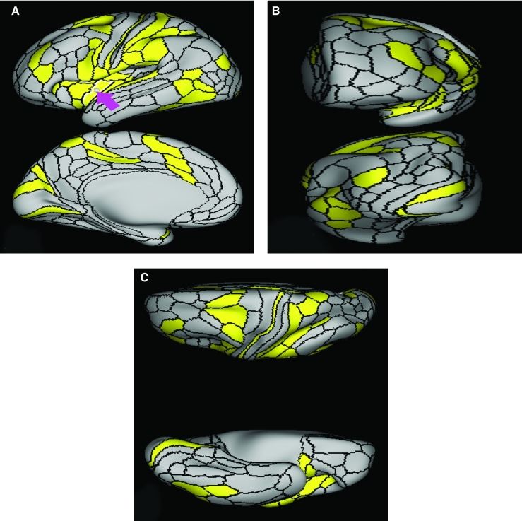
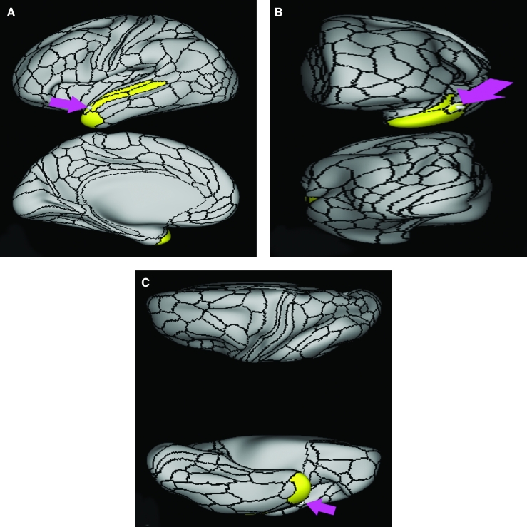
Area AAIC demonstrates functional connectivity to areas AVI, MI, Pir, 47s, and PoI2 in the insula (Figure 5).
FIGURE 5.
Functional connectivity of AAIC demonstrated on an inflated left hemisphere. A, Lateral and medial views. B, Rostral and caudal views. C, Dorsal and ventral views. Parcellations with the strongest functional connectivity are shown in yellow. Pink arrows designate the parcellation of interest.
What are its white matter connections?
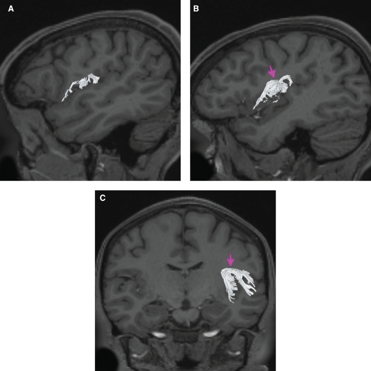
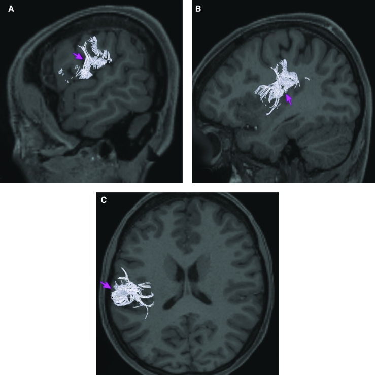
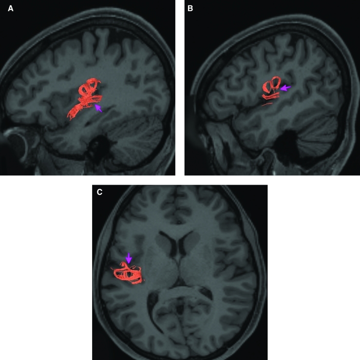
Area AAIC is structurally connected to local parcellations. Local short association bundles connect with Pir, 47s, AVI, PoI2, and MI (Figure 6).
FIGURE 6.
Structural connectivity of AAIC in the left hemisphere, shown on T1-weighted MR images. Sagittal views of A, medial and C, lateral planes. B, Coronal view. Pink: white matter tracts of AAIC demonstrating connections with local parcellations. Pink arrows designate the parcellation of interest.
What is known about its function?
Area AAIC is a newly described area of the brain and was parcellated from the anterior insula. The anterior insula is suggested to have a role in sensation and control of autonomic nervous system processes as well as playing a role in human awareness, self-recognition, time perception, and perceptual decision making.5,6 Area AAIC was parcellated from areas MI and AVI in the anterior insula based on functional activity differences between regions related to motor, arithmetic, auditory language, and semantic tasks.7
Area AVI
Where is it?
Area AVI (anterior ventral insula) is located in anterior superior apex of the insula.
What are its borders?
Area AVI borders area 47l anteriorly, areas 47s and AAIC inferiorly, MI posteriorly, and FOP4, and FOP5 superiorly.
What is its functional connectivity?
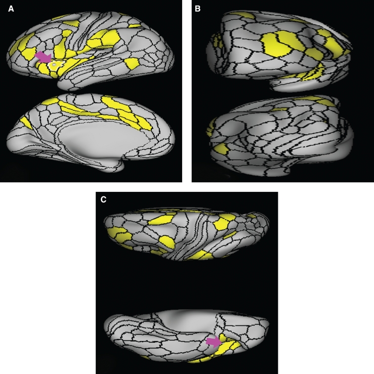
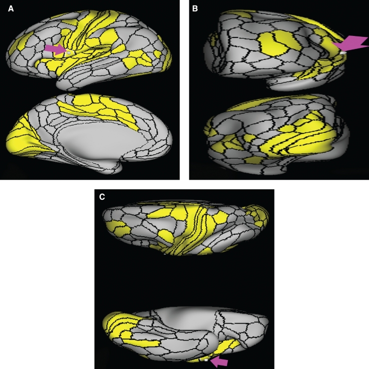
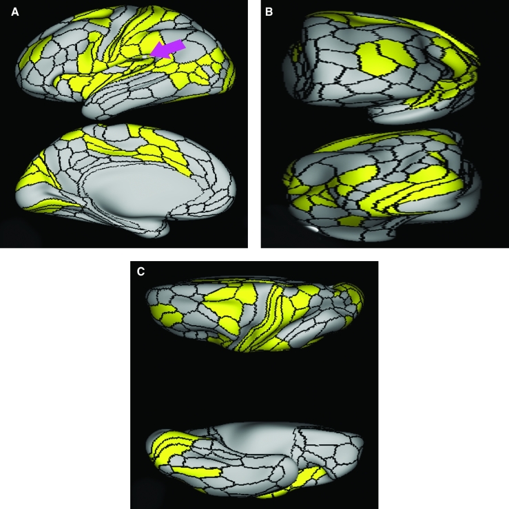
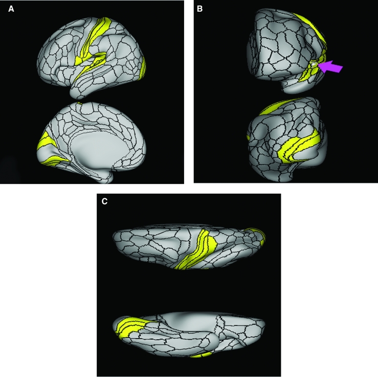
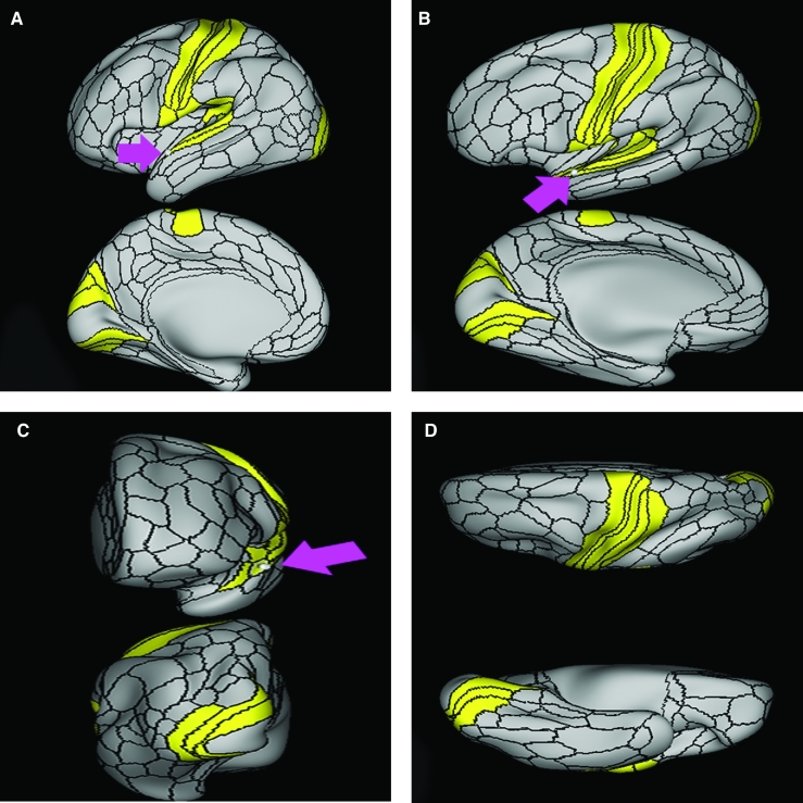
Area AVI demonstrates functional connectivity to areas 6ma and 6r in the premotor regions, areas 44, p47r, 8C, a9-46v, p9-46v, and 9-46d in the lateral frontal lobe, areas 8BM d32, and a32prime in the medial frontal lobe, areas FOP4 and FOP5 in the superior insula opercular regions, areas AAIC and MI in the lower opercula and Heschl's gyrus regions, areas TE2p, PHA3, and PHT in the temporal lobe, areas LIPd, PF, PFm, and IP2 in the lateral parietal lobe, and area V1 in the medial occipital lobe (Figure 7).
FIGURE 7.
Functional connectivity of AVI demonstrated on an inflated left hemisphere. A, Lateral and medial views. B, Rostral and caudal views. C, Dorsal and ventral views. Parcellations with the strongest functional connectivity are shown in yellow. Pink arrows designate the parcellation of interest.
What are its white matter connections?
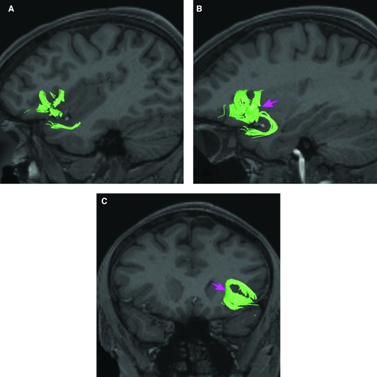
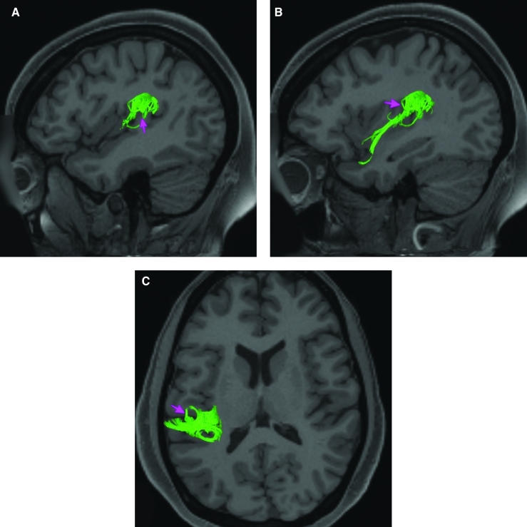
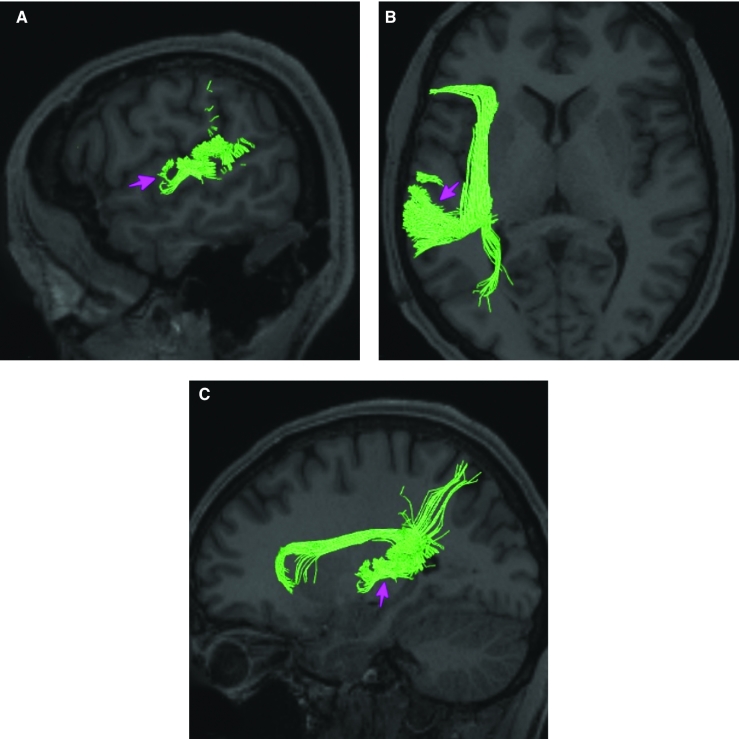
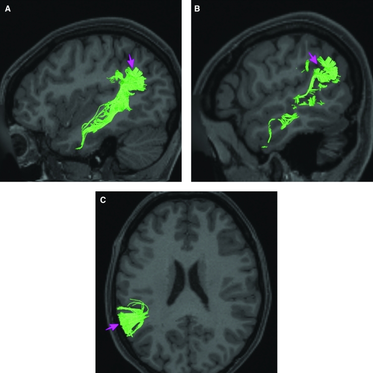
Area AVI is structurally connected to local parcellations. Local short association bundles connect to insular parcellations 47s, 47l, AAIC, FOP4, FOP5, and MI, and temporal pole parcellations TGd (Figure 8).
FIGURE 8.
Structural connectivity of AVI in the left hemisphere, shown on T1-weighted MR images. Sagittal views of A, lateral and B, medial planes. C, Coronal view. Green: white matter tracts of AVI demonstrating connections with local parcellations. Pink arrows designate the parcellation of interest.
What is known about its function?
Area AVI is a newly described area of the brain and was parcellated from the anterior insula. The anterior insula is suggested to have a role in sensation and control of autonomic nervous system processes as well as playing a role in human awareness, self-recognition, time perception, and perceptual decision making.5,6 Area AVI was parcellated from areas MI and AAIC in the anterior insula based on functional activity differences between regions related to motor, arithmetic, auditory language, and semantic tasks.7
Area MI
Where is it?
Area MI (middle insula) is located in the posterior superior portion of the short insular gyrus.
What are its borders?
Area MI borders AVI anteriorly, POI2 posteriorly, AAIC inferiorly, and FOP3 and FOP4 superiorly.
What is its functional connectivity?
Area MI demonstrates functional connectivity to areas SCEF, FEF, PEF, 6ma, and 6r in the premotor regions, areas IFSa, 46, and 9-46d in the lateral frontal lobe, areas 5mv, 23c, a24prime, p24prime, a32prime, and p32prime in the medial frontal lobe, areas OP4, 43, PFcm, FOP1, FOP3, FOP4, and FOP5 in the superior insula opercular regions, areas 52, PI, AVI, AAIC, PoI1, and PoI2 in the lower opercula and Heschl's gyrus regions, area PHT in the temporal lobe, areas LIPd, PF, PFm, PFop, and PFt in the lateral parietal lobe, area 7am in the medial parietal lobe, and area V6 in the dorsal visual stream area (Figure 9).
FIGURE 9.
Functional connectivity of MI demonstrated on an inflated left hemisphere. A, Lateral and medial views. B, Rostral and caudal views. C, Dorsal and ventral views. Parcellations with the strongest functional connectivity are shown in yellow. Pink arrows designate the parcellation of interest.
What are its white matter connections?
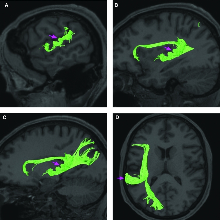
Area MI is structurally connected with the arcuate/SLF, frontal aslant tract, and local parcellations. Some individuals have connections to the parietal and occipital lobe though these tracts are inconsistent. Connections to the superior frontal gyrus through the FAT project superior to 8BL, 6ma, and SFL. Superior temporal gyrus connections are portions of the arcuate/SLF and course from MI, medial and superior to the insula to end laterally at A4 and A5. Local short association bundles connect with PoI1, 47s, Pir, AAIC, AVI, FOP3, FOP4, and FOP5 (Figure 10).
FIGURE 10.
Structural connectivity of MI in the left hemisphere, shown on T1-weighted MR images. Sagittal views of A, lateral, B, medial, and C, far medial planes. D, Coronal view. Orange: white matter tracts of MI demonstrating connections with the arcuate/SFL, frontal aslant tract, and local parcellations. Pink arrows designate the parcellation of interest.
What is known about its function?
Area MI is a newly described area of the brain and was parcellated from the anterior insula. The anterior insula is suggested to have a role in sensation and control of autonomic nervous system processes as well as playing a role in human awareness, self-recognition, time perception, and perceptual decision making.5,6 Area MI was parcellated from areas AAIC and AVI in the anterior insula based on functional activity differences between regions related to motor, arithmetic, auditory language, and semantic tasks.7
Area FOP5
Where is it?
Area FOP5 (frontal operculum 5) is located on the undersurface of the opercular portions of pars triangularis of the IFG.
What are its borders?
Area FOP5 is principally the underside of area 45, though parts of area 44 and area 47L are on its exterior surface. AVI is inferior to it and FOP4 is posterior to it.
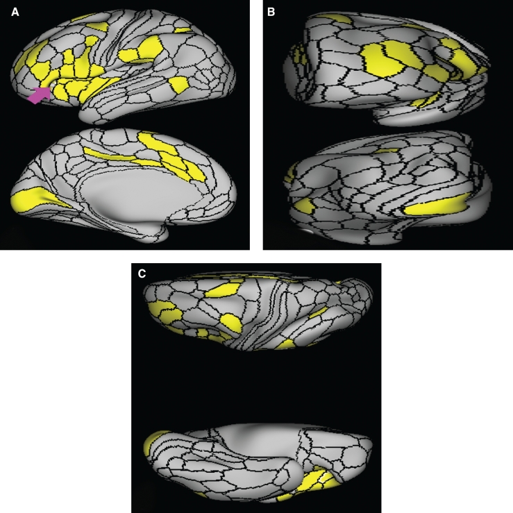
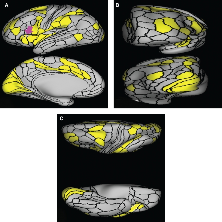
What is its functional connectivity?
Area FOP5 demonstrates functional connectivity to areas SCEF, FEF, PEF, 6ma, and 6r in the premotor regions, areas 44, 45, IFSa, IFja, 46, and 9-46d in the lateral frontal lobe, areas 23c, a24prime, p24prime, a32prime, and p32prime in the medial frontal lobe, areas 43, PFcm, FOP3, and FOP4 in the superior insula opercular regions, areas AVI, MI, PoI1, and PoI2 in the lower opercula and Heschl's gyrus regions, area PHT in the temporal lobe, areas LIPd, PF, and PFop in the lateral parietal lobe, area 7am in the medial parietal lobe, and area V1 in the medial occipital lobe (Figure 11).
FIGURE 11.
Functional connectivity of FOP5 demonstrated on an inflated left hemisphere. A, Lateral and medial views. B, Rostral and caudal views. C, Dorsal and ventral views. Parcellations with the strongest functional connectivity are shown in yellow. Pink arrows designate the parcellation of interest.
What are its white matter connections?
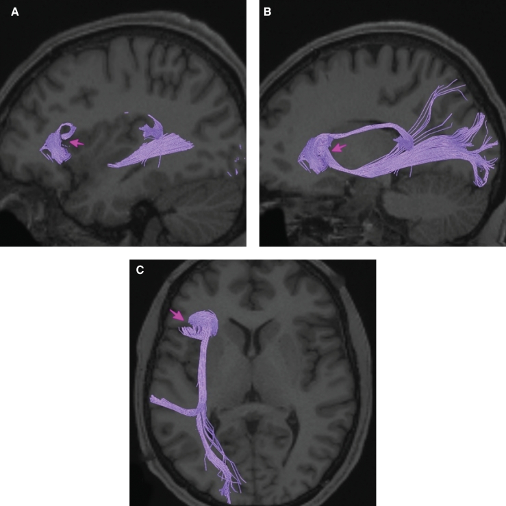
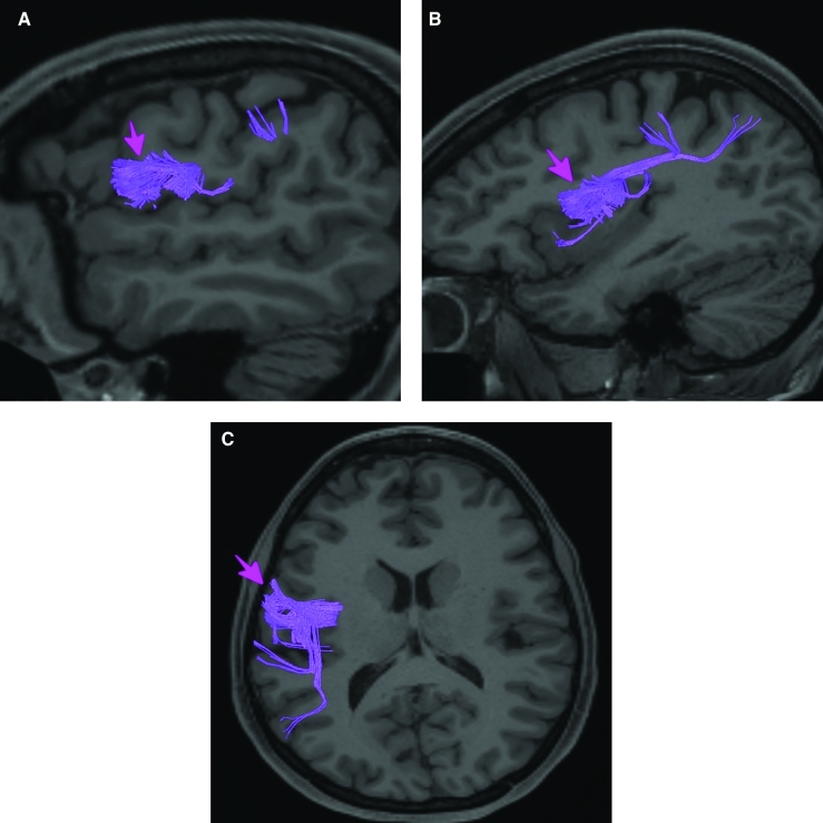
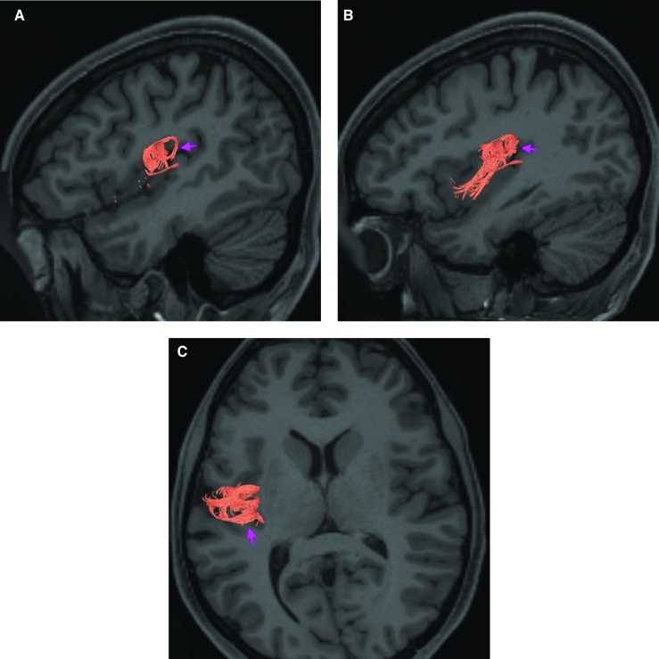
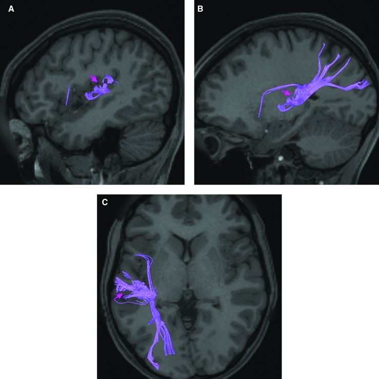
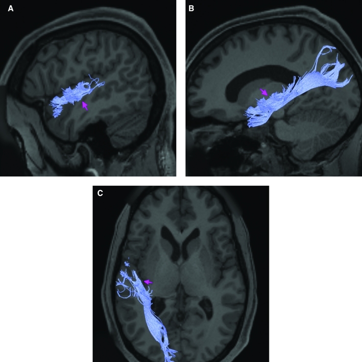
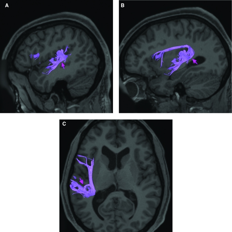
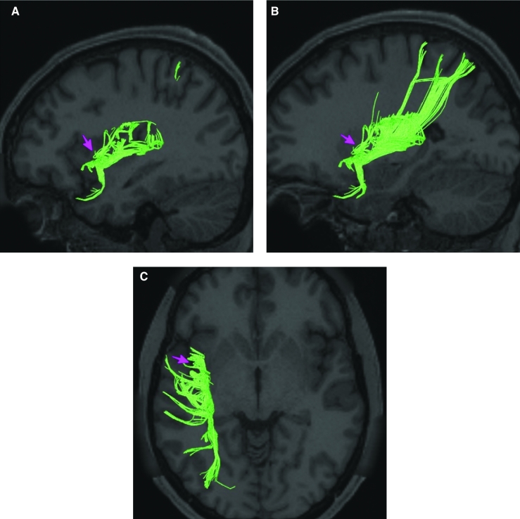
Area FOP5 is structurally connected to the IFOF and arcuate/SLF. The IFOF courses from occipital lobe parcellations V1, V2 V3, and parietal area MIP, through the extreme/external capsule to turn laterally to FOP5. FOP5 has arcuate/SLF fibers projecting posteriorly above the insula and turning laterally to end at the posterior insula and planum temporale parcellations RI and LBelt. Local short association fibers connect with 45, FOP4, and AVI (Figure 12). White matter tracts from FOP5 in the right hemisphere have less consistent connections with the arcuate/SLF (Figure 12).
FIGURE 12.
Structural connectivity of FOP5 in the left hemisphere, shown on T1-weighted MR images. Sagittal views of A, lateral and B, medial planes. C, Axial view. Purple: white matter tracts of FOP5 demonstrating connections with the IFOF and the arcuate/SFL. Pink arrows designate the parcellation of interest.
What is known about its function?
Area FOP5 is a newly described area of the brain that was parcellated from the frontal operculum. The frontal operculum plays a key role in the initiation of language and lexical retrieval required for language learning.8,9 Area FOP5 was parcellated from FOP4 based on functional activity differences between these 2 regions. Specifically, FOP5 showed greater activity in motor tasks during which individuals squeezed their left and right toes, tapped their left and right fingers, and moved their tongue.7 Area FOP5 was parcellated from area 44 based on functional activity differences in motor cue and semantic tasks.7
Area FOP4
Where is it?
Area FOP4 (frontal operculum 4) is located on the inner surface of the pars opercularis of the IFG.
What are its borders?
Area FOP4 borders area 44 on its exterior surface as well as a small border with area 6r posterosuperiorly. Its posterior border is made up of FOP1 and FOP3. Its inferior border is with MI and AVI. Its anterior border is with FOP5.
What is its functional connectivity?
Area FOP4 demonstrates functional connectivity to areas SCEF, FEF, PEF, 6v, 6a, 6ma, and 6r in the premotor regions, areas IFSa, 46, and 9-46d in the lateral frontal lobe, areas 5mv, 23c, 24dv, a24prime, p24prime, a32prime, and p32prime in the medial frontal lobe, areas 43, PFcm, OP4, FOP3, and FOP5 in the superior insula opercular regions, areas AVI, MI, PI, 52, PoI1, and PoI2 in the lower opercula and Heschl's gyrus regions, area PHT in the temporal lobe, areas 7AL, 7PL, AIP, LIPv, LIPd, PF, PFt, PGp, and PFop in the lateral parietal lobe, area V6 in the dorsal visual stream area, area 7am and DVT in the medial parietal lobe, and areas V1, V2, and V3 in the medial occipital lobe (Figure 13).
FIGURE 13.
Functional connectivity of FOP4 demonstrated on an inflated left hemisphere. A, Lateral and medial views. B, Rostral and caudal views. C, Dorsal and ventral views. Parcellations with the strongest functional connectivity are shown in yellow. Pink arrows designate the parcellation of interest.
What are its white matter connections?
Area FOP4 is structurally connected to the frontal aslant tract and the arcuate/SLF. Connections from FOP4 with the frontal aslant tract project superior to the superior frontal gyrus to end at parcellations 6ma and SFL. Arcuate/SLF fibers project posteriorly above the insula, curving around the termination of the sylvain fissure to end at TGd and TE1a. From the arcuate/SLF there are also projections to the superior temporal gyrus that end at A4 and A5. Local short association fibers connect with AVI, FOP3, FOP5, and MI (Figure 14). White matter connections from FOP4 in the right hemisphere have less consistent connections with the arcuate/SLF.
FIGURE 14.
Structural connectivity of FOP4 in the left hemisphere, shown on T1-weighted MR images. Sagittal views of A, lateral, B, medial, and C, far medial planes. D, Coronal view. Light blue: white matter tracts of FOP4 demonstrating connections with the frontal aslant tract and the arcuate/SFL. Pink arrows designate the parcellation of interest.
What is known about its function?
Area FOP4 is a newly described area of the brain that was parcellated from the frontal operculum. The frontal operculum plays a key role in the initiation of language and lexical retrieval required for language learning.8,9 Area FOP4 was parcellated from areas AVI and FOP1 based on differences in functional activity in motor-based tasks.7
Area FOP3
Where is it?
Area FOP3 (frontal operculum 3) is located in the superior circular sulcus of the insula, just anterior to the junction of this sulcus with the superior aspects of the long and short gyri. FOP3 sits just between these 2 junctions, slightly deviated toward the short gyri.
What are its borders?
Area FOP3 borders FOP1 and FOP4 superiorly. FOP2 is its posterior boundary and FOP4 is its anterior boundary. Its inferior boundary is with MI.
What is its functional connectivity?
Area FOP3 demonstrates functional connectivity to areas SCEF, 6v, 6ma, and 6r in the premotor regions, areas 46 and 9-46d in the lateral frontal lobe, areas 5mv, 23c, 24dv, a24prime, and p32prime in the medial frontal lobe, areas 43, PFcm, OP4, FOP1, FOP2, FOP3, and FOP5 in the superior insula opercular regions, areas MI, 52, PoI1, and PoI2 in the lower opercula and Heschl's gyrus regions, area PHT in the temporal lobe, areas 7AL, PF, and PFop in the lateral parietal lobe, areas V3a V6 in the dorsal visual stream area, and area V2 in the medial occipital lobe (Figure 15).
FIGURE 15.
Functional connectivity of FOP3 demonstrated on an inflated left hemisphere. A, Lateral and medial views. B, Rostral and caudal views. C, Dorsal and ventral views. Parcellations with the strongest functional connectivity are shown in yellow. Pink arrows designate the parcellation of interest.
What are its white matter connections?
Area FOP3 is structurally connected to the frontal aslant tract and local parcellations. Portions of the frontal aslant connect with FOP3, the fibers project superiorly to the superior frontal gyrus to end at parcellations 6ma and SFL. Local short association bundles connect with FOP2, FOP4, FOP5, MI, PoI2, and Ig (Figure 16).
FIGURE 16.
Structural connectivity of FOP3 in the left hemisphere, shown on T1-weighted MR images. Sagittal views of A, lateral and B, medial planes. C, Coronal view. Orange: white matter tracts of FOP3 demonstrating connections with the frontal aslant track and local parcellations. Pink arrows designate the parcellation of interest.
What is known about its function?
Area FOP3 is a newly described area of the brain that was parcellated from the frontal operculum. The frontal operculum plays a key role in the initiation of language and lexical retrieval required for language learning.8,9 Area FOP3 was separated from FOP4-based functional activity differences during motor cue and arithmetic tasks.7
Area FOP2
Where is it?
Area FOP2 (frontal operculum 2) lies in the superior circular sulcus of the insula. It is a small area which sits just anterior to the junction of the long insular gyri and the circular sulcus.
What are its borders?
Area FOP2 borders FOP1 and FOP3 on its anterior surface and area 43 on its superior surface. Its inferior and posterior surfaces are made up of IG and to a lesser extent PoI2.
What is its functional connectivity?
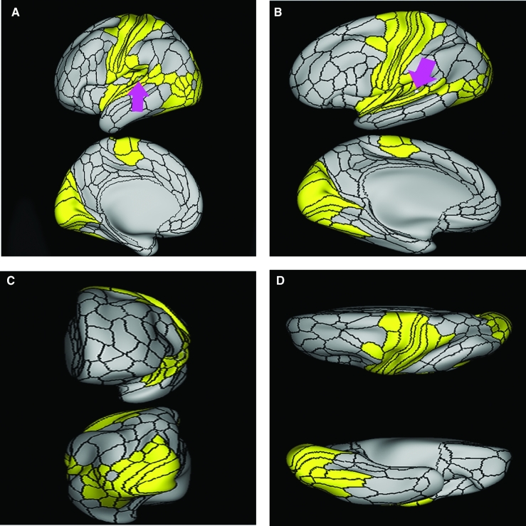
Area FOP2 demonstrates functional connectivity to areas 1, 2, 3a, and 3b in the sensory strip, area 4 in the motor strip, areas SCEF, 6v, 6d, 6a, 6mp, and 6r in the premotor regions, areas 43, PFcm, IG, OP4, OP1, FOP1, and FOP3 in the superior insula opercular regions, areas PoI1 and PoI2 in the lower opercula and Heschl's gyrus regions, areas 7AL, 7PC, AIP, PFt, and PFop in the lateral parietal lobe, and area FST in the lateral occipital lobe (Figure 17).
FIGURE 17.
Functional connectivity of FOP2 demonstrated on an inflated left hemisphere. A, Lateral and medial views. B, Oblique views demonstrating the operculum. C, Dorsal and ventral views. D, Rostral and caudal views. Parcellations with the strongest functional connectivity are shown in yellow. Pink arrows designate the parcellation of interest.
What are its white matter connections?
Area FOP2 is structurally connected to local parcellations. Local short association bundles are connected to FOP3, FOP4, FOP1, OP1, and OP2-3 (Figure 18).
FIGURE 18.
Structural connectivity of FOP2 in the left hemisphere, shown on T1-weighted MR images. Sagittal views of A, lateral and B, medial planes. C, Coronal view. White: white matter tracts of FOP2 demonstrating connections with local parcellations. Pink arrows designate the parcellation of interest.
What is known about its function?
Area FOP2 is a newly described area of the brain that was parcellated from the frontal operculum. The frontal operculum plays a key role in the initiation of language and lexical retrieval required for language learning.8,9 Area FOP2 was separated from FOP3 based on functional activity differences during arithmetic and auditory language tasks.7 FOP2 was also separated from FOP1 based on object interaction tasks.7
Area FOP1
Where is it?
Area FOP1 (frontal operculum 1) is a tiny area on the undersurface of the posterior portions of the pars opercularis of the IFG.
What are its borders?
Area FOP1 borders areas 6r and 43 superiorly. It has a small anterior border with FOP4 and its inferior borders are with FOP2 and FOP3.
What is its functional connectivity?
Area FOP1 demonstrates functional connectivity to area 2 in the sensory strip, areas SCEF, FEF, 6v, 6ma, 6mp, and 6r in the premotor regions, areas 46 and 9-46d in the lateral frontal lobe, areas 5mv, 23c, 24dv, a24prime, and p32prime in the medial frontal lobe, areas 43, PFcm, OP4, FOP2, FOP3, and FOP4 in the superior insula opercular regions, areas MI, 52, PoI1, and PoI2 in the lower opercula and Heschl's gyrus regions, area PHT in the temporal lobe, areas 7AL, PF, and PFop in the lateral parietal lobe, and areas V3a and V6 in the dorsal visual stream area (Figure 19).
FIGURE 19.
Functional connectivity of FOP1 demonstrated on an inflated left hemisphere. A, Lateral and medial views. B, Oblique views demonstrating the operculum. C, Dorsal and ventral views. D, Rostral and caudal views. Parcellations with the strongest functional connectivity are shown in yellow. Pink arrows designate the parcellation of interest.
What are its white matter connections?
Area FOP1 is structurally connected to portions of the frontal aslant tract and arcuate/SLF. While the majority has connections to these white matter tracts, they are not consistent across all individuals. Frontal aslant fibers project superiorly from FOP1 to the superior frontal gyrus to end at SFL. Connections with the arcuate/SLF course above the insula, wrapping around the termination of Sylvian fissure to end at inferior temporal gyrus area TE1a. From the arcuate/SLF there are also connections to the inferior parietal lobule to PF. Local short association bundles connect with FOP2, FOP3, FOP4, and FOP5 (Figure 20). Majority of connections from FOP1 in the right hemisphere are local.
FIGURE 20.
Structural connectivity of FOP1 in the left hemisphere, shown on T1-weighted MR images. Sagittal views of A, lateral, B, medial, and C, far medial planes. D, Coronal view. Purple: white matter tracts of FOP1 demonstrating connections with portions of the frontal aslant tract, and the arcuate/SFL. Pink arrows designate the parcellation of interest.
What is known about its function?
Area FOP1 is located in the posterior operculum. This region of the cortex demonstrates activity during imagined abstract movement from a third person perspective.10 It has also been proposed that the posterior operculum is an important area for perception of pain, and may act as a messenger for transmitting somatosensory information to the limbic system.11
Area 43
Where is it?
Area 43 is found on the anterior portions of the subcentral gyrus (where the precentral and postcentral gyri meet just below the central sulcus). It involves the lateral surface of that operculum as well as the inferior surface which faces the Sylvian fissure.
What are its borders?
Area 43 borders area 6r anteriorly and OP4 posteriorly. Its superior border includes area 6v, as well as areas 4 and 3a. Its inferior borders include FOP1 and FOP2.
What is its functional connectivity?
Area 43 demonstrates functional connectivity to areas 1, 2, 3a, and 3b in the sensory strip, area 4 in the motor strip, areas SCEF, FEF, PEF, 6ma, 6mp, 6r, and 6v in the premotor regions, areas a24prime, p24prime, p32prime, 24dd, 24dv, 5mv, and 23c in the middle cingulate regions, areas 46 and 9-46d in the lateral frontal lobe, areas IG, OP4, OP2-3, OP1, PFcm, FOP1, FOP3, FOP4, and FOP5 in the superior insula opercular regions, areas STV, LBelt, PBelt, MBelt, A1, TA2, PI, A4, MI, STV, 52, RI, PoI1, and PoI2 in the lower opercula and Heschl's gyrus regions, area PHT in the temporal lobe, areas PF, and 7AL in the lateral parietal lobe, area DVT in the medial parietal lobe, areas V1, V2, V3, and V4 in the medial occipital lobe, areas V3a, V3b, V6, V6a, and V7 of the dorsal visual stream, areas V8 and FFC of the ventral visual stream, and areas LO3, TPOJ2, MST, and FST of the lateral occipital lobe (Figure 21).
FIGURE 21.
Functional connectivity of 43 demonstrated on an inflated left hemisphere. A, Lateral and medial views. B, Rostral and caudal views. C, Dorsal and ventral views. Parcellations with the strongest functional connectivity are shown in yellow. Pink arrows designate the parcellation of interest.
What are its white matter connections?
Area 43 is structurally connected to the superior parietal lobe and local parcellations. Parietal lobe connections project posterior form 43 to end at PFt, PFm, and PFcm. Local short association bundles connect with 6v, 6r, OP4, 3a, 4, FOP1, FOP2, and MI (Figure 22).
FIGURE 22.
Structural connectivity of 43 in the left hemisphere, shown on T1-weighted MR images. Sagittal views of A, lateral and B, medial planes. C, Axial view. Purple: white matter tracts of 43 demonstrating connections with the superior parietal lobe and local parcellations. Pink arrows designate the parcellation of interest.
What is known about its function?
It has been proposed that area 43 is functionally correlated with area 41,7 which has been implicated in motor activities related to swallowing.12,13
Parietal Apex Regions
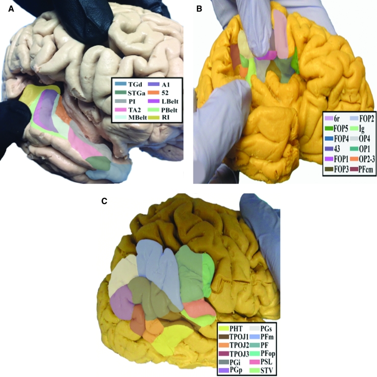
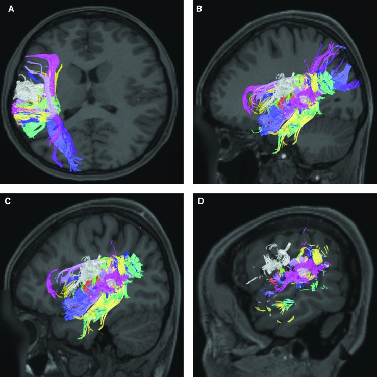
The parietal apex is one of the most difficult areas of the cortex to visualize in the cerebrum as it is tightly packed, and not one into which we make frequent forays given its eloquent anatomy. This section includes discussion of the regions of the parietal opercula including the 3 OP areas, the supramarginal gyrus areas, the retroinsular area, and the areas involving Heschl's gyrus. The specific parcellations that comprise this region include Ig, OP4, OP2-3, OP1, PFcm, RI, 52, A1, MBelt, LBelt, PSL, and STV. The anatomic location of these parcellations is shown on a cadaver brain in Figure 23. This region has consistent white matter connections with the arcuate/SLF, middle longitudinal fasciculus, anterior insular areas, and local parcellations. The combined tractography of these parcellations is shown in Figure 24.
FIGURE 23.
Anatomical location of parietal apex parcellations shown on the left hemisphere of a cadaver brain. A, Oblique view with a widening of Heschel's gyrus to show extensions of parcellations. B, Inferolateral view of parietal opercular regions. C, Lateral view of the parcellation comprising the inferior parietal lobule.
FIGURE 24.
Combined structural connectivity of the parietal apex parcellations in the left hemisphere, shown on T1-weighted MR images. A, Axial view. Sagittal views of B, medial, C, lateral, and D, far lateral planes.
Area Ig
Where is it?
Area Ig (insula granular cortex) is at the superior portion of the long insular gyri. It is the posterior superior most part of the insula proper.
What are its borders?
Area Ig borders PoI1 and PoI2 inferiorly, FOP2 anteriorly, OP2-3 and a bit of OP4 superiorly, and RI and area 52 posteriorly.
What is its functional connectivity?
Area Ig demonstrates functional connectivity to areas 1, 3a, and 3b of the sensory strip, area 4 of the motor strip, and areas FOP2, OP2-3, OP4, 42, 52, and PoI2 in the insula opercular region (Figure 25).
FIGURE 25.
Functional connectivity of Ig demonstrated on an inflated left hemisphere. A, Lateral and medial views. B, Rostral and caudal views. C, Dorsal and ventral views. Parcellations with the strongest functional connectivity are shown in yellow. Pink arrows designate the parcellation of interest.
What are its white matter connections?
Area Ig is structurally connected to local parcellations and MI. From Ig there are anterior fibers that project to MI. Local short association bundles are connected with A1, MBelt, FOP2, OP1, OP2-3, OP4, PoI2, and PoI1 (Figure 26).
FIGURE 26.
Structural connectivity of Ig in the left hemisphere, shown on T1-weighted MR images. Sagittal views of A, lateral and B, medial planes. C, Axial view. Green: white matter tracts of Ig demonstrating connections with local parcellations and MI. Pink arrows designate the parcellation of interest.
What is known about its function?
Area Ig has been shown to be activated in response to cutaneous stimuli.14 This area has also been shown to respond to different stimuli related to taste and is thought to be the gustatory center of the brain.15
Area OP4
Where is it?
Area OP4 (operculum 4) is located in the posterior portion of the subcentral gyrus, which is the opercular portion of the sensory and motor cortices. It folds onto the inferior surface to face the Sylvian fissure. It appears slightly on the lateral surface of the operculum, but is mostly located on the undersurface
What are its borders?
Area OP4 borders 43 anteriorly, and the parietal areas (discussed elsewhere) PFcm and PFop posteriorly. Its superior border is with postcentral areas 1, 3a, and 3b. Its interior (inferior) border is with OP1, OP2-3, and a small part of IG.
What is its functional connectivity?
Area OP4 demonstrates functional connectivity to areas 1, 2, 3a, and 3b in the sensory strip, area 4 in the motor strip, areas SCEF, FEF, 6mp, 6r, 6d, and 6v in the premotor regions, areas a24prime, p32prime, 24dd, 24dv, 5mv, and 23c in the middle cingulate regions, area 46 in the lateral frontal lobe, areas IG, 43, OP2-3, OP1, PFcm, FOP1, FOP2, FOP3, and FOP4 in the superior insula opercular regions, areas STV, LBelt, PBelt, MBelt, A1, TA2, PI, A4, A5, TA2, MI, STV, 52, RI, PoI1, and PoI2 in the lower opercula and Heschl's gyrus regions, areas PF, PFop, PFt, AIP, 7PC, and 7AL in the lateral parietal lobe, area V3 in the medial occipital lobe, areas V6, V6a, and V7 of the dorsal visual stream, areas FFC of the ventral visual stream, and areas LO3, TPOJ2, TPOJ3, MT, MST, and FST of the lateral occipital lobe (Figure 27).
FIGURE 27.
Functional connectivity of OP4 demonstrated on an inflated left hemisphere. A, Lateral and medial views. B, Rostral and caudal views. C, Dorsal and ventral views. Parcellations with the strongest functional connectivity are shown in yellow. Pink arrows designate the parcellation of interest.
What are its white matter connections?
Area OP4 is structurally connected to local parcellations. Local short association bundles connect with 43, PFcm, PFop, Ig, LBelt, OP1, OP2-3, OP4, 1, 3a, 3b, RI, PoI1, and PoI2 (Figure 28).
FIGURE 28.
Structural connectivity of OP4 in the left hemisphere, shown on T1-weighted MR images. Sagittal views of A, lateral and B, medial planes. C, Axial view. White: white matter tracts of OP4 demonstrating connections with local parcellations. Pink arrows designate the parcellation of interest.
What is known about its function?
Area OP4 is a part of the posterior opercular cortex. This region is involved in processes required for sensory motor functions, such as integrating sensory responses into motor actions, and is crucial in tangible object recognition and manipulation.16 Area OP4 was differentiated from areas OP1 and OP2-3 based on differences in activity on fMRI during motor and arithmetic tasks.7
Area OP2-3
Where is it?
Area OP2-3 (operculum part 2 and 3) is a small area located in the posterior superior most part of the circular sulcus of the insula.
What are its borders?
Area OP2-3 borders OP 4 and OP1 superiorly, RI posteriorly, and IG anterosuperiorly.
What is its functional connectivity?
Area OP2-3 demonstrates functional connectivity to areas 1, 2, 3a, and 3b in the sensory strip, area 4 in the motor strip, areas IG, 43, OP4, OP1, and PFcm in the superior insula opercular regions, areas PBelt, A4, and RI in the lower opercula and Heschl's gyrus regions, area PFop in the lateral parietal lobe, areas V2 and V3 in the medial occipital lobe, and areas V6, andV3a of the dorsal visual stream (Figure 29).
FIGURE 29.
Functional connectivity of OP2-3 demonstrated on an inflated left hemisphere. A, Lateral and medial views. B, Rostral and caudal views. C, Dorsal and ventral views. Parcellations with the strongest functional connectivity are shown in yellow. Pink arrows designate the parcellation of interest.
What are its white matter connections?
Area OP2-3 is structurally connected to anterior insula areas and local parcellations. Anterior projections from OP2-3 end at anterior insula parcellations MI, AAIC, and PoI2. Local short association bundles connect with A1, Ig, LBelt, MBelt, and OP4 (Figure 30).
FIGURE 30.
Structural connectivity of OP2-3 in the left hemisphere, shown on T1-weighted MR images. Sagittal views of A, lateral and B, medial planes. C, Axial view. Orange: white matter tracts of OP2 demonstrating connections with the anterior insula areas and local parcellations. Pink arrows designate the parcellation of interest.
What is known about its function?
Area OP2-3 is a newly described region originating in the posterior operculum. These 2 areas are located within the Sylvian fissure and are so closely intertwined that it is difficult to distinguish between them.17 There is evidence that the posterior operculum plays a role in receiving input from somatosensory stimuli.18 Area OP2-3 was separated from areas Ig and OP1 based on differences in activity on fMRI during motor and arithmetic tasks.7
Area OP1
Where is it?
Area OP1 (operculum 1) is located on the undersurface of the anterior portion of the superior supramarginal gyrus.
What are its borders?
Area OP1 is small and diamond shaped. Its anterior superior border is OP4. Its anterior inferior border is OP2-3. Its posterior superior border is PFcm. Its posterior inferior border is RI.
What is its functional connectivity?
Area OP1 demonstrates functional connectivity to areas 1, 2, 3a, and 3b in the sensory strip, area 4 in the motor strip, areas 6d, 6v, and 6mp in the premotor areas, areas 5L, 24dd, 24dv, and SCEF in the medial cingulate areas, areas 43, OP4, OP2-3, FOP2, and PFcm, in the superior insula opercular regions, areas PBelt, A4, 52, and RI in the lower opercula and Heschl's gyrus regions, areas 7AL and PFop in the lateral parietal lobe, and areas MST and FST in the lateral occipital lobe (Figure 31).
FIGURE 31.
Functional connectivity of OP1 demonstrated on an inflated left hemisphere. A, Lateral and medial views. B, Rostral and caudal views. C, Dorsal and ventral views. Parcellations with the strongest functional connectivity are shown in yellow. Pink arrows designate the parcellation of interest.
What are its white matter connections?
Area OP1 is structurally connected to local parcellations, the anterior insula and the temporal pole. Fibers from OP1 project anteriorly to the anterior insula ending at parcellation MI and continue to the temporal pole at parcellation TGd. Local short association bundles connect with PFcm, RI, OP2-3, OP4, and 43 (Figure 32). There are less consistent connections from OP1 to the temporal pole (Figure 32).
FIGURE 32.
Structural connectivity of OP1 in the left hemisphere, shown on T1-weighted MR images. Sagittal views of A, lateral and B, medial planes. C, Axial view. Green: white matter tracts of OP1 demonstrating connections with local parcellations, the anterior insula, and the temporal pole. Pink arrows designate the parcellation of interest.
What is known about its function?
Area OP1 is also part of the posterior opercular cortex. This area likely contributes to somatosensory processing tasks such as recognition of pain, tactile attention, and working memory.17 It has also been suggested that area OP1 plays an important role in bimanual task activities.16 Area OP1 was differentiated from OP2-3 based on decreased levels of fMRI activity during working memory tasks.7
Area PFcm
Where is it?
Area PFcm (parietal F, region cm) is located in the superior portion of the supramarginal gyrus. It is primarily located on the opercular cleft of this part of the gyrus.
What are its borders?
Area PFcm borders parietal regions PF and PFop superiorly. Its anteroinferior border is OP1. RI is its inferior border. Its posterior border is PSL.
What is its functional connectivity?
Area PFcm demonstrates functional connectivity to area OP4 demonstrates functional connectivity to areas 1, 2, 3a, and 3b in the sensory strip, areas SCEF, FEF, PEF, 6mp, 6r, 6a, and 6v in the premotor regions, areas 24dv, a24prime, p24prime, p32prime, 24dd, 24dv, 5mv, and 23c in the middle cingulate regions, areas 9-46d and 46 in the lateral frontal lobe, areas 43, OP4 OP2-3, OP1, PFcm, FOP1, FOP2, FOP3, FOP4, and FOP5 in the superior insula opercular regions, areas LBelt, PBelt, MBelt, A1, TA2, PI, A4, TA2, MI, STV, 52, RI, PoI1, and PoI2 in the lower opercula area and Heschl's gyrus regions, PHT in the temporal lobe, areas PF, PFop, PFt, PGp, AIP, 7PC, and 7AL in the lateral parietal lobe, areas 7am and DVT in the medial parietal lobe, areas V2 and V3 in the medial occipital lobe, area V3a of the dorsal visual stream, areas FFC of the ventral visual stream, and areas LO3, TPOJ1, TPOJ2, TPOJ3, MST, and FST of the lateral occipital lobe (Figure 33).
FIGURE 33.
Functional connectivity of PFcm demonstrated on an inflated left hemisphere. A, Lateral and medial views. B, Rostral and caudal views. C, Dorsal and ventral views. Parcellations with the strongest functional connectivity are shown in yellow. Pink arrows designate the parcellation of interest.
What are its white matter connections?
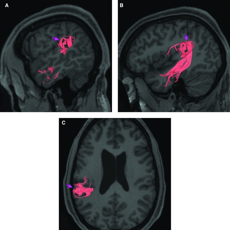
Area PFcm is structurally connected to portions of the arcuate/SLF. Arcuate/SLF connection wraps around the termination of the Sylvian fissure with inferior projections through the temporal lobe to end at inferior and middle temporal gyrus parcellations TE1a, STSva and TE2a. Local short association bundles are connected with OP1, OP4, PFop, PBelt, PF, and RI (Figure 34). There are less consistent inferior connections from PFcm in the right hemisphere through the temporal lobe.
FIGURE 34.
Structural connectivity of PFcm in the left hemisphere, shown on T1-weighted MR images. Sagittal views of A, lateral and B, medial planes. C, Axial view. Red: white matter tracts of PFcm demonstrating connections with portions of the arcuate/SFL. Pink arrows designate the parcellation of interest.
What is known about its function?
Area PFcm is a subdivision of the inferior parietal cortex. The inferior parietal cortex is believed to be important in processing language with regard to language vocabulary, semantics, articulation, and working memory.19 Area PFcm was differentiated from area PSL based on lower levels of activity on fMRI during arithmetic and auditory story tasks.7 Area PFcm was also differentiated from area RI based on higher levels of activity on fMRI during motor cue tasks.7
Area RI
Where is it?
Area RI (retroinsular cortex) is located in the retroinsular cortex, which itself is located anterior superior to the long gyri of the insula. It is located at the deep termination of Heschl's gyrus at its superomedial termination.
What are its borders?
Area RI is slanted from anterior-inferior to posterior superior. On its anterior inferior border, it meets IG, OP2-3, and OP1. On its exterior border, it meets PFcm and PSL. On its deep inferoposterior border, it joins with A1, Mbelt, Lbelt, and Pbelt, the Heschl's gyrus regions.
What is its functional connectivity?
Area RI demonstrates functional connectivity to areas 1, 2, 3a, and 3b in the sensory strip, area 4 in the motor strip, areas FEF, 6mp, and 6v in the premotor regions, areas 43, OP4 OP2-3, OP1, and PFcm in the superior insula opercular regions, areas LBelt, PBelt, MBelt, A1, TA2, PI, A4, A5, TA2, STV, and 52 in the lower opercula area and Heschl's gyrus regions, area PFop in the lateral parietal lobe, areas V2, V3, and V4 in the medial occipital lobe, areas V6, V3b, V3a, and V7 of the dorsal visual stream, areas FFC of the ventral visual stream, and areas LO3, TPOJ1, TPOJ2, TPOJ3, MT, MST V4t, and FST of the lateral occipital lobe (Figure 35).
FIGURE 35.
Functional connectivity of RI demonstrated on an inflated left hemisphere. A, Lateral and medial views. B, Rostral and caudal views. C, Dorsal and ventral views. Parcellations with the strongest functional connectivity are shown in yellow. Pink arrows designate the parcellation of interest.
What are its white matter connections?
Area RI is structurally connected to portions of the arcuate/SLF, anterior insula, and local parcellations. Arcuate/SLF fibers project anterior form RI to inferior frontal gyrus parcellations 44 and 6r. Insular fibers course anterior to insular parcellations PoI2 and PoI1. Local short association bundles are connected with A1, OP1, OP4, PBelt, and A4 (Figure 36).
FIGURE 36.
Structural connectivity of RI in the left hemisphere, shown on T1-weighted MR images. Sagittal views of A, lateral and B, medial planes. C, Axial view. Yellow: white matter tracts of RI demonstrating connections with portions of the arcuate/SFL, anterior insula, and local parcellations. Pink arrows designate the parcellation of interest.
What is known about its function?
Area RI is an area of the early auditory cortex. Area RI is connected to various somatosensory areas and receives somatosensory input.20 As a result, it is believed that this region plays a role in receiving auditory-somatosensory communications from the auditory cortex in conjunction with the granular insula.20,21
Area 52
Where is it?
Area 52 is located in the posterior edge of the lower limb of the circular sulcus of the insula. It lies between Heschl's gyrus and the long gyri of the insula.
What are its borders?
Area 52 borders MBelt laterally, and PoI1 and IG medially. Its posterosuperior limit ends in OP2-3, and its anteroinferior limit ends in PI.
What is its functional connectivity?
Area 52 demonstrates functional connectivity to areas 1, 2, 3a, and 3b in the sensory strip, areas 24dv, p32prime, a24prime, 23c and 5mv in the middle cingulate areas, areas SCEF and FEF in the premotor regions, area 46 in the lateral frontal lobe, areas 43, IG, FOP1, FOP3, FOP4, OP4, OP1, and PFcm in the superior insula opercular regions, areas MI, PoI1, PoI2, LBelt, PBelt, MBelt, A1, TA2, PI, A4, and STV in the lower opercula area and Heschl's gyrus regions, area PHT in the temporal lobe, areas DVT, PF, and PFop in the parietal lobe, areas V2, V3, and V4 in the medial occipital lobe, areas V6 and V3a of the dorsal visual stream, and area TPOJ2 of the lateral occipital lobe (Figure 37).
FIGURE 37.
Functional connectivity of 52 demonstrated on an inflated left hemisphere. A, Lateral and medial views. B, Rostral and caudal views. C, Dorsal and ventral views. Parcellations with the strongest functional connectivity are shown in yellow. Pink arrows designate the parcellation of interest.
What are its white matter connections?
Area 52 is structurally connected to local parcellations. In some individuals fibers connect to parietal and occipital lobes, however, these tracts are inconsistent. Local short association bundles connect anteriorly to PoI1 and PoI2, and to surrounding parcellations A1, Ig, MBelt, OP1, OP2-3, PoI1, PoI2, and TA2 (Figure 38).
FIGURE 38.
Structural connectivity of 52 in the left hemisphere, shown on T1-weighted MR images. Sagittal views of A, medial and B, lateral planes. C, Axial view. Orange: white matter tracts of 52 demonstrating connections with local parcellations. Pink arrows designate the parcellation of interest.
What is known about its function?
Area 52 is an understudied region on the insular-opercular cortex. No specific functional capabilities have been discussed in the literature for this region. However, area 52 was parcellated from MBelt in the Human Connectome Project study based on differences in activity on fMRI during arithmetic and auditory story tasks.7
Area A1
Where is it?
Area A1 (primary auditory cortex) is located in superior central portion of Heschl's gyrus.
What are its borders?
Area A1 is largely surrounded by the MBelt and Lbelt. Its deep surface borders the RI area.
What is its functional connectivity?
Area A1 demonstrates functional connectivity to areas 2, 3a, and 3b in the sensory strip, areas 43, PFcm, and OP4 in the superior opercular region, areas A4, MBelt, PBelt, LBelt, RI, and 52 in the inferior insula opercular region, and areas V1, V2, V3, and V4 in the medial occipital lobe (Figure 39).
FIGURE 39.
Functional connectivity of A1 demonstrated on an inflated left hemisphere. A, Lateral and medial views. B, Rostral and caudal views. C, Dorsal and ventral views. Parcellations with the strongest functional connectivity are shown in yellow. Pink arrows designate the parcellation of interest.
What are its white matter connections?
Area A1 is structurally connected to the parietal lobe, occipital lobe, inferior frontal gyrus, and local parcellations. Parietal connections are likely portions of the middle longitudinal fasciculus. Fibers from A1 project posterior to parietal parcellations LIPv and 7PC, and to occipital parcellations V3B, V3CD, and V4. Fibers that project anteriorly to the inferior frontal gyrus connect to 44. Local short association bundles connect with 52, LBelt, MBelt, PBelt, and MI (Figure 40).
FIGURE 40.
Structural connectivity of A1 in the left hemisphere, shown on T1-weighted MR images. Sagittal views of A, lateral and B, medial planes. C, Axial view. Purple: white matter tracts of A1 demonstrating connections with the parietal lobe, occipital lobe, inferior frontal gyrus, and local parcellations. Pink arrows designate the parcellation of interest.
What is known about its function?
Area A1, also known as the primary auditory cortex, receives input from the ventral division of the medial geniculate complex and contains a tonotopic map which then interprets input received from the cochlea.22,23
Area MBelt
Where is it?
Area MBelt (medial belt) runs on the medial surfaces of Heschl's gyrus.
What are its borders?
Area MBelt borders A1 on its lateral surface and IG and area 52 on its medial surface. Its superior edge borders RI and its inferior edge borders TA2.
What is its functional connectivity?
Area MBelt demonstrates functional connectivity to areas 1, 2, 3a, and 3b in the sensory strip, areas 43, PFcm, and OP4 in the superior opercular region, areas PoI1, A1, A4, Pbelt, LBelt, Ta2, PI, RI, and 52 in the inferior insula opercular region, and areas V1, V2, V3, and V4 in the medial occipital lobe (Figure 41).
FIGURE 41.
Functional connectivity of Mbelt demonstrated on an inflated left hemisphere. A, Lateral and medial views. B, Rostral and caudal views. C, Dorsal and ventral views. Parcellations with the strongest functional connectivity are shown in yellow. Pink arrows designate the parcellation of interest.
What are its white matter connections?
Area MBelt is structurally connected with the middle longitudinal fasciculus. Fibers from the middle longitudinal fasciculus project posterior just lateral to the lateral ventricle to occipital and parietal lobes to end at parcellations V3, V3A, V6, V6A, IPS1, and DVT. The majority of short association bundles connect anterior to A4, TA2, STGa, and locally to PBelt and LBelt (Figure 42).
FIGURE 42.
Structural connectivity of MBelt in the left hemisphere, shown on T1-weighted MR images. Sagittal views of A, lateral and B, medial planes. C, Axial view. Light Blue: white matter tracts of MBelt demonstrating connections with the middle longitudinal fasciculus. Pink arrows designate the parcellation of interest.
What is known about its function?
Area MBelt is a newly described area of the brain parcellated from the auditory cortex. Area MBelt was differentiated from area 52 based on differences in activity on fMRI during arithmetic and auditory story tasks.7 In addition, MBelt was differentiated from PBelt based on differences in activity on fMRI during arithmetic, auditory story, and object recognition tasks.7
Area LBelt
Where is it?
Area LBelt (lateral belt) is located on the lateral surface of Heschl's gyrus.
What are its borders?
Area LBelt borders A1 and part of Mbelt medially and Pbelt laterally. Its deep surface abuts RI.
What is its functional connectivity?
Area LBelt demonstrates functional connectivity to areas 1, 2, 3a, and 3b in the sensory strip, areas 43, PFcm, OP1, and OP4 in the superior opercular region, areas PoI1, PoI2, A1, A4, A5, MBelt, STV, Ta2, PI, RI, and 52 in the inferior insula opercular region, and areas V1, V2, V3, and V4 in the medial occipital lobe (Figure 43).
FIGURE 43.
Functional connectivity of Lbelt demonstrated on an inflated left hemisphere. A, Lateral and medial views. B, Rostral and caudal views. C, Dorsal and ventral views. Parcellations with the strongest functional connectivity are shown in yellow. Pink arrows designate the parcellation of interest.
What are its white matter connections?
Area LBelt is structurally connected to the arcuate/SLF. Arcuate/SLF fibers wrap around the termination of the sylvain fissure ending at inferior frontal gyrus and insula parcellations 44 and MI. Local short association bundles include the temporal terminations of the arcuate/SLF to parcellations MBelt, PBelt, A4, and A5 (Figure 44). White matter tracts from LBelt in the right hemisphere have more insula projections from the arcuate/SLF.
FIGURE 44.
Structural connectivity of LBelt in the left hemisphere, shown on T1-weighted MR images. Sagittal views of A, lateral and B, medial planes. C, Axial view. Purple: white matter tracts of LBelt demonstrating connections with the arcuate/SFL. Pink arrows designate the parcellation of interest.
What is known about its function?
Area LBelt is a newly described area of the brain parcellated from the auditory cortex. Area LBelt was differentiated from area PBelt and RI based on differences in activity on fMRI during arithmetic, auditory story, and social interaction tasks.7
Area PBelt
Where is it?
Area PBelt (parabelt complex) is located on the superior surface of the inferior portion of the supramarginal gyrus. It lies in the small region between the lateral edge of Heschl's gyrus and the opercular cleft of the inferior SMG.
What are its borders?
Area PBelt borders Lbelt and Mbelt medially and A4 laterally. Its deep surface borders with RI.
What is its functional connectivity?
Area PBelt demonstrates functional connectivity to areas 1, 2, 3a, and 3b in the sensory strip, area 4 in the motor strip, area 24dd in the paracingulate areas, areas FEF, 6d, 6v, and 6mp in the premotor areas, areas 43, PFcm, OP1, OP2-3, and OP4 in the superior opercular region, areas A1, A4, A5 Mbelt, LBelt, PoI1, PoI2, STV, Ta2, PI, RI, and 52 in the inferior insula opercular region, areas PFop and 7PC in the parietal lobe, areas V1, V2, V3, and V4 in the medial occipital lobe, areas V6, V67a, V7, V3a, and V3b in the dorsal visual stream, areas V8, FFC, Pit, and VVC in the ventral visual stream, and areas LO2, LO3, V3cd, FST, MT, MST, V4t, TPOJ1, and TPOJ2 in the lateral occipital lobe (Figure 45).
FIGURE 45.
Functional connectivity of PBelt demonstrated on an inflated left hemisphere. A, Lateral and medial views. B, Oblique views demonstrating the operculum. C, Rostral and caudal views. D, Dorsal and ventral views. Parcellations with the strongest functional connectivity are shown in yellow. Pink arrows designate the parcellation of interest.
What are its white matter connections?
Area PBelt is structurally connected to the middle longitudinal fasciculus and arcuate/SLF. Arcuate/SLF fibers wrap around the termination of the Sylvian fissure to end at the inferior frontal gyrus at parcellation 45 and FOP5. Fibers from the middle longitudinal fasciculus project posterior just lateral to the lateral ventricle to occipital and parietal lobes to end at parcellations MIP, LIPv, and IP1. Local short association bundles connect with A1, LBelt, MBelt, PFcm, PSL, A4, A5, and TPOJ1 (Figure 46).
FIGURE 46.
Structural connectivity of PBelt in the left hemisphere, shown on T1-weighted MR images. Sagittal views of A, lateral and C, medial planes. B, Axial view. Yellow: white matter tracts of PBelt demonstrating connections with the middle longitudinal fasciculus and the arcuate/SFL. Pink arrows designate the parcellation of interest.
What is known about its function?
Area PBelt is a newly described area of the brain parcellated from the auditory cortex. Area PBelt was differentiated from areas A4 and LBelt based on differences in activity on fMRI during arithmetic, auditory story, and motor cue tasks.7
Area PSL
Where is it?
Area PSL (perisylvian language area) is located in the supramarginal gyrus. It is located at the apex of the posterior Sylvian fissure, in the lower portion of this posterior part of the SMG.
What are its borders?
Area PSL borders STV inferiorly, PFm posteriorly, PF superiorly, and PFcm anteriorly. It borders RI on its internal surface.
What is its functional connectivity?
Area PSL demonstrates functional connectivity to area SCEF in the paracingulate areas, area 55b in the premotor areas, areas IFJa, 9–46d, 44, and 45 in the lateral frontal lobe, areas STV and A5 in the inferior insula opercular region, areas STSda, STSdp, and STSvp in the temporal lobe, areas V2, V3, and V4 in the medial occipital lobe, areas V6, V67a, V7, V3a, and V3b in the dorsal visual stream, areas V8, FFC, Pit and VVC in the ventral visual stream, and area TPOJ1 in the lateral occipital lobe (Figure 47).
FIGURE 47.
Functional connectivity of PSL demonstrated on an inflated left hemisphere. A, Lateral and medial views. B, Rostral and caudal views. C, Dorsal and ventral views. Parcellations with the strongest functional connectivity are shown in yellow. Pink arrows designate the parcellation of interest.
What are its white matter connections?
Area PSL is structurally connected to the arcuate/SLF. Arcuate/SLF fibers project anteriorly from PSL above the insula to end at 6r, and inferiorly from PSL through the temporal lobe to end at TE1a, STSdp, STSva, and STSvp. Local short association bundles connect with PBelt, STV, RI, A5, and PF (Figure 48).
FIGURE 48.
Structural connectivity of PSL in the left hemisphere, shown on T1-weighted MR images. Sagittal views of A, lateral and B, medial planes. C, Axial view. Light Pink: white matter tracts of PSL demonstrating connections with the arcuate/SFL. Darker pink arrows designate the parcellation of interest.
What is known about its function?
Area PSL is a newly described area of the brain parcellated from the temporo-parieto-occipital junction (TPOJ). This area of the brain is thought to play a role in higher cognitive functions such as essential information processing, motional control, and control of cognitive functions.24 The TPOJ is also believed to be associated with special human cognitive functions such as generation of language, visuospatial attention, and assimilation of audiovisual information.24 Area PSL was differentiated from areas PFcm and RI based on differences in fMRI activity during arithmetic and auditory story tasks.7
Area STV
Where is it?
Area STV (superior temporal visual area) is on the inferior posterior, and straddles across the posterior part of the superior temporal sulcus to have its posterior aspect on the anterior angular gyrus.
What are its borders?
Area STV borders PSL superiorly, and TPOJJ1 inferiorly. Its posterior boundaries are made up of PGi and a smaller portion of PFm. Its primary anterior border is with A4.
What is its functional connectivity?
Area STV demonstrates functional connectivity to areas 1, 2, 3a, and 3b in the sensory strip, area 4 in the motor strip, areas FEF and 55b in the premotor areas, areas 43, PFcm, and OP4 in the superior opercular region, areas PSL, A4, A5 Pbelt, LBelt, PoI1, PI, RI, and 52 in the inferior insula opercular region, areas STSda and STSdp in the temporal lobe, areas PCV in the parietal lobe, areas V2, V3, and V4 in the medial occipital lobe, area V6 in the dorsal visual stream, area FFC in the ventral visual stream, and areas TPOJ3 and TPOJ2 in the lateral occipital lobe (Figure 49).
FIGURE 49.
Functional connectivity of STV demonstrated on an inflated left hemisphere. A, Lateral and medial views. B, Rostral and caudal views. C, Dorsal and ventral views. Parcellations with the strongest functional connectivity are shown in yellow. Pink arrows designate the parcellation of interest.
What are its white matter connections?
Area STV is structurally connected to the temporal portion of the arcuate/SLF. Fibers from the STV course through the temporal lobe to end at middle temporal gyrus parcellations TE1a, STSdp, STSda, STSva, and STSvp. Local short association fibers connect with PSL, STV, PGi, PFm, and PFcm (Figure 50).
FIGURE 50.
Structural connectivity of STV in the left hemisphere, shown on T1-weighted MR images. Sagittal views of A, medial and B, lateral planes. C, Axial view. Yellow: white matter tracts of STV demonstrating connections with the temporal lobe and local parcellations. Pink arrows designate the parcellation of interest.
What is known about its function?
Area STV is a newly described area of the brain parcellated from the TPOJ. This area of the brain is thought to play a role in higher cognitive functions such as essential information processing, emotional control, and control of cognitive functions.24 The TPOJ is also believed to be associated with special human cognitive functions such as generation of language, visuospatial attention, and assimilation of audiovisual information.24 Area PSL was differentiated from area STV based on differences in fMRI activity during arithmetic and auditory story tasks.7
Temporal “Hypotenuse” Regions
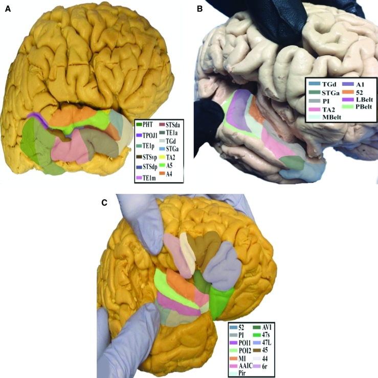
These regions are somewhat simpler to grasp once the remaining insular regions have been studied. They consist of 2 posterior insular regions which make up the long insular gyri and which follow the anteroinferior to posterosuperior path of this region, including the temporal opercular regions of the STG, the planum polare of the temporal lobe, and the inferior portions of the circular sulcus of the insula. The specific parcellations that comprise the temporal insula include A4, A5, PI, PoI1, PoI2, STGa, and TA2. The anatomic location of these parcellations is shown on a cadaver brain in Figure 51. This region has consistent white matter connections with the SLF/arcuate, inferior longitudinal fasciculus, middle longitudinal fasciculus, and temporal pole. The combined tractography of these parcellations is shown in Figure 52.
FIGURE 51.
Anatomical location of temporal “hypotenuse” parcellations shown on the left hemisphere of a cadaver brain. A, Lateral view of the superior temporal sulcus. B, Oblique view with a widening of the lateral sulcus to visualize superior temporal lobe parcellations. C, Lateral view with widening of the lateral sulcus to visualize frontal opercular and insular regions adjacent to the temporal lobe.
FIGURE 52.
Combined structural connectivity of the temporal “hypotenuse” parcellations in the left hemisphere, shown on T1-weighted MR images. Sagittal views of A, lateral, B, medial, and C, far medial planes. D, Axial view.
Area PoI1
Where is it?
Area PoI1 (posterior insula 1) is on the posterior long gyrus of the insula.
What are its borders?
Area PoI1 borders PoI2 anteriorly, and areas 52 and PI inferolaterally. Its superior border is with IG.
What is its functional connectivity?
Area PoI1 demonstrates functional connectivity to area 2 in the sensory strip, areas SCEF, FEF, 6a, 6ma, and 6r in the premotor regions, areas IFSa, 46, and 9-46d in the lateral frontal lobe, areas 5mv, 23c, 24dv, a24prime, p24prime, and p32prime in the medial frontal lobe, areas OP4, 43, PFcm, FOP1, FOP2, FOP3, FOP4, and FOP5 in the superior insula opercular regions, areas Pir, 52, PI, MI, MBelt, PBelt, A4, STV, and PoI2 in the lower opercula and Heschl's gyrus regions, area PHT in the temporal lobe, areas 7AL, PF, PGp, PFop, and PFt in the lateral parietal lobe, areas DVT and 7am in the medial parietal lobe, areas V2 and V3 in the medial occipital lobe, areas V3a and V6 in the dorsal visual stream area, and area FST in the lateral occipital lobe (Figure 53).
FIGURE 53.
Functional connectivity of Pol1 demonstrated on an inflated left hemisphere. A, Lateral and medial views. B, Oblique view. C, Rostral and caudal views. D, Dorsal and ventral views. Parcellations with the strongest functional connectivity are shown in yellow. Pink arrows designate the parcellation of interest.
What are its white matter connections?
Area PoI1 is structurally connected to the occipital and parietal lobe through the middle longitudinal fasciculus and to local parcellations. Fibers from PoI1 project posterior deep to the supramarginal gyrus ending at parietal and occipital parcellations 7AL, 7PC V3A, V3B, V6, and V6A. The majority of local short association bundles project posteriorly to parcellations PoI1, Ig, MBelt, PBelt, FOP2, OP2-3, OP1, and MI (Figure 54).
FIGURE 54.
Structural connectivity of PoI1 in the left hemisphere, shown on T1-weighted MR images. Sagittal views of A, lateral and B, medial planes. C, Axial view. Pink: white matter tracts of PoI1 demonstrating connections with the occipital lobe, parietal lobe, and local parcellations. Pink arrows designate the parcellation of interest.
What is known about its function?
Area PoI1 is a newly described area parcellated from the posterior insula. The posterior insula is anatomically and functionally connected with primary and secondary somatosensory and motor cortices, and takes part in processing various sensory inputs.25 Area PoI1 was differentiated from area PoI2 based on differences in fMRI activity during motor-based tasks.7
Area PoI2
Where is it?
Area PoI2 (posterior insula 2) is on the anterior of the 2 long insular gyri.
What are its borders?
Area PoI2 borders PoI1 posterior and inferiorly. Its superior border is IG, and small portions of FOP2 and FOP3. It shares an anterior border with MI and a small portion of AAIC.
What is its functional connectivity?
Area PoI2 demonstrates functional connectivity to areas 2 and 3a in the sensory strip, areas SCEF, FEF, 6a, 6ma, 6v, and 6r in the premotor regions, areas IFSa and 46 in the lateral frontal lobe, areas 5mv, 23c, 24dv, a24prime, p24prime, and p32prime in the medial frontal lobe, areas OP4, 43, IG, PFcm, FOP1, FOP2, FOP3, FOP4, and FOP5 in the superior insula opercular regions, areas Pir, 52, PI, MI, PBelt, A4, AAIC, and PoI1 in the lower opercula and Heschl's gyrus regions, areas TE2p and PHT in the temporal lobe, areas 7AL, AIP, LIPd, PF, PGp, PFop, and PFt in the lateral parietal lobe, area7am in the medial parietal lobe, area V2 in the medial occipital lobe, areas V6 in the dorsal visual stream area, and areas TPOJ2, PH, and FST in the lateral occipital lobe (Figure 55).
FIGURE 55.
Functional connectivity of Pol2 demonstrated on an inflated left hemisphere. A, Lateral and medial views. B, Rostral and caudal views. C, Dorsal and ventral views. Parcellations with the strongest functional connectivity are shown in yellow. Pink arrows designate the parcellation of interest.
What are its white matter connections?
Area PoI2 is structurally connected to the entorhinal cortex, local parcellations, and to the parietal lobe through the middle longitudinal fasciculus. Fibers from PoI2 project posterior deep to the supramarginal gyrus ending at parietal parcellations 2, 7PC, 7PL, LIPd, LIPv, and VIP. PoI2 has inferior projections to PeEc of the entorhinal cortex and TGd of the temporal pole. The majority of local short association bundles project posteriorly to parcellations PoI1, Ig, MBelt, PBelt, FOP2, OP2-3, OP1, and MI (Figure 56). Fibers from PoI2 in the right hemisphere have less consistent projections to the entorhinal cortex.
FIGURE 56.
Structural connectivity of PoI2 in the left hemisphere, shown on T1-weighted MR images. Sagittal views of A, lateral and B, medial planes. C, Axial view. Green: white matter tracts of PoI2 demonstrating connections with the parietal lobe, entorhinal cortex and local parcellations. Pink arrows designate the parcellation of interest.
What is known about its function?
Area PoI2 is a newly described area parcellated from the posterior insula. The posterior insula is anatomically and functionally connected with primary and secondary somatosensory and motor cortices and takes part in processing various sensory inputs.25 Area PoI2 was differentiated from area AAIC based on differences in fMRI activity during motor-based tasks.7
Area PI
Where is it?
Area PI (parainsular area) is in the anterior inferior circular sulcus of the insula.
What are its borders?
Area PI borders PoI1 medially, and TA2, STGa, and Mbelt laterally. Its posterior border is with area 52, and its anterior border is with Pir and TGd.
What is its functional connectivity?
Area PI demonstrates functional connectivity to areas PoI1, PoI2, MBelt, PBelt, 43, PFcm, RI, 52, FOP4, OP4, TA2, and STGa in the insula opercular regions, and PFop in the parietal lobe (Figure 57).
FIGURE 57.
Functional connectivity of Pl demonstrated on an inflated left hemisphere. A, Lateral and medial views. B, Oblique views demonstrating the operculum. C, Rostral and caudal views. D, Dorsal and ventral views. Parcellations with the strongest functional connectivity are shown in yellow. Pink arrows designate the parcellation of interest.
What are its white matter connections?
Area PI is structurally connected to the temporal pole, occipital lobe, and local parcellations. This area's structural connectivity is difficult to distinguish due to the proximity of the area to the extreme and external capsule. Occipital connections project through the temporal lobe to end at parcellation V1. Connections to the temporal pole course inferiorly to end at area TGd. Local short asscoation bundles connect with PoI1, PoI2, Ig, and OP2-3 (Figure 58).
FIGURE 58.
Structural connectivity of PI in the left hemisphere, shown on T1-weighted MR images. Sagittal views of A, lateral and B, medial planes. C, Axial view. White: white matter tracts of PI demonstrating connections with the temporal pole, occipital lobe, and local parcellations. Pink arrows designate the parcellation of interest.
What is known about its function?
Area PI plays a role in internal regulatory processes and in the processing of somatosensory, visual, and motor stimuli.25 There is also evidence that suggests area PI is activated when the body assesses its physiological condition when individuals are subject to heat.6
Area A4
Where is it?
Area A4 (auditory area 4) is on the superior face of the posterior half of the STG. It occupies this portion of the gyrus posterior to its junction with Heschl's gyrus.
What are its borders?
Area A4 borders A5 laterally and PBelt medially. Its posterior borders are with PSL and STV. Its anterior border is with MBelt and TA2.
What is its functional connectivity?
Area A4 demonstrates functional connectivity to areas 1, 2, 3a, and 3b in the sensory strip, area 4 in the motor strip, areas SCEF, FEF, 6mp, and 6v in the premotor regions, areas 5m, 5l, and 24dd in the medial frontal lobe, areas 43, PFcm, OP4, OP2-3, and OP1 in the superior insula opercular regions, areas STV, 52, RI, TA2, PI, MI, PBelt, MBelt, LBelt, A1, A5, PoI1, and PoI2 in the lower opercula and Heschl's gyrus regions, areas 7PC and PFop in the lateral parietal lobe, areas V2, V3, and V4 in the medial occipital lobe, areas V6, V6a, V7, and V3a in the dorsal visual stream area, areas V8, FFC, VVC, and PIT in the ventral visual stream regions, and areas TPOJ1, TPOJ2, LO1, LO3, MT, MST, PH, and FST in the lateral occipital lobe (Figure 59).
FIGURE 59.
Functional connectivity of A4 demonstrated on an inflated left hemisphere. A, Lateral and medial views. B, Rostral and caudal views. C, Dorsal and ventral views. Parcellations with the strongest functional connectivity are shown in yellow. Pink arrows designate the parcellation of interest.
What are its white matter connections?
Area A4 is structurally connected to the arcuate/SLF and middle longitudinal fasciculus. Arcuate/SLF fibers wrap around the termination of the Sylvian fissure from A4 to inferior frontal gyrus parcellations 45 and FOP5. The middle longitudinal fasciculus projects posterior from A4 to run just lateral to the lateral ventricle ending at occipital and parietal lobe parcellations V2, V3, V6, V7, MIP, LIPd, LIPv, and IP1. Local short association bundles connect with A5, PFop, RI, STV, LBelt, MBelt, PBelt, and PFcm (Figure 60).
FIGURE 60.
Structural connectivity of A4 in the left hemisphere, shown on T1-weighted MR images. Sagittal views of A, lateral, B, medial, and C, far medial planes. D, Axial view showing projections to the contralateral hemisphere. Green: white matter tracts of A4 demonstrating connections with the arcuate/SFL and the middle longitudinal fasciculus. Pink arrows designate the parcellation of interest.
What is known about its function?
Area A4 is a newly described area of the brain parcellated from the auditory association cortex. There is evidence that this region of the brain processes perceptual and conceptual acoustic sounds.26 Area A4 was differentiated from area TA2 based on differences in fMRI activity during arithmetic and auditory story tasks.7
Area A5
Where is it?
Area A5 (auditory area 5) is a thin anterior-posterior running strip on the superior lateral surface of the posterior portions of the STG.
What are its borders?
Area A5 shares its primary medial border with A4. Its primary posterior border is with TPOJ1. Its lateral borders are with STSdp and STSda (discussed with the temporal lobe section). Its anterior border is with TA2.
What is its functional connectivity?
Area A5 demonstrates functional connectivity to areas 1, 2, 3a, and 3b in the sensory strip, area 4 in the motor strip, area OP4 in the superior insula opercular regions, areas STV, RI, STGa, TA2, PBelt, LBelt, A4, and PSL in the lower opercula and Heschl's gyrus regions, areas STSda and STSdp in the temporal lobe, area FFC in the ventral visual stream regions, and areas TPOJ1 and MT in the lateral occipital lobe (Figure 61).
FIGURE 61.
Functional connectivity of A5 demonstrated on an inflated left hemisphere. A, Lateral and medial views. B, Rostral and caudal views. C, Dorsal and ventral views. Parcellations with the strongest functional connectivity are shown in yellow. Pink arrows designate the parcellation of interest.
What are its white matter connections?
Area A5 is structurally connected to the arcuate/SLF and middle longitudinal fasciculus. Arcuate/SLF fibers wrap around the termination of the Sylvian fissure from A5 to inferior frontal gyrus parcellation 44. The middle longitudinal fasciculus projects posterior from A5 to run just lateral to the lateral ventricle to end at intraparietal sulcus parcellations V6 and V6A. Local short association bundles connect with A4, STSda, and STSdp (Figure 62).
FIGURE 62.
Structural connectivity of A5 in the left hemisphere, shown on T1-weighted MR images. Sagittal views of A, lateral, B, medial, and C, far medial planes. D, Axial view. Green: white matter tracts of A5 demonstrating connections with the arcuate/SFL and the middle longitudinal fasciculus. Pink arrows designate the parcellation of interest.
What is known about its function?
Area A5 is a newly described area of the brain parcellated from the auditory association cortex. There is evidence that this region of the brain processes perceptual and conceptual acoustic sounds.26 Area A5 was differentiated from area TA2 based on differences in fMRI activity during arithmetic, auditory story, and social interaction tasks.7
Area STGa
Where is it?
Area STGa (superior temporal gyrus region a) is located on the anterior superior surface of the superior temporal gyrus with no extension onto the lateral surface.
What are its borders?
Area STGa borders PI medially, TA2 anterormedially, STSda posterolaterally, and TGd anteriorly.
What is its functional connectivity?
Area STGa demonstrates functional connectivity to A5 and Pi in the insula opercular region, and areas TGd, STSda, and STSdp in the temporal lobe (Figure 63).
FIGURE 63.
Functional connectivity of STGa demonstrated on an inflated left hemisphere. A, Lateral and medial views. B, Rostral and caudal views. C, Dorsal and ventral views. Parcellations with the strongest functional connectivity are shown in yellow. Pink arrows designate the parcellation of interest.
What are its white matter connections?
Area STGa is structurally connected to the inferior longitudinal fasciculus and local parcellations. Inferior longitudinal fasciculus fibers project posterior from STGa coursing through the temporal lobe to end at occipital lobe parcellations V1, V2, V3, V3A, and V3CD. Local short association bundles connect with TA2 and STSda (Figure 64).
FIGURE 64.
Structural connectivity of STGa in the left hemisphere, shown on T1-weighted MR images. Sagittal views of A, lateral and B, medial planes. C, Axial view. Light Blue: white matter tracts of STGa demonstrating connections with the inferior longitudinal fasciculus and local parcellations. Pink arrows designate the parcellation of interest.
What is known about its function?
Area STGa is a newly described area of the brain parcellated from the auditory association cortex. There is evidence that this region of the brain processes perceptual and conceptual acoustic sounds.26 Area STGa was differentiated from areas TGd and PI based on differences in fMRI activity during arithmetic, auditory story, and social interaction tasks.7
Area TA2
Where is it?
Area TA2 (temporal region A, area 2) is a region of the medial planum polare anterior to the anterolateral extent of Heschl's gyrus.
What are its borders?
Area TA2 borders area PI medially, STGa and area A5 laterally, and temporal area TGd anteriorly. Its posterior border is with the anterior edges of A4 and Mbelt.
What is its functional connectivity?
Area TA2 demonstrates functional connectivity to areas 1, 2, 3a, and 3b in the sensory strip, area 4 in the motor strip, areas OP4, 43, PFcm, A4, A5, MBelt, PBelt, LBelt, RI, and PI in the insula opercular regions, areas V2, V3, and V4 in the medial occipital lobe, and area V6 of the dorsal visual stream (Figure 65).
FIGURE 65.
Functional connectivity of TA-2 demonstrated on an inflated left hemisphere. A, Lateral and medial views. B, Oblique views demonstrating the operculum. C, Rostral and caudal views. D, Dorsal and ventral views. Parcellations with the strongest functional connectivity are shown in yellow. Pink arrows designate the parcellation of interest.
What are its white matter connections?
Area TA2 is structurally connected to the inferior longitudinal fasciculus and local parcellations. Inferior longitudinal fasciculus fibers project posterior from TA2 coursing through the temporal lobe to end at occipital lobe parcellations V1, V2, V3, and V6A. Local short association fibers connect with 52, MBelt, A5, PI, and STGa (Figure 66).
FIGURE 66.
Structural connectivity of TA2 in the left hemisphere, shown on T1-weighted MR images. Sagittal views of A, lateral and B, medial planes. C, Axial view. Yellow: white matter tracts of TA2 demonstrating connections with the inferior longitudinal fasciculus and local parcellations. Pink arrows designate the parcellation of interest.
What is known about its function?
Area TA2 is believed to be a transition zone between nonprimary and primary auditory areas of the auditory cortex. There is evidence suggesting that this area is activated when perceiving auditory stimuli, as each hemisphere recognizes different complex acoustic stimulation with the left auditory cortex processing quickly changing sounds of speech and the right activating more with the tone of speech.27,28 This area of the brain shares features with cortical sensory areas, but contains fewer granular cells than the primary auditory cortex.27
DISCUSSION
It goes without saying that the insula and opercular cortices have a complex anatomy with numerous overlapping systems. This is expected in an area sitting at the junction of 3 lobes of the brain. In addition to the well-known presence of the early auditory areas, our subjective assessment of the connectivity patterns of these regions suggests that there are at least 4 other clusters of interconnected areas with common patterns of connectivity among their members. This gives a total of 5 key clusters. As with all other attempts to group cortical parcellations, the brain frustrates our efforts to simplify it. For example, many areas are probably parts of multiple clusters. Regardless, some simplification is necessary to make sense of the insular-opercular cortex. A brief summary of each area is as follows. Of note, area STGa has a connectivity pattern which suggests that it is better grouped with the lateral temporal lobe areas, such as the superior temporal sulcus areas, than with any of these clusters. As such, it is discussed in greater detail in Chapter 6 in relationship to other temporal areas.
Frontal Opercular Cluster
Areas like FOP4, FOP5, AVI, and probably AAIC form an interconnected cluster which seems to be more comparable to neighboring areas 44 and 45 than the rest of the areas in this chapter. While these regions have some connectivity to the rest of the insula, their functional connectivity patterns are similar to frontal areas, and their structural connections are mainly through the IFOF and SLF/arcuate system.
Frontal Aslant Tract Cluster
We named this cluster based on the observation that the frontal aslant tract forms a greater part of the long-range outflow from these areas. The putative hubs of this cluster, such as MI, FOP1, or FOP3, feed directly into the FAT, while FOP2 seems to be linked locally to this cluster. Connections to supplementary motor areas like 6ma and SFL suggest a role for these areas in speech, motor planning, and task selection.29-33 The functional characteristics of these areas based on task-based fMRI from the HCP parcellation study strongly suggest that these areas play some role in the production motor cue and speech production.7 It is also interesting to note that these areas, especially MI, demonstrate functional connectivity similar to a pattern we would expect with the so-called salience network, which has been implicated as the intermediate network when switching from default mode to central executive functioning.34 This is interesting, but needs further validation.
Parietal Opercular Cluster
These areas are grouped together based on their mutual interconnectedness and their general output into the lateral SLF/arcuate networks. These are among the most functionally connected areas of the cerebral cortex. Areas like PFcm, 43, and OP4 have functional connectivity involving numerous diffuse areas of the brain. This is interesting as the parietal opercular cleft of the supramarginal gyrus does not command as much respect in the neurosurgical community compared to other areas of the opercula. It is not commonly found during brain mapping procedures, and so it is interesting to ask why these areas are so highly connected.
The members of this cluster seem to signal mainly through 2 highly connected hubs: PFcm, which sends fibers to the posterior temporal lobe and aspects of the parietotemporal junction via the arcuate system (in addition to its connections to the auditory network), and area 43, which connects to the parietal lobe via the lateral opercular portions of the SLF. It should also be noted that areas OP1, OP4, and Ig are mostly locally interconnected, but show some evidence of interconnectedness with the auditory system.
Supramarginal Cluster
The supramarginal cluster, which includes LBelt, PBelt, RI, STV, PSL, A4, and A5, is united by the unique trait that they all seem to have long-range connections into the SLF/arcuate system, as well as other white matter bundles such as the middle longitudinal fasciculus. The fact that some of these areas are putatively in the auditory network or are highly interconnected to the frontal operculum or dorsal visual stream highlights the complexity of the anatomy of the supramarginal gyrus and the challenges we face in understanding it.
Why not group areas like A4, A5, LBelt, and PBelt which are immediately adjacent to Heschl's gyrus and are functionally part of the auditory network? Certainly this could be argued as there appears to be substantial functional overlap between these areas and the auditory network. We decided not to do this because, despite their names, their individual connections to the SLF/arcuate complex raise the idea that these areas may be playing a different role than areas like A1 or MBelt which connect locally or into the middle longitudinal fasciculus.
Auditory Cluster
The auditory areas are complex both anatomically and functionally. As noted above, these areas are interconnected with the supramarginal gyrus cluster, and an argument can be made that some of these areas are the hubs of both clusters. However, in this grouping, the auditory cluster is mainly connected locally and with the MdLF. Areas like MBelt, POI1, and POI2 seem to have more distant connections and “hubness,” while areas like 52, A1, OP2-3, TA2, Pir, and PI do not seem to have these traits.
What About the Inferior Insular Stem?
In Chapter 6, we discussed inferior insular stem in more detail; however, it is of such critical importance to neurosurgery that it is also discussed here. It seems likely that some aspect of the inferior insula is critical for the maintenance of semantic function. We make this claim based on personal experience, and that of others, who have noted language problems resulting from cutting in the left insula.35 Given that this is the position of the IFOF, it has been thought that this tract plays some role in semantic function.36-38 This is a helpful explanation; however, our work has failed to find a plausible mechanism by which IFOF is transmitting semantic information to the language motor areas in the frontal lobe. Notably, we have found little evidence that any part of the temporal lobe gives fibers into the IFOF, and given that the occipital termini of this tract can be removed in an occipital lobectomy without causing anomia, it is not certain the path by which visual-semantic-speech motor transfer occurs.
Area PI represents the only area for which we have found any evidence for feeding into the IFOF between the occipital and frontal lobes. It sits on the cortex in the inferior insula, and it is interesting to think of this area as playing a semantic role. However, the literature does not provide any support for this, and the functional connectivity of PI is mostly to local, adjacent parcellations. Technically this area is small and thin, and so it is conceivable that our tractography is simply picking up IFOF running deep to PI. Ultimately this is a key question which needs to be addressed in future studies.
Disclosures
Synaptive Medical assisted in the funding of all 18 chapters of this supplement. No other funding sources were utilized in the production or submission of this work.
Acknowledgments
Data were provided [in part] by the Human Connectome Project, WU Minn Consortium (Principal Investigators: David Van Essen and Kamil Ugurbil; 1U54MH091657) funded by the 16 NIH Institutes and Centers that support the NIH Blueprint for Neuroscience Research; and by the McDonnell Center for Systems Neuroscience at Washington University. We would also like to thank Brad Fernald, Haley Harris, and Alicia McNeely of Synaptive Medical for their assistance in constructing the network figures for Chapter 18 and for coordinating the completion and submission of this supplement.
REFERENCES
- 1. Tanriover N, Rhoton AL Jr, Kawashima M, Ulm AJ, Yasuda A. Microsurgical anatomy of the insula and the sylvian fissure. J Neurosurg. 2004;100(5):891-922. [DOI] [PubMed] [Google Scholar]
- 2. Türe U, Yaşargil DCH, Al-Mefty O, Yaşargil MG. Topographic anatomy of the insular region. J Neurosurg. 1999;90(4):720-733. [DOI] [PubMed] [Google Scholar]
- 3. Johnson DM, Illig KR, Behan M, Haberly LB. New features of connectivity in piriform cortex visualized by intracellular injection of pyramidal cells suggest that “primary” olfactory cortex functions like “association” cortex in other sensory systems. J Neurosci. 2000;20(18):6974-6982. [DOI] [PMC free article] [PubMed] [Google Scholar]
- 4. Vaughan DN, Jackson GD. The piriform cortex and human focal epilepsy. Front Neurol. 2014;5:259. [DOI] [PMC free article] [PubMed] [Google Scholar]
- 5. Nelson SM, Dosenbach NU, Cohen AL, Wheeler ME, Schlaggar BL, Petersen SE. Role of the anterior insula in task-level control and focal attention. Brain Struct Funct. 2010;214(5-6):669-680. [DOI] [PMC free article] [PubMed] [Google Scholar]
- 6. Craig AD. How do you feel–now? The anterior insula and human awareness. Nat Rev Neurosci. 2009;10(1):59-70. [DOI] [PubMed] [Google Scholar]
- 7. Glasser MF, Coalson TS, Robinson ECet al. A multi-modal parcellation of human cerebral cortex. Nature. 2016;536(7615):171-178. [DOI] [PMC free article] [PubMed] [Google Scholar]
- 8. Li Y, Yao H, Lin Pet al. Frequency-dependent altered functional connections of default mode network in Alzheimer's disease. Front Aging Neurosci. 2017;9:259. [DOI] [PMC free article] [PubMed] [Google Scholar]
- 9. Steinmetz H, Seitz RJ. Functional anatomy of language processing: neuroimaging and the problem of individual variability. Neuropsychologia. 1991;29(12):1149-1161. [DOI] [PubMed] [Google Scholar]
- 10. Binkofski F, Buccino G. Motor functions of the Broca's region. Brain Lang. 2004;89(2):362-369. [DOI] [PubMed] [Google Scholar]
- 11. Sawamoto N, Honda M, Okada Tet al. Expectation of pain enhances responses to nonpainful somatosensory stimulation in the anterior cingulate cortex and parietal operculum/posterior insula: an event-related functional magnetic resonance imaging study. J Neurosci. 2000;20(19):7438-7445. [DOI] [PMC free article] [PubMed] [Google Scholar]
- 12. Ertekin C, Aydogdu I. Neurophysiology of swallowing. Clin Neurophysiol. 2003;114(12):2226-2244. [DOI] [PubMed] [Google Scholar]
- 13. Martin RE, Goodyear BG, Gati JS, Menon RS. Cerebral cortical representation of automatic and volitional swallowing in humans. J Neurophysiol. 2001;85(2):938-950. [DOI] [PubMed] [Google Scholar]
- 14. Nieuwenhuys R. The insular cortex: a review. Prog Brain Res. 2012;195:123-163. [DOI] [PubMed] [Google Scholar]
- 15. Mizoguchi N, Kobayashi M, Muramoto K. Integration of olfactory and gustatory chemosignals in the insular cortex. J Oral Biosci. 2016;58(3):81-84. [Google Scholar]
- 16. Eickhoff SB, Jbabdi S, Caspers Set al. Anatomical and functional connectivity of cytoarchitectonic areas within the human parietal operculum. J Neurosci. 2010;30(18):6409-6421. [DOI] [PMC free article] [PubMed] [Google Scholar]
- 17. Eickhoff SB, Schleicher A, Zilles K, Amunts K. The human parietal operculum. I. Cytoarchitectonic mapping of subdivisions. Cereb Cortex. 2006;16(2):254-267. [DOI] [PubMed] [Google Scholar]
- 18. Garcia-Larrea L. The posterior insular-opercular region and the search of a primary cortex for pain. Neurophysiol Clin/Clin Neurophysiol. 2012;42(5):299-313. [DOI] [PubMed] [Google Scholar]
- 19. Barbeau EB, Chai XJ, Chen JKet al. The role of the left inferior parietal lobule in second language learning: an intensive language training fMRI study. Neuropsychologia. 2017;98:169-176. [DOI] [PubMed] [Google Scholar]
- 20. Chudler EH, Nahin RL, Kenshalo DR Jr. Distribution and size of GABAergic neurons in area 7b and the retroinsular cortex of the monkey. Brain Res. 1989;481(2):383-387. [DOI] [PubMed] [Google Scholar]
- 21. Hackett TA. Anatomic organization of the auditory cortex. Handb Clin Neurol. 2015;129:27-53. [DOI] [PubMed] [Google Scholar]
- 22. Pickles JO. Auditory pathways: anatomy and physiology. Handb Clin Neurol. 2015;129:3-25. [DOI] [PubMed] [Google Scholar]
- 23. Purves D, Augustine G, Fitzpatrick Det al. Neuroscience. 4: e uppl. Sinauer Associates, Massachusetts, USA; 2012. [Google Scholar]
- 24. Wu Y, Sun D, Wang Y, Wang Y, Wang Y. Tracing short connections of the temporo-parieto-occipital region in the human brain using diffusion spectrum imaging and fiber dissection. Brain Res. 2016;1646:152-159. [DOI] [PubMed] [Google Scholar]
- 25. Chen X, Duan M, He Het al. Functional abnormalities of the right posterior insula are related to the altered self-experience in schizophrenia. Psychiatry Res Neuroimaging. 2016;256:26-32. [DOI] [PubMed] [Google Scholar]
- 26. Trumpp NM, Kliese D, Hoenig K, Haarmeier T, Kiefer M. Losing the sound of concepts: damage to auditory association cortex impairs the processing of sound-related concepts. Cortex. 2013;49(2):474-486. [DOI] [PubMed] [Google Scholar]
- 27. Morosan P, Rademacher J, Schleicher A, Amunts K, Schormann T, Zilles K. Human primary auditory cortex: cytoarchitectonic subdivisions and mapping into a spatial reference system. Neuroimage. 2001;13(4):684-701. [DOI] [PubMed] [Google Scholar]
- 28. Burton H, Firszt JB, Holden T, Agato A, Uchanski RM. Activation lateralization in human core, belt, and parabelt auditory fields with unilateral deafness compared to normal hearing. Brain Res. 2012;1454:33-47. [DOI] [PMC free article] [PubMed] [Google Scholar]
- 29. Cunnington R, Bradshaw JL, Iansek R. The role of the supplementary motor area in the control of voluntary movement. Hum Mov Sci. 1996;15(5):627-647. [Google Scholar]
- 30. Gelmers HJ. Non-paralytic motor disturbances and speech disorders: the role of the supplementary motor area. J Neurol Neurosurg Psychiatry. 1983;46(11):1052-1054. [DOI] [PMC free article] [PubMed] [Google Scholar]
- 31. Johns P. Chapter 13 - Parkinson's disease. Clinical Neuroscience: Churchill Livingstone, New York; 2014:163-179. [Google Scholar]
- 32. Quiñones-Hinojosa A. Schmidek and Sweet Operative Neurosurgical Techniques: Indications, Methods, and Results. 6th ed. 2006. Saunders Publishing, New York. [Google Scholar]
- 33. Ramachandran VS. Encyclopedia of the Human Brain. Vol. 12002. academic press, Cambridge Mass. [Google Scholar]
- 34. Goulden N, Khusnulina A, Davis NJet al. The salience network is responsible for switching between the default mode network and the central executive network: replication from DCM. Neuroimage. 2014;99:180-190. [DOI] [PubMed] [Google Scholar]
- 35. Sughrue M, Bonney PA, Burks JDet al. Multilobar insular-involving gliomas: results with hyperaggressive resection. Cureus. 2017;9(8):e1623. [DOI] [PMC free article] [PubMed] [Google Scholar]
- 36. Duffau H, Moritz-Gasser S, Mandonnet E. A re-examination of neural basis of language processing: proposal of a dynamic hodotopical model from data provided by brain stimulation mapping during picture naming. Brain Lang. 2014;131:1-10. [DOI] [PubMed] [Google Scholar]
- 37. Duffau H, Gatignol P, Mandonnet E, Peruzzi P, Tzourio-Mazoyer N, Capelle L. New insights into the anatomo-functional connectivity of the semantic system: a study using cortico-subcortical electrostimulations. Brain. 2005;128(4):797-810. [DOI] [PubMed] [Google Scholar]
- 38. Chang EF, Raygor KP, Berger MS. Contemporary model of language organization: an overview for neurosurgeons. J Neurosurg. 2015;122(2):250-261. [DOI] [PubMed] [Google Scholar]


