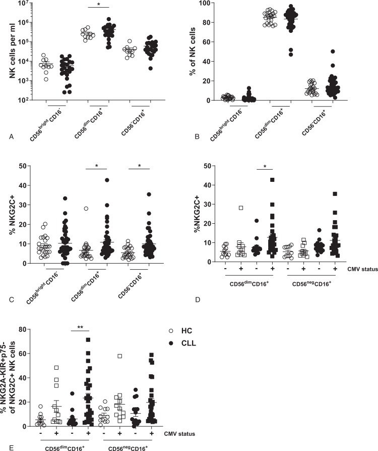
Figure 1.
Expansion of “CMV-specific” NKG2C+ NK cells in CLL patients. Subset composition of NK cells from HC (n = 22) or CLL (n = 41) was analyzed by flow cytometry. (A) Absolute number of NK cell subsets in peripheral blood of HC and CLL. (B) Relative subset distribution of NK cells. (C) Expression of NKG2C on NK cells of HC and CLL patients. (D) Expression of NKG2C on NK cells of HC and CLL patients stratified by CMV serology. (E) Frequency of NKG2C+ NK cells with a matured (NKG2A-KIR+p75-) phenotype. Bars indicate mean + SEM.∗ p < 0.05; ∗∗p < 0.01 (One-Way ANOVA).

