Younger age at diagnosis often reflects a greater influence of genetic factors in disease. Type 1 diabetes, which develops most frequently in childhood but can also present in adult life, is a prime candidate to explore the relationships among risk loci, age at diagnosis, and genetic contribution to disease. Type 1 diabetes genetic risk scores (GRS) (calculated as the weighted sum of alleles statistically associated with type 1 diabetes present in a given individual) are inversely correlated with age at diagnosis. Studies have suggested age-related heterogeneity in the association of established risk alleles with type 1 diabetes, although no consistent pattern has developed. Further investigations into the genetic factors that influence age of clinical onset may refine our understanding of type 1 diabetes pathogenesis and provide opportunities for individualized preventive therapies.
It has long been recognized that genetics plays a role in determining age at type 1 diabetes diagnosis. For monozygotic twins, concordance for type 1 diabetes increases with younger age at diagnosis in the index twin (1). HLA genotypes that confer risk for type 1 diabetes are more prevalent among subjects with younger age at clinical onset (2). GRS combining information from type 1 diabetes–associated HLA and non-HLA loci predict progression from single to multiple autoantibody positivity in individuals under age 35 years but not in older participants (3). A genome-wide analysis by Inshaw et al. (4) showed that rs9273363 single-nucleotide polymorphism (SNP) (tagging the HLA DQB1*03:02 haplotype) and the 6q22.33 region, which contains the genes encoding protein tyrosine phosphatase receptor κ (PTPRK) and thymocyte-expressed molecule involved in selection (THEMIS), are associated with younger age at diagnosis, although not with type 1 diabetes overall. Additional chromosomal regions reported to influence age at type 1 diabetes diagnosis are interleukin-2 (IL2) (4q27, rs2069763) and renalase (RNLS) (10q23.31, rs10509540) (5). On the other end of the spectrum, adult-onset autoimmune diabetes displays weaker associations with HLA and stronger associations with INS and loci associated with type 2 diabetes (6). A major concern with all of these studies is whether the observed differences with age reflect underlying differences in pathogenesis or lack of statistical power due to limited samples sizes for older-onset subjects.
In the current issue of Diabetes Care, Inshaw et al. (7) describe the most comprehensive evaluation of the relationship between type 1 diabetes risk loci and age at diagnosis published to date. They used extensive resources of genotyped case and control subjects from prior studies, affording sufficient statistical power to evaluate individual risk loci even with modest effect sizes. Their findings bring together several disparate observations regarding type 1 diabetes: 1) the aforementioned skewed age distribution at diagnosis; 2) distinct age-specific histologic phenotypes related to islet autoimmune infiltration (8), which they used to partition their population (i.e., <7, 7–13, and ≥13 years); and 3) heterogeneity in the autoimmune factors of type 1 diabetes, raising the possibility of additional diabetogenic mechanisms in some individuals (9).
The authors compared age subgroups for the strength of the associations between type 1 diabetes and HLA (8 class II, 9 class I) and 55 non-HLA loci, selected based on their prior data (4). In the youngest category, the highest risk HLA DR3-DQ2/DR4-DQ8 diplotype, as well as A*2402 alleles and B39*06, were more common, while the protective DR15-DQ6 (DRB1*15:01-DQB1*06:02) and DR7-DQ3 (DRB1*07:01-DQB1*03:03) were least common. While the results for HLA were not unexpected, the novelty of the study lies in several non-HLA regions that were differentially associated between the youngest and the oldest categories. Existing fine mapping data and colocalization with whole-blood expression quantitative trait loci studies revealed likely causal variants in these regions. Most of the credible candidate causal genes are involved in T and/or B cell biology: interleukin-2 receptor α (IL2RA), interleukin-10 (IL10), THEMIS, Ikaros family zinc finger 3 (IKZF3)/ORMDL sphingolipid biosynthesis regulator 3 (ORMDL3)/gasdermin B (GSDMB), and cathepsin H (CTSH). However, a few of the candidate causal genes identified may have effects on the target organ. Most notable among these is Gli-similar protein 3 (GLIS3), a transcription factor that regulates insulin gene expression as well as β-cell development, survival, and proliferation. GLIS3 variants have been involved in neonatal diabetes, type 1 diabetes, and type 2 diabetes (10). Unlike most candidate risk loci for type 1 diabetes, GLIS3 has not been linked to other autoimmune disorders, suggesting target organ–specific effects. However, since GLIS3 variants increase β-cell susceptibility to apoptosis and cytokine-induced β-cell death, contributing to β-cell fragility (11), an intriguing possibility is that GLIS3 variants could magnify the aggressive autoimmune attack on β-cells characteristic of younger children. The mechanisms underlying the involvement of the CTSH and IKZF3 loci are more ambiguous. CTSH is ubiquitously expressed, including in β-cells. Allelic variation at the index SNP in the region, rs3825932, confers differential susceptibility to β-cell apoptosis, hinting at multiple underlying pathogenic mechanisms. For IKZF3, which is clearly immune-related in function, the direction of the risk in the current study is opposite of that reported for other autoimmune disorders such as asthma. Overall, one should keep in mind that, while fine mapping and colocalization can hint at plausible candidate genes, additional work is required to definitely determine the causal gene or genes in a given disease-associated region.
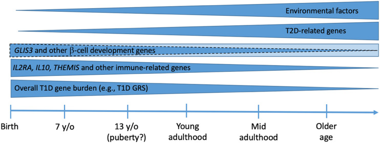
Genetics may provide a useful window into the puzzling age differences in the epidemiology, clinical characteristics, immunology, and histopathology of type 1 diabetes (12). Most of the genes that Inshaw et al. (7) found preferentially associated with early-childhood type 1 diabetes work in the immune system (13) (Fig. 1). Accordingly, islet autoimmunity most often appears early in life (14), and its progression to clinical type 1 diabetes is faster and more likely in the youngest children (15). T-cell responses to islet antigens differ by age; for instance, the secretion of regulatory cytokine interleukin-10 by CD4+ T-cells increases with age at type 1 diabetes presentation (16). Aggressive autoimmunity in younger children could lead to faster and more profound loss of β-cells, explaining more rapid progression through preclinical type 1 diabetes stages (17), higher incidence of type 1 diabetes diagnosis (18), and poorer β-cell function (and consequently more frequent diabetic ketoacidosis) at diagnosis in children than in adults (19). After type 1 diabetes onset, β-cell function falls faster in younger individuals (20), and the partial remission period is shorter in young children. On the other extreme of the spectrum, adult-onset autoimmune diabetes has a lower burden of type 1 diabetes–associated genes, milder autoimmunity (as reflected by single autoantibody positivity), and a characteristic slow decline in β-cell function. Histopathology indicates that individuals who develop type 1 diabetes later in life maintain higher numbers of insulin-containing islets and their insulitis is less aggressive and different from that in younger people (8). Inshaw et al. (7) leveraged histopathological differences by age to define age categories.
Figure 1.
Genetic and environmental influences combine and interact to cause type 1 diabetes. Their strength and relative contribution determine rate of progression through preclinical stages and, thus, the age at clinical onset of type 1 diabetes. The burden of type 1 diabetes–associated genes (as measured by type 1 diabetes GRS) is highest in young children who develop clinical type 1 diabetes. In particular, genes related to the immune function (e.g., IL2RA, THEMIS, etc.) are associated with very early-onset type 1 diabetes and characteristically aggressive histopathology. GLIS3 variants, which have been associated with very early-onset type 1 diabetes in the study by Inshaw et al. (7), is also involved in type 2 and monogenic diabetes. Studies in adult-onset type 1 diabetes have found a higher burden of type 2 diabetes genes. Twin studies support that environmental factors are less important at younger ages of onset. Interactions at various levels (gene-gene, gene-environment) have been described and could modify the relative importance of factors. T1D, type 1 diabetes; T2D, type 2 diabetes; y/o, years old.
The majority of the age-related type 1 diabetes risk loci identified by Inshaw et al. (7) act on the immune system. This observation reinforces the concept of a stronger autoimmune component of type 1 diabetes in younger children. In individuals with milder islet autoimmunity, the development of clinical type 1 diabetes may depend upon additional influences. The “threshold hypothesis” proposed that clinical diabetes develops when the combination of diabetogenic genetic and environmental factors exceeds a threshold (21). There is evidence that environmental factors cooperating with genes not directly associated with autoimmunity may help to initiate islet autoimmunity (22). The relationship of obesity with type 1 diabetes has been demonstrated (23). Type 2 diabetes–associated transcription factor 7-like 2 (TCF7L2) genetic variants are more frequent in type 1 diabetes with single autoantibody positivity (24) or lacking high-risk HLA alleles (25), implying that type 2 diabetes risk factors may contribute to type 1 diabetes pathogenesis in a subset of cases with fewer markers of islet autoimmunity (9). Therefore, the relative influence of immune and nonimmune genetic and environmental factors, and the pathways to diabetes, can be expected to vary by onset age. While most of the variants tested by Inshaw et al. (7) are in regions involved in the immune function, studies on genes implicated in glucose metabolism may uncover additional age-driven differences.
The physiopathologic differences underlying age-related heterogeneity could be leveraged therapeutically. Some of the immunomodulatory agents are more effective at preventing type 1 diabetes or its progression in children than in adults (17,26,27). The current report raises the possibility of a “precision medicine” approach to type 1 diabetes interventions, using either individual SNP genotypes or a GRS to identify subjects whose disease might have a stronger autoimmune nature, warranting more aggressive immunomodulatory therapies.
The study by Inshaw et al. (7) is encouraging but also highlights areas for further work. The current study uses a targeted genotyping approach with the ImmunoChip platform and could be productively expanded via genome-wide genotyping; the prior article by Inshaw et al. (4) identified loci that are associated with age at diagnosis but not disease overall, and thus targeted genotyping focusing on disease-associated SNPs may be insufficient. Replication studies are needed in populations of non-European origin (28,29), since the authors used a rather homogeneous population from the U.K. The older age category had the smallest sample size (and most likely the greatest heterogeneity), and thus the results from this study need to be replicated in cohorts well powered for older participants. Only a minority of cases were adult-onset type 1 diabetes, which limited the ability to analyze this distinct subset separately. Finally, the mechanistic implications could be further explored in existing longitudinal cohorts to understand the influence of individual variants on specific preclinical stages of type 1 diabetes, e.g., appearance of islet autoimmunity, progression to multiple autoantibody positivity, and development of clinical disease. Better understanding of the age effect in type 1 diabetes, its underlying mechanisms, and whether it is a continuum or there are discrete categories could reveal opportunities for prediction and prevention.
Article Information
Funding. M.J.R. acknowledges support from National Institutes of Health grants R01 DK121843-01, U01 DK103180-01, and U54 DK118638-01. P.C. acknowledges support from grants R01 DK106718, R01 DK116954, DP3 DK111906, and P01 AI042288.
Duality of Interest. P.C. reports relationships with Amgen (stockholder) and Canon, Illumina, and 10X Genomics (event support for the University of Florida Genetics Institute). No other potential conflicts of interest relevant to this article were reported.
Footnotes
See accompanying article, p. 169.
References
- 1.Redondo MJ, Yu L, Hawa M, et al. Heterogeneity of type I diabetes: analysis of monozygotic twins in Great Britain and the United States. Diabetologia 2001;44:354–362 [DOI] [PubMed] [Google Scholar]
- 2.Howson JM, Rosinger S, Smyth DJ, Boehm BO, Todd JA; ADBW-END Study Group . Genetic analysis of adult-onset autoimmune diabetes. Diabetes 2011;60:2645–2653 [DOI] [PMC free article] [PubMed] [Google Scholar]
- 3.Redondo MJ, Geyer S, Steck AK, et al.; Type 1 Diabetes TrialNet Study Group . A type 1 diabetes genetic risk score predicts progression of islet autoimmunity and development of type 1 diabetes in individuals at risk. Diabetes Care 2018;41:1887–1894 [DOI] [PMC free article] [PubMed] [Google Scholar]
- 4.Inshaw JRJ, Walker NM, Wallace C, Bottolo L, Todd JA. The chromosome 6q22.33 region is associated with age at diagnosis of type 1 diabetes and disease risk in those diagnosed under 5 years of age. Diabetologia 2018;61:147–157 [DOI] [PMC free article] [PubMed] [Google Scholar]
- 5.Howson JM, Cooper JD, Smyth DJ, et al.; Type 1 Diabetes Genetics Consortium . Evidence of gene-gene interaction and age-at-diagnosis effects in type 1 diabetes. Diabetes 2012;61:3012–3017 [DOI] [PMC free article] [PubMed] [Google Scholar]
- 6.Mishra R, Chesi A, Cousminer DL, et al.; Bone Mineral Density in Childhood Study . Relative contribution of type 1 and type 2 diabetes loci to the genetic etiology of adult-onset, non-insulin-requiring autoimmune diabetes. BMC Med 2017;15:88. [DOI] [PMC free article] [PubMed] [Google Scholar]
- 7.Inshaw JRJ, Cutler AJ, Crouch DJM, Wicker LS, Todd JA. Genetic variants predisposing most strongly to type 1 diabetes diagnosed under age 7 years lie near candidate genes that function in the immune system and in pancreatic β-cells. Diabetes Care 2020;43:169–177. [DOI] [PMC free article] [PubMed] [Google Scholar]
- 8.Leete P, Willcox A, Krogvold L, et al. Differential insulitic profiles determine the extent of β-cell destruction and the age at onset of type 1 diabetes. Diabetes 2016;65:1362–1369 [DOI] [PubMed] [Google Scholar]
- 9.Redondo MJ, Evans-Molina C, Steck AK, Atkinson MA, Sosenko J. the influence of type 2 diabetes-associated factors on type 1 diabetes. Diabetes Care 2019;42:1357–1364 [DOI] [PMC free article] [PubMed] [Google Scholar]
- 10.Wen X, Yang Y. Emerging roles of GLIS3 in neonatal diabetes, type 1 and type 2 diabetes. J Mol Endocrinol 2017;58:R73–R85 [DOI] [PubMed] [Google Scholar]
- 11.Liston A, Todd JA, Lagou V. Beta-cell fragility as a common underlying risk factor in type 1 and type 2 diabetes. Trends Mol Med 2017;23:181–194 [DOI] [PubMed] [Google Scholar]
- 12.Leete P, Mallone R, Richardson SJ, Sosenko JM, Redondo MJ, Evans-Molina C. The effect of age on the progression and severity of type 1 diabetes: potential effects on disease mechanisms. Curr Diab Rep 2018;18:115. [DOI] [PMC free article] [PubMed] [Google Scholar]
- 13.Onengut-Gumuscu S, Chen WM, Burren O, et al.; Type 1 Diabetes Genetics Consortium . Fine mapping of type 1 diabetes susceptibility loci and evidence for colocalization of causal variants with lymphoid gene enhancers. Nat Genet 2015;47:381–386 [DOI] [PMC free article] [PubMed] [Google Scholar]
- 14.Krischer JP, Lynch KF, Lernmark Å, et al.; TEDDY Study Group . Genetic and environmental interactions modify the risk of diabetes-related autoimmunity by 6 years of age: the TEDDY study. Diabetes Care 2017;40:1194–1202 [DOI] [PMC free article] [PubMed] [Google Scholar]
- 15.Krischer JP, Liu X, Lernmark Å, et al.; TEDDY Study Group . the influence of type 1 diabetes genetic susceptibility regions, age, sex, and family history on the progression from multiple autoantibodies to type 1 diabetes: a TEDDY study report. Diabetes 2017;66:3122–3129 [DOI] [PMC free article] [PubMed] [Google Scholar]
- 16.Arif S, Tree TI, Astill TP, et al. Autoreactive T cell responses show proinflammatory polarization in diabetes but a regulatory phenotype in health. J Clin Invest 2004;113:451–463 [DOI] [PMC free article] [PubMed] [Google Scholar]
- 17.Wherrett DK, Chiang JL, Delamater AM, et al.; Type 1 Diabetes TrialNet Study Group . Defining pathways for development of disease-modifying therapies in children with type 1 diabetes: a consensus report. Diabetes Care 2015;38:1975–1985 [DOI] [PMC free article] [PubMed] [Google Scholar]
- 18.Karvonen M, Viik-Kajander M, Moltchanova E, Libman I, LaPorte R, Tuomilehto J. Incidence of childhood type 1 diabetes worldwide. Diabetes Mondiale (DiaMond) Project Group. Diabetes Care 2000;23:1516–1526 [DOI] [PubMed] [Google Scholar]
- 19.Sosenko JM, Geyer S, Skyler JS, et al. The influence of body mass index and age on C-peptide at the diagnosis of type 1 diabetes in children who participated in the Diabetes Prevention Trial-Type 1. Pediatr Diabetes 2018;19:403–409 [DOI] [PMC free article] [PubMed] [Google Scholar]
- 20.Hao W, Gitelman S, DiMeglio LA, Boulware D, Greenbaum CJ; Type 1 Diabetes TrialNet Study Group . Fall in C-peptide during first 4 years from diagnosis of type 1 diabetes: variable relation to age, HbA1c, and insulin dose. Diabetes Care 2016;39:1664–1670 [DOI] [PMC free article] [PubMed] [Google Scholar]
- 21.Wasserfall C, Nead K, Mathews C, Atkinson MA. The threshold hypothesis: solving the equation of nurture vs nature in type 1 diabetes. Diabetologia 2011;54:2232–2236 [DOI] [PMC free article] [PubMed] [Google Scholar]
- 22.Rich SS, Concannon P. Role of type 1 diabetes-associated SNPs on autoantibody positivity in the Type 1 Diabetes Genetics Consortium: overview. Diabetes Care 2015;38(Suppl. 2):S1–S3 [DOI] [PMC free article] [PubMed] [Google Scholar]
- 23.Ferrara CT, Geyer SM, Liu YF, et al.; Type 1 Diabetes TrialNet Study Group . Excess BMI in childhood: a modifiable risk factor for type 1 diabetes development? Diabetes Care 2017;40:698–701 [DOI] [PMC free article] [PubMed] [Google Scholar]
- 24.Redondo MJ, Geyer S, Steck AK, et al.; Type 1 Diabetes TrialNet Study Group . TCF7L2 genetic variants contribute to phenotypic heterogeneity of type 1 diabetes. Diabetes Care 2018;41:311–317 [DOI] [PMC free article] [PubMed] [Google Scholar]
- 25.Redondo MJ, Grant SF, Davis A, Greenbaum C; T1D Exchange Biobank . Dissecting heterogeneity in paediatric type 1 diabetes: association of TCF7L2 rs7903146 TT and low-risk human leukocyte antigen (HLA) genotypes. Diabet Med 2017;34:286–290 [DOI] [PubMed] [Google Scholar]
- 26.Rigby MR, Harris KM, Pinckney A, et al. Alefacept provides sustained clinical and immunological effects in new-onset type 1 diabetes patients. J Clin Invest 2015;125:3285–3296 [DOI] [PMC free article] [PubMed] [Google Scholar]
- 27.Pescovitz MD, Greenbaum CJ, Krause-Steinrauf H, et al.; Type 1 Diabetes TrialNet Anti-CD20 Study Group . Rituximab, B-lymphocyte depletion, and preservation of beta-cell function. N Engl J Med 2009;361:2143–2152 [DOI] [PMC free article] [PubMed] [Google Scholar]
- 28.Zhu M, Xu K, Chen Y, et al. Identification of novel T1D risk loci and their association with age and islet function at diagnosis in autoantibody-positive T1D individuals: based on a two-stage genome-wide association study. Diabetes Care 2019;42:1414–1421 [DOI] [PubMed] [Google Scholar]
- 29.Onengut-Gumuscu S, Chen WM, Robertson CC, et al.; SEARCH for Diabetes in Youth; Type 1 Diabetes Genetics Consortium . Type 1 diabetes risk in African-ancestry participants and utility of an ancestry-specific genetic risk score. Diabetes Care 2019;42:406–415 [DOI] [PMC free article] [PubMed] [Google Scholar]


