



Abstract
Objective:
To identify achieved oxygen saturations (SpO2) associated with increased risk of severe retinopathy of prematurity (ROP).
Design:
Secondary analysis of the SUPPORT randomized controlled trial. SpO2 was recorded up to 36 weeks’ postmenstrual age. Saturations through 9 postnatal weeks were explored graphically, and logistic regression models were created to predict severe ROP.
Setting:
20 centers of the Eunice Kennedy Shriver National Institute of Child Health and Human Development Neonatal Research Network.
Patients:
984 surviving infants of 24–27 weeks’ gestational age born 2005–2009.
Interventions:
SUPPORT targeted SpO2 to a lower (85–89%) or higher (91–95%) range through 36 weeks’ postmenstrual age or off respiratory support.
Main Outcome Measures:
Severe ROP defined as threshold ROP, ophthalmologic surgery, or bevacizumab treatment.
Results:
There were statistically significant interactions between duration of oxygen supplementation and percentage of time in certain achieved saturation ranges. Specifically, for infants who spent at least 2 weeks on oxygen in postnatal weeks 1–5, a higher percentage of time at 91–96% SpO2 was associated with increased odds of severe ROP. For infants who spent at least 3 weeks on oxygen in postnatal weeks 6–9, a higher percentage of time at 97–100% SpO2 was associated with increased odds of severe ROP. Other significant risk factors were lower gestational age and birth weight, non-Hispanic white versus black race, prospectively defined severe illness, late onset sepsis or meningitis, and clinical center.
Conclusions:
Among extremely preterm survivors to discharge, the association between SpO2 and severe ROP depended on the timing and duration of oxygen supplementation.
Clinicaltrials.gov identifier for SUPPORT: NCT00233324
Introduction
Retinopathy of prematurity (ROP) is a cause of visual disabilities in preterm infants. ROP risk increases with decreasing gestational age (GA), and an association between ROP and unrestricted oxygen use in preterm infants was established in the 1950s.1 In 2005–2009, the SUPPORT trial randomized 1316 infants of 24 0/7 weeks to 27 6/7 weeks GA to oxygen saturation target ranges of 85–89% or 91–95% (with suggested alarms set at 84% and 96% in both groups).2 Among survivors to discharge, the lower target group had a reduced risk of severe ROP (relative risk 0.52, 95% confidence interval (CI) 0.37 – 0.73, p<0.001), and shorter duration of oxygen supplementation (mean 59.8 vs. 67.4 days, p<0.001),2 however, the lower target group had an unexpected increase in mortality.2 The Benefits of Oxygen Saturation Targeting II (BOOST II) trials in Australia and the United Kingdom were terminated early for similar mortality findings. Those trials also reported a decrease in ROP with a lower target range.3
Previous data had suggested that oxygen saturations in the upper part of the recommended and generally accepted range at the time (85 – 95%)4 might increase the risk of ROP relative to the lower part of the range.5–7 Although a multicenter observational study published in 1977 did not find an association between partial pressure of oxygen (PaO2) concentrations and retinopathy,8 a single center cohort study in the 1980s using transcutaneous oxygen monitoring supported an association between retinopathy and arterial oxygen levels ≥ 80 mmHg.9 Since then, pulse oximetry has largely supplanted transcutaneous oxygen tension for continuous clinical monitoring of oxygenation.
SUPPORT hypothesized that ROP would be decreased by targeting oxygen saturations at 85–89% compared to 91–95%. However, oxygen targets are difficult to maintain in critically ill infants due to their labile clinical status. Thus, as expected,10 actual saturations while on supplemental oxygen differed from the targets, and the achieved saturations in the two randomized groups overlapped.2 This study tested the hypothesis that there were specific achieved oxygen saturations associated with increased risk of severe ROP.
Subjects and Methods
This was a secondary analysis of SUPPORT trial data with prespecified outcomes. Institutional Review Board approval was obtained for SUPPORT at all sites. Infants were examined by ophthalmologists trained in the diagnosis of ROP beginning by 31 to 33 weeks’ postmenstrual age (PMA) and continuing until the severe ROP outcome was reached or resolution occurred. 2,11 Resolution was defined as fully vascularized retinas or immature vessels in zone 3 on two consecutive examinations in each eye. Threshold ROP (called “Type 1 ROP” by the Early Treatment of Retinopathy Cooperative Group12,13) was defined as any of the following: (a) zone 1: stage 3 ROP, without plus disease (i.e., two or more quadrants of dilated veins and tortuous arteries in the posterior pole), or plus disease with any stage of ROP; (b) zone 2: plus disease with stage 2 or 3 ROP. Surgical ophthalmologic intervention included laser therapy, cryotherapy, scleral buckling, or vitrectomy. Severe ROP was defined as threshold ROP, ophthalmologic surgery, or bevacizumab treatment.
Respiratory support data, including mode of support and fraction of inspired oxygen, were collected on study forms. Through February 2006, these data were collected every 8 hours during the first 14 days of life and once a day from 15 days through 36 weeks’ PMA or death, transfer, or discharge, whichever occurred first. After February 2006, the data were collected every two hours for the first 14 days and every 6 hours thereafter to facilitate monitoring of treatment adherence.
Treatment assignment was masked using specially designed pulse oximeters with skewed display algorithms such that, for both treatment groups, oxygen saturation (SpO2) values in the target range were displayed as 88–92% (a maximum variation of 3% from the actual value); 2 recommended alarm settings were 84% and 96% in both groups. Oximeter averaging time was set to 16 seconds, and sensitivity was set to normal mode. Displayed SpO2 was sampled every 10 seconds, and display values were transformed to actual SpO2 for analysis. Where there was not one-to-one correspondence between display and actual values,14 quadratic and cubic spline interpolation methods were used.
Study pulse oximeters were discontinued at 36 weeks’ PMA or when the infant was in room air and off ventilatory support or continuous positive airway pressure for 72 hours, whichever occurred earlier. However, if respiratory support was resumed prior to 36 weeks’ PMA, the study oximeter was placed back on the infant. Since saturations in room air cannot be controlled, this analysis included SpO2 data collected only during oxygen supplementation, identified based on whether the infant was receiving oxygen at the closest time point for which respiratory support data were collected on daily study forms. Saturation data collected after the ophthalmologic outcome were excluded from this analysis.
Demographic and neonatal characteristics of infants with and without severe ROP were compared using Student’s t tests and chi-square tests. Kaplan-Meier survival curves were generated for the probability of oxygen supplementation through 12 postnatal weeks. Hours on oxygen with various SpO2 values were estimated by postnatal week, assuming SpO2 remained constant over the 10-second sampling interval. Graphs were used to identify periods when SpO2 differed between infants with and without severe ROP, and informed a logistic regression model to predict severe ROP based on percent of time while on oxygen at specific SpO2 values during selected postnatal age intervals. Because infants born at 27 weeks’ GA had SpO2 data only through 9 postnatal weeks, only data through week 9 were used in modeling for all infants. The model included total hours on supplemental oxygen during the time interval, and demographic and neonatal characteristics. When infants were not receiving oxygen support, time on oxygen at specific SpO2 values was set to zero for modeling purposes. Demographic and neonatal characteristics were selected prospectively and included known risk factors for ROP and comorbidities impacting supplemental oxygen use. Model covariates were clinical center, sex, race and ethnicity, GA, birth weight, antenatal steroid exposure, severe illness (defined prospectively as FiO2 >0.4 and ventilator use for >8 consecutive hours in the first 14 days of life), periventricular leukomalacia (PVL) (increased echogenicity or cysts in the periventricular region), grade III or IV intraventricular hemorrhage (severe IVH), Bell’s stage II or III necrotizing enterocolitis (NEC), and culture-positive late-onset sepsis or meningitis. Comorbidity diagnoses after the ophthalmologic outcome were excluded. Similar analyses used weeks’ PMA in place of weeks’ postnatal age. Statistical tests were 2-sided and p values < 0.05 were considered statistically significant. Analysis were performed using SAS 9.4, SAS Institute Inc., Cary, North Carolina.
Results
SUPPORT enrolled 1,316 infants, and 1,079 survived to discharge. Among survivors, 984 (92%) had an ophthalmologic outcome. Of these, 132/984 (13%) had severe ROP, including 22 infants who did not have threshold ROP but were treated with ophthalmologic surgery or bevacizumab. Ninety-five percent (932/984) of infants with outcome data also had SpO2 data (128/132 [97%] with severe ROP and 804/852 [94%] without). Missing SpO2 data resulted from technical issues such as lost or corrupted files. Infants without SpO2 data had higher GA and birth weights and none had NEC (Supplemental Table S1).
Infants with severe ROP had lower GA and birth weights, and were more likely to have PVL, severe IVH, late onset sepsis or meningitis, and severe illness compared to infants without severe ROP (Table 1). Based on data from respiratory support forms, 97% (128/132) of infants with severe ROP received oxygen through the 6th postnatal week (Figure 1); 2 others were transferred before 6 weeks, and subsequent respiratory support was unknown. In comparison, 68% (577/852) of infants without severe ROP still received oxygen at 6 weeks. Of the 288 infants who were not confirmed to be on oxygen at 6 weeks, only 4 (1.4%) developed severe ROP.
Table 1.
Characteristics of infants with and without severe ROP
| Characteristic | Severe ROP (N=132) |
No severe ROP (N=852) |
P value | |
|---|---|---|---|---|
| Perinatal Characteristics | ||||
| Gestational age at birth (weeks), Mean ± SD | 25.4 ± 0.9 | 26.4 ± 1.0 | <0.001 | |
| Birth weight (grams), Mean ± SD | 715.1 ± 142.4 | 871.9 ± 186.9 | <0.001 | |
| Male, N (%) | 74 (56.1) | 447 (52.5) | 0.44 | |
| Race/ethnicity, N (%) | Non-Hispanic Black | 41 (31.1) | 329 (38.6) | 0.30 |
| Non-Hispanic White | 57 (43.2) 27 (20.5) |
334 (39.2) 161 (18.9) |
||
| Hispanic | ||||
| Other/unknown | 7 (5.3) | 28 (3.3) | ||
| Any antenatal steroids, N (%) | 129 (97.7) | 814 (95.5) | 0.24 | |
| Comorbidities | ||||
| Severe illness (FiO2>.4 and on a ventilator for >8 consecutive hours) in 1st 14 days, N (%) | 83 (62.9) | 191 (22.4) | <0.001 | |
| PVL prior to ROP determination, N (%) | 12 (9.1) | 35 (4.1) | 0.013 | |
| IVH grade 3–4 prior to ROP determination, N (%) | 19 (14.4) | 76 (8.9) | 0.048 | |
| NEC prior to ROP determination, N (%) | 16 (12.1) | 69 (8.1) | 0.13 | |
| LOS/meningitis prior to ROP determination, N (%) | 68 (51.5) | 248 (29.1) | <0.001 | |
ROP = Retinopathy of prematurity; SD = Standard deviation; FiO2 = Fraction of inspired oxygen; PVL = Periventricular leukomalacia; IVH = Intraventricular hemorrhage; NEC = Bell’s stage II or III necrotizing enterocolitis; LOS = Late-onset sepsis
Figure 1.

Survival curve for probability of oxygen supplementation through 12 postnatal weeks, by severe ROP.
Among infants receiving supplemental oxygen, those with severe ROP spent more total hours per infant per week at 91–96% SpO2, particularly in weeks 1–5, and more hours at 97–100% SpO2, particularly in weeks 6–9, compared to those without severe ROP (Figure 2, Supplemental Figures S1a, S1b, and S2). Total hours per infant per week on oxygen at 97–100% SpO2 increased over the first 5 weeks in both groups (Figure 3). After 5 weeks, there was increased separation between infants with and without severe ROP in hours at 97–100% SpO2 (Figure 3). In week 6, infants with severe ROP spent a median of 24 hours (interquartile range [IQR] 17 to 44 hours) at 97–100% SpO2 compared to 18 hours (IQR 9 to 33 hours) for infants without severe ROP. Infants with more than 20 hours (the overall median) versus 20 or fewer hours at 97–100% SpO2 in week 6 had lower GA and birthweight, were more likely to be non-Hispanic white compared to non-Hispanic black race, and were more likely to have had severe illness in the first 2 postnatal weeks (Supplemental Table S2). In week 9, infants with severe ROP spent a median of 22 hours (IQR 12 to 37 hours) at 97–100% SpO2 compared to 18 hours (IQR 9 to 33 hours) for infants without severe ROP.
Figure 2.

Total hours per week each infant spent at each oxygen saturation value during time on supplemental oxygen in postnatal weeks 1–5 and 6–9, by severe ROP.
Figure 3.
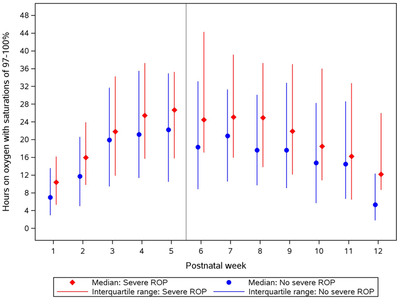
Total hours with saturations of 97–100% for each infant during time on supplemental oxygen, by postnatal week and severe ROP.
Based on the graphical findings, a logistic regression model was created that (a) accounted separately for time on oxygen at 1–5 weeks and 6–9 weeks, because nearly all infants with ROP were still on oxygen at 6 weeks, and (b) examined the specific saturation ranges that differed during those postnatal weeks between infants with and without severe ROP. The model predicted severe ROP based on total hours of oxygen supplementation at 1–5 weeks, the percentage of that time with SpO2 of 91–96%, and their interaction; total hours of oxygen supplementation at 6–9 weeks, the percentage of that time with SpO2 of 97–100%, and their interaction; and prespecified perinatal risk factors. The interaction terms were significant at the p<0.05 level, indicating that the association between severe ROP and the specific SpO2 ranges depended on the duration of time spent on supplemental oxygen (Supplemental Table S3). For infants who spent at least 2 weeks on oxygen in postnatal weeks 1–5, a higher percentage of time at saturations of 91–96% was associated with increased odds of severe ROP (Figure 4). For infants who spent at least 3 weeks on oxygen in postnatal weeks 6–9, a higher percentage of time at saturations of 97–100% was associated with increased odds of severe ROP (Figure 4).
Figure 4.
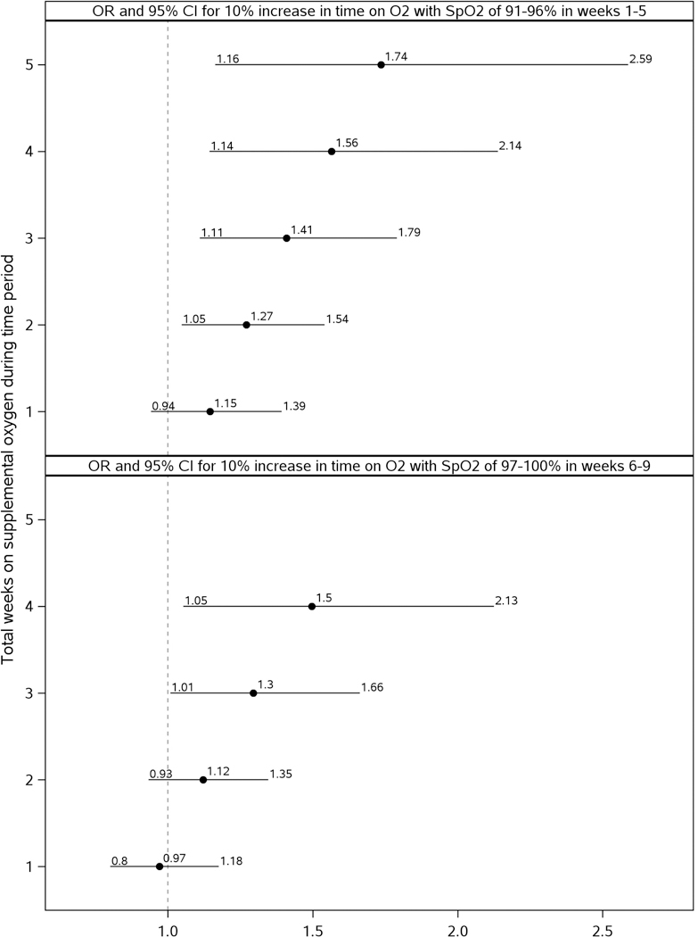
Results of logistic regression model to predict severe ROP. Adjusted odds ratios (OR) and 95% confidence intervals (CI) are shown for the effect of percentage of time on oxygen with saturations of 91–96% during postnatal weeks 1–5, and percentage of time on oxygen with saturations of 97–100% during postnatal weeks 6–9. Because there were significant interactions in the logistic regression model, ORs and CIs for the effect of the percentage of time spent in the saturation ranges are presented separately for infants spending 1, 2, 3, 4, or 5 weeks on supplemental oxygen during the specified postnatal time period.
Other risk factors significantly associated with severe ROP were lower GA and birthweight, non-Hispanic white compared to non-Hispanic black race, severe illness, late onset sepsis or meningitis, and clinical center (Supplemental Table S3). Analyses using PMA in place of postnatal age did not demonstrate the same strength of association between time at SpO2 values and severe ROP (data not shown).
Discussion
Previous large studies of oxygen saturation levels and ROP reported targeted, rather than achieved, saturations.5,6 We found that the relationship between achieved saturations and severe ROP depended on the timing and duration of supplemental oxygen. A greater percentage of time on oxygen at 91–96% SpO2 in weeks 1–5 was associated with severe ROP for infants who spent at least 2 weeks on oxygen. The SUPPORT primary analysis found an unexpected increase in mortality with saturation targets below 91%.2 Together, these findings imply that increased severe ROP may be an unavoidable consequence of targeting higher saturations to avoid mortality. However, this study also found that infants in room air by 6 weeks had a very low incidence of severe ROP (1.4%). Thus, infants healthy enough to be off oxygen by 6 weeks may have low risk of severe ROP regardless of earlier saturations. For infants still receiving oxygen 3 or more weeks past the 5-week mark, the association between severe ROP and a higher percentage of time at 97–100% SpO2 in weeks 6–9 may point to a modifiable risk factor, particularly given the increased stability of most infants past 5 weeks.
ROP proceeds in two phases (I: vasoconstrictive; II: vasoproliferative), during which similar oxygen saturations may have different effects. Data suggest that supplemental oxygen poses a risk during phase I, prior to vasoproliferation.15 ROP phase II begins to develop after 32 weeks’ PMA but has a wide range of onset.15 The STOP-ROP study found that an SpO2 target of 96–99% implemented at a mean (± standard deviation) PMA of 35.4 ± 2.5 weeks did not increase ROP severity for infants with prethreshold ROP.16 We found an association between severe ROP and exposure to 97–100% SpO2 in weeks 6 through 9, or 30–33 weeks through 33–36 weeks PMA, earlier than the average age at which infants in STOP-ROP were randomized. Extremely preterm infants may be more vulnerable to harm from higher saturations during this earlier period.
In a single center study of pulse oximeter data continuously recorded through 8 weeks of age, hyperoxemic events >95% oxygen saturation for 10 or more seconds were less frequent in week 2 and weeks 4–8 in infants requiring laser treatment for ROP compared to those with no ROP or mild ROP.17 The authors speculated that this was because infants not requiring laser treatment spent more time in room air where oxygen saturations of 94–100% are common.17 The HOPE-ROP study suggests that spontaneous high saturations (>94%) in room air do not have the same detrimental effect as high saturations during oxygen support.18 Consistent with this, we found that infants who were in room air by 6 weeks had a very low incidence of severe ROP.
While some studies have found that timing of ROP onset corresponds with PMA,19,20 both SUPPORT and a Swedish study of 22–26 week GA infants found it to be more closely associated with postnatal age.11,21 In a previous analysis, the postnatal age of ROP onset in SUPPORT did not differ between lower and higher GA infants, and infants of lower GA had onset at earlier postmenstrual age.11 This is consistent with the weaker association in this study between severe ROP and SpO2 measured at specific weeks’ PMA compared to postnatal weeks.
In SUPPORT, severe ROP was not diagnosed prior to 6.4 weeks of age,11 consistent with other studies that found onset of Type 1 ROP or need for treatment did not occur before 6 or 8 weeks, respectively.20,22 In this study, exposure to 97–100% SpO2 increased over the first 5 postnatal weeks for infants with and without severe ROP, which may be due to an improvement in the infants’ clinical condition over time or other factors. Exposure to 97–100% SpO2 decreased more quickly in weeks 6–12 in infants without severe ROP. Observational studies have found an association between duration of oxygen supplementation and severe ROP.23,24 It is possible that infants on respiratory support longer are more likely to receive oxygen during a window of increased risk from higher saturations.
Saturations >96% are above the range typically recommended, either before SUPPORT4 or subsequently. Guidance for oxygen targeting in extremely low birth weight infants published in 2016 concluded that “a target saturation range of 90% to 95% may be safer than 85% to 89% at least for some infants. However, the ideal oxygen saturation range for extremely low birth weight infants remains unknown.”25 It is challenging to keep infants within a target range for a variety of reasons. First, less stable infants may be maintained at higher saturations to avoid intermittent hypoxic episodes. Second, studies supporting an association between higher saturation targets during ROP phase II and decreased progression to severe ROP 15,26,27 have led some NICUs to routinely increase saturation targets for the most extremely preterm infants from around 33 weeks PMA until the retina has stabilized. This practice may lead to decreased concern about higher saturations even before that. Third, bedside personnel may be less consistent, and staffing ratios may decrease, with increasing postnatal age of the infants. A study of infants enrolled in BOOST II at The Royal Women’s Hospital Melbourne found that upper alarm limits on study oximeters were correctly set on 79.8% of the days prior to 32 weeks’ PMA in contrast with 65.6% in weeks 32–36.28 Though SUPPORT recommended alarm settings of 84% and 96%, the alarms could be changed at the discretion of the healthcare team, and data on actual alarm settings were not collected. These factors and unknown others may have contributed to some infants spending more time at higher saturations beginning around 6 weeks.
Strengths of this study include prospective collection of detailed neonatal, respiratory, and SpO2 data on a large cohort of extremely preterm infants, and rigorous monitoring and evaluation of the ophthalmologic outcome. The study included 20 clinical centers which increases its generalizability over single-site studies. Because sites differ with respect to unmeasured characteristics, clinical center was included as a covariate in statistical modeling to obtain risk factor estimates that are averaged over the centers.
A limitation is that saturations were only recorded through 36 weeks’ PMA; thus, data after 9 weeks are not available for infants of higher GA. In order to include consistent data for all infants, only data through 9 weeks (33 weeks PMA for 24 week GA infants) were used in statistical modeling. It may be that higher saturations while on oxygen beyond 9 postnatal weeks are associated with severe ROP, but that was not tested. Infants spending more time with higher saturations may also have more desaturations, but oxygen fluctuations were not examined in this analysis.
Identification of oximeter data during oxygen supplementation may be imprecise, since it is based on respiratory data collected at 2–6 hour intervals. Since infants with low oxygen requirement can go in and out of oxygen supplementation, inclusion of time on room air may have increased or decreased the estimated number of hours with a high SpO2. Missing saturation data may have an impact on results, though this impact is expected to be small since most missing SpO2 data occurred in infants with higher gestational ages and birth weights who did not have severe ROP.
It is possible that the data for actual SpO2 values of 91–96% are influenced by the transformation from masked to actual values. However, in both the lower- and higher-target saturation groups, SpO2 values of 97% and higher were not masked (actual values were displayed on the study oximeters and captured in the resulting datasets). Thus, we do not expect that the oximeter masking had a substantial impact on our results regarding saturations of 97–100%. A previously published characteristic of the study oximeters reduced the frequency of saturations of 87–90% and may have impacted the data used in this study.29
In conclusion, the relationship between achieved oxygen saturations and severe ROP in this study depended on the timing and duration of oxygen supplementation. For infants requiring oxygen past 5 postnatal weeks, the percentage of time spent with saturations of 97–100% may be a modifiable risk factor for severe ROP, particularly given the increased stability of most infants past 5 weeks.
Supplementary Material
What is already known on this topic:
Retinopathy of prematurity (ROP) was associated with unrestricted oxygen use in the 1950s. However, the specific oxygen saturations associated with development of severe ROP in extremely preterm infants are unknown.
What this study adds:
Among infants of 24–27 weeks’ gestational age, the relationship between achieved oxygen saturations and severe ROP depended on the timing and duration of oxygen supplementation. For infants requiring oxygen past 5 postnatal weeks, the percentage of time spent with saturations of 97–100% may be a modifiable risk factor.
ACKNOWLEDGEMENTS
The National Institutes of Health, the Eunice Kennedy Shriver National Institute of Child Health and Human Development (NICHD), and the National Heart, Lung, and Blood Institute (NHLBI) provided grant support for the Neonatal Research Network’s SUPPORT Trial. While NICHD staff did have input into the study design, conduct, analysis, and manuscript drafting, the comments and views of the authors do not necessarily represent the views of the NICHD.
Participating NRN sites collected data and transmitted it to RTI International, the data coordinating center (DCC) for the network, which stored, managed and analyzed the data for this study. On behalf of the NRN, Drs. Abhik Das (DCC Principal Investigator) and Marie Gantz (DCC Alternate Principal Investigator and Senior Statistician) had full access to all of the data in the study, and with the NRN Center Principal Investigators, take responsibility for the integrity of the data and accuracy of the data analysis.
FUNDING/SUPPORT
The National Institutes of Health, the Eunice Kennedy Shriver National Institute of Child Health and Human Development (NICHD), and the National Heart, Lung, and Blood Institute (NHLBI) provided grant support for the Neonatal Research Network’s SUPPORT Trial. While NICHD staff did have input into the study design, conduct, analysis, and manuscript drafting, the comments and views of the authors do not necessarily represent the views of the NICHD.
Author contributions were as follows:
Conception and design: Gantz, Carlo, Das
Acquisition, analysis, or interpretation of the data: All authors
Drafting of the manuscript: Gantz, Carlo
Critical revision of the manuscript: All authors
Statistical analysis: Gantz, Das
We are indebted to our medical and nursing colleagues and the infants and their parents who agreed to take part in this study. The following investigators, in addition to those listed as authors, participated in this study:
NRN Steering Committee Chairs: Alan H. Jobe, MD PhD, University of Cincinnati (2003–2006); Michael S. Caplan, MD, University of Chicago, Pritzker School of Medicine (2006–2011).
Alpert Medical School of Brown University and Women & Infants Hospital of Rhode Island (U10 HD27904) - William Oh, MD; Betty R. Vohr, MD; Angelita M. Hensman, RN BSN; Bonnie E. Stephens, MD; Barbara Alksninis, PNP; Dawn Andrews, RN; Kristen Angela, RN; Susan Barnett, RRT; Bill Cashore, MD; Melinda Caskey, MD; Kim Francis, RN; Dan Gingras, RRT; Regina A. Gargus, MD FAAP; Katharine Johnson, MD; Shabnam Lainwala, MD; Theresa M. Leach, MEd CAES; Martha R. Leonard, BA BS; Sarah Lillie, RRT; Kalida Mehta; James R. Moore, MD; Lucy Noel; Suzy Ventura; Rachel V. Walden; Victoria E. Watson, MS CAS.
Case Western Reserve University, Rainbow Babies & Children’s Hospital (U10 HD21364, M01 RR80) - Avroy A. Fanaroff, MD; Deanne E. Wilson-Costello, MD; Bonnie S. Siner, RN; Arlene Zadell RN; Julie DiFiore, BS; Monika Bhola, MD; Harriet G. Friedman, MA; Gulgun Yalcinkaya, MD.
Cincinnati Children’s Hospital Medical Center, University of Cincinnati Hospital, and Good Samaritan Hospital (U10 HD27853, M01 RR8084) - Edward F. Donovan, MD; Vivek Narendran, MD MRCP; Kimberly Yolton, PhD; Kate Bridges, MD; Barbara Alexander, RN; Cathy Grisby, BSN CCRC; Marcia Worley Mersmann, RN CCRC; Holly L. Mincey, RN BSN; Jody Hessling, RN; Teresa L. Gratton, PA.
Duke University School of Medicine, University Hospital, Alamance Regional Medical Center, and Durham Regional Hospital (U10 HD40492, M01 RR30) - Ronald N. Goldberg, MD; C. Michael Cotten, MD MHS; Ricki F. Goldstein, MD; Patricia Ashley, MD; Kathy J. Auten, MSHS; Kimberley A. Fisher, PhD FNP-BC IBCLC; Katherine A. Foy, RN; Sharon F. Freedman, MD; Kathryn E. Gustafson, PhD; Melody B. Lohmeyer, RN MSN; William F. Malcolm, MD; David K. Wallace, MD MPH.
Emory University, Children’s Healthcare of Atlanta, Grady Memorial Hospital, and Emory Crawford Long Hospital (U10 HD27851, UL1 RR25008, M01 RR39) - Barbara J. Stoll, MD; Susie Buchter, MD; Anthony J. Piazza, MD; David P. Carlton, MD; Ira Adams-Chapman, MD; Linda Black, MD; Ann M. Blackwelder, RNC BS MS; Sheena Carter, PhD; Elisabeth Dinkins, PNP; Sobha Fritz, PhD; Ellen C. Hale, RN BS CCRC; Amy K. Hutchinson, MD; Maureen Mulligan LaRossa, RN; Gloria V. Smikle, PNP MSN.
Eunice Kennedy Shriver National Institute of Child Health and Human Development - Stephanie Wilson Archer, MA.
Indiana University, University Hospital, Methodist Hospital, Riley Hospital for Children, and Wishard Health Services (U10 HD27856, M01 RR750) - Brenda B. Poindexter, MD MS; Anna M. Dusick, MD FAAP; James A. Lemons, MD; Leslie D. Wilson, BSN CCRC; Faithe Hamer, BS; Ann B. Cook, MS; Dianne E. Herron, RN; Carolyn Lytle, MD MPH; Heike M. Minnich, PsyD HSPP.
McGovern Medical School at The University of Texas Health Science Center at Houston and Children’s Memorial Hermann Hospital (U10 HD21373) - Kathleen A. Kennedy, MD MPH; Jon E. Tyson, MD MPH; Patricia W. Evans, MD; Nora I. Alaniz, BS; Patricia Evans, MD; Beverly Foley Harris, RN BSN; Charles Green, PhD; Margarita Jiminez, MD MPH; Anna E. Lis, RN BSN; Sarah Martin, RN BSN; Georgia E. McDavid, RN; Brenda H. Morris, MD; Margaret L. Poundstone, RN BSN; Stacy Reddoch, BA; Saba Siddiki, MD; Patti L. Pierce Tate, RCP; Laura L. Whitely, MD; Sharon L. Wright, MT (ASCP).
National Heart, Lung, and Blood Institute - Mary Anne Berberich, PhD; Carol J. Blaisdell, MD; Dorothy B. Gail, PhD; James P. Kiley, PhD.
RTI International (U10 HD36790) - W. Kenneth Poole, PhD; Lisa A. Wrage, MPH; Jamie E. Newman, PhD MPH; Betty K. Hastings; Jeanette O’Donnell Auman, BS; Carolyn Petrie Huitema, MS; James W. Pickett II, BS; Dennis Wallace, PhD; Kristin M. Zaterka-Baxter, RN BSN.
Stanford University and Lucile Packard Children’s Hospital (U10 HD27880, UL1 RR25744, M01 RR70) - Krisa P. Van Meurs, MD; David K. Stevenson, MD; Susan R. Hintz, MD MS Epi; M. Bethany Ball, BS CCRC; Barbara Bentley, PsychD MSEd; Elizabeth F. Bruno, PhD; Alexis S. Davis, MD MS; Maria Elena DeAnda, PhD; Anne M. DeBattista, RN, PNP; Jean G. Kohn, MD MPH; Melinda S. Proud, RCP; Renee P. Pyle, PhD; Nicholas H. St. John, PhD; Hali E. Weiss, MD.
Tufts Medical Center, Floating Hospital for Children (U10 HD53119, M01 RR54) - Ivan D. Frantz III, MD; Elisabeth C. McGowan, MD; John M. Fiascone, MD; Anne Furey, MPH; Brenda L. MacKinnon, RNC; Ellen Nylen, RN BSN; Ana Brussa, MS OTR/L; Cecelia Sibley, PT MHA.
University of Alabama at Birmingham Health System and Children’s Hospital of Alabama (U10 HD34216, M01 RR32) - Namasivayam Ambalavanan, MD; Myriam Peralta-Carcelen, MD MPH; Monica V. Collins, RN BSN MaEd; Shirley S. Cosby, RN BSN. Vivien A. Phillips, RN BSN; Kirstin J. Bailey, PhD; Fred J. Biasini, PhD; Maria Hopkins, PhD; Kristen C. Johnston, MSN CRNP; Sara Krzywanski, MS; Kathleen G. Nelson, MD; Cryshelle S. Patterson, PhD; Richard V. Rector, PhD; Leslie Rodriguez, PhD; Amanda Soong, MD; Sally Whitley, MA OTR-L FAOTA; Sheree York, PT DPT MS PCS.
University of California - San Diego Medical Center and Sharp Mary Birch Hospital for Women (U10 HD40461) - Maynard R. Rasmussen, MD; Paul R. Wozniak, MD; Yvonne E. Vaucher, MD MPH; Kathy Arnell, RNC; Rene Barbieri-Welge; Ayala Ben-Tall; Renee Bridge, RN; Clarence Demetrio, RN; Martha G. Fuller, RN MSN; Elaine Ito; Meghan Lukasik; Deborah Pontillo; Donna Posin, OTR/L MPA; Cheryl Runyan; James Wilkes; Paul Zlotnik.
University of Iowa Children’s Hospital (U10 HD53109, UL1 RR24979, M01 RR59) - Edward F. Bell, MD; John A. Widness, MD; Michael J. Acarregui, MD; Jonathan M. Klein, MD; Tarah T. Colaizy, MD MPH; Karen J. Johnson, RN BSN; Diane L. Eastman, RN CPNP MA.
University of Miami, Holtz Children’s Hospital (U10 HD21397, M01 RR16587) - Shahnaz Duara, MD; Charles R. Bauer, MD; Ruth Everett-Thomas, RN MSN; Maria Calejo, MEd; Alexis N. Diaz, BA; Silvia M. Frade Eguaras, BA; Andrea Garcia, MA; Kasey Hamlin-Smith, PhD; Michelle Harwood Berkowits, PhD; Sylvia Hiriart-Fajardo, MD; Elaine O. Mathews, RN; Helina Pierre, BA; Arielle Riguard, MD; Alexandra Stroerger, BA.
University of New Mexico Health Sciences Center (U10 HD53089, M01 RR997) - Kristi L. Watterberg, MD; Robin K. Ohls, MD; Janell Fuller, MD; Julie Rohr, MSN RNC CNS; Conra Backstrom Lacy, RN; Jean Lowe, PhD; Rebecca Montman, BSN.
University of Rochester Medical Center, Golisano Children’s Hospital (U10 HD40521, M01 RR44) - Dale L. Phelps, MD; Nirupama Laroia, MD; Gary David Markowitz, MD; Gary J. Myers, MD; Linda J. Reubens, RN CCRC; Diane Hust, MS RN CS; Lisa Augostino; Julie Babish Johnson, MSW; Erica Burnell, RN; Rosemary L. Jensen; Emily Kushner, MA; Joan Merzbach, LMSW; Kelley Yost, PhD.
University of Texas Southwestern Medical Center at Dallas, Parkland Health & Hospital System, and Children’s Medical Center Dallas (U10 HD40689, M01 RR633) - Pablo J. Sánchez, MD; Charles R. Rosenfeld, MD; Walid A. Salhab, MD; Roy J. Heyne, MD; Sally S. Adams, MS RN CPNP; James Allen, RRT; Laura Grau, RN; Alicia Guzman; Gaynelle Hensley, RN; Elizabeth T. Heyne, PsyD PA-C; Melissa H. Lepps, RN; Linda A. Madden, RN CPNP; Melissa Martin, RN; Nancy A. Miller, RN; Janet S. Morgan, RN; Araceli Solis, RRT; Lizette E. Torres, RN; Catherine Twell Boatman, MS CIMI; Diana M Vasil, RNC-NIC; Kerry Wilder, RN.
University of Utah Medical Center, Intermountain Medical Center, LDS Hospital, and Primary Children’s Medical Center (U10 HD53124, M01 RR64) - Anna Bodnar, MD; Shawna Baker, RN; Karie Bird, RN; Jill Burnett, RN; Laura Cole, RN; Karen A. Osborne, RN BSN CCRC; Cynthia Spencer, RNC; Mike Steffens, PhD; Kimberlee Weaver-Lewis, RN BSN; Karen Zanetti, RN.
Wake Forest University, Baptist Medical Center, Brenner Children’s Hospital, and Forsyth Medical Center (U10 HD40498, M01 RR7122) - T. Michael O’Shea, MD MPH; Robert G. Dillard, MD; Lisa K. Washburn, MD; Nancy J. Peters, RN CCRP; Barbara G. Jackson, RN BSN; Korinne Chiu, MA; Deborah Evans Allred, MA LPA; Donald J. Goldstein, PhD; Raquel Halfond, MA; Carroll Peterson, MA; Ellen L. Waldrep, MS; Cherrie D. Welch, MD MPH; Melissa Whalen Morris, MA; Gail Wiley Hounshell, PhD.
Wayne State University, Hutzel Women’s Hospital, and Children’s Hospital of Michigan (U10 HD21385) - Seetha Shankaran, MD; Beena G. Sood, MD MS; Athina Pappas, MD; Rebecca Bara, RN BSN; Elizabeth Billian, RN MBA; Laura A. Goldston, MA; Mary Johnson, RN BSN.
Yale University, Yale-New Haven Children’s Hospital, and Bridgeport Hospital (U10 HD27871, UL1 RR24139, M01 RR125) - Richard A. Ehrenkranz, MD; Vineet Bhandari, MD DM; Harris C. Jacobs, MD; Pat Cervone, RN; Patricia Gettner, RN; Monica Konstantino, RN BSN; JoAnn Poulsen, RN; Janet Taft, RN BSN; Christine G. Butler, MD; Nancy Close, PhD; Walter Gilliam, PhD; Sheila Greisman, RN; Elaine Romano, MSN; Joanne Williams, RN BSN.
Retinopathy of Prematurity Adjudication Committee - Gary David Markowitz, MD, University of Rochester; Amy K. Hutchinson, MD, Emory University; David K. Wallace, MD MPH, Duke University; Sharon F. Freedman, MD, Duke University.
Footnotes
DISCLOSURES
The authors have no relevant disclosures.
References
- 1.Duc G, Sinclair JC. Oxygen administration In: Sinclair JC, Bracken MB, eds. Effective Care of the Newborn Infant. New York: Oxford University Press Inc; 1992. p. 178–94. [Google Scholar]
- 2.SUPPORT Study Group of the Eunice Kennedy Shriver NICHD Neonatal Research Network, Carlo WA, Finer NN, Walsh MC, Rich W, Gantz MG, et al. Target ranges of oxygen saturation in extremely preterm infants. N Engl J Med. 2010;362(21):1959–69. [DOI] [PMC free article] [PubMed] [Google Scholar]
- 3.Stenson B, Brocklehurst P, Tarnow-Mordi W; U.K. BOOST II trial; Australian BOOST II trial; New Zealand BOOST II trial. Increased 36-week survival with high oxygen saturation target in extremely preterm infants. N Engl J Med. 2011;364(17):1680–2. [DOI] [PubMed] [Google Scholar]
- 4.American Academy of Pediatrics, American College of Obstetricians and Gynecologists. Guidelines for perinatal care 6th ed Elk Grove Village (IL): AAP; Washington, DC: ACOG; 2007. p.261–262. [Google Scholar]
- 5.Tin W, Milligan DW, Pennefather P, Hey E. Pulse oximetry, severe retinopathy, and outcome at one year in babies of less than 28 weeks gestation. Arch Dis Child Fetal Neonatal Ed 2001;84:F106–10. [DOI] [PMC free article] [PubMed] [Google Scholar]
- 6.Chow LC, Wright KW, Sola A, and the CSMC Oxygen Administration Study Group. Can changes in clinical practice decrease the incidence of severe retinopathy of prematurity in very low birth weight infants? Pediatrics. 2003;111:339–45. [DOI] [PubMed] [Google Scholar]
- 7.Anderson G, Benitz WE, Madan A. Retinopathy of prematurity and pulse oximetry: A national survey of recent practices. J Perinatol. 2004;24:164–8. [DOI] [PubMed] [Google Scholar]
- 8.Kinsey VE, Arnold HJ, Kalina RE, et al. PaO2 levels and retrolental fibroplasia: a report of the cooperative study. Pediatrics. 1977;60:655–68. [PubMed] [Google Scholar]
- 9.Flynn JT, Bancalari E, Snyder ES, et al. A cohort study of transcutaneous oxygen tension and the incidence and severity of retinopathy of prematurity. N Engl J Med. 1992;326:1050–4. [DOI] [PubMed] [Google Scholar]
- 10.Hagadorn JI, Furey AM, Nghiem TH, Schmid CH, Phelps DL, Pillers DA, Cole CH, AVIOx Study Group. Achieved versus intended pulse oximeter saturation in infants born less than 28 weeks’ gestation: the AVIOx study. Pediatrics, 2006;188(4):1574–82. [DOI] [PubMed] [Google Scholar]
- 11.Kennedy KA, Wrage LA, Higgins RD, Finer NN, Carlo WA, Walsh MC, Laptook AR, Faix RG, Yoder BA, Schibler K, Gantz MG, Das A, Newman NS, Phelps DL, SUPPORT Study Group of the NICHD Neonatal Research Network. Evaluating retinopathy of prematurity screening guidelines for 24–27 week gestational age infants. J Perinatol. 2014;34(4):311–318. [DOI] [PMC free article] [PubMed] [Google Scholar]
- 12.Hardy RJ, Good WV, Dobson V, et al. Multicenter trial of early treatment for retinopathy of prematurity: study design. Control Clin Trials. 2004; 25:311–25. [DOI] [PubMed] [Google Scholar]
- 13.Early Treatment for Retinopathy of Prematurity Cooperative Group. Revised indications for the treatment of retinopathy of prematurity: results of the early treatment for retinopathy of prematurity randomized trial. Arch Ophthalmol. 2003;121:1684–94. [DOI] [PubMed] [Google Scholar]
- 14.Schmidt B, Roberts RS, Whyte RK, Asztalos EV, Poets C, Rabi Y, Solimano A, Nelson H, for the Canadian Oxygen Trial Group. Impact of study oximeter masking algorithm on titration of oxygen therapy in the Canadian Oxygen Trial. J Pediatr. 2014; 165:666–71. [DOI] [PubMed] [Google Scholar]
- 15.Smith LE, Hard AL, Hellstrom A. The biology of retinopathy of prematurity: How knowledge of pathogenesis guides treatment. Clin Perinatol. 2013;40(2):201–214. [DOI] [PMC free article] [PubMed] [Google Scholar]
- 16.The STOP-ROP Multicenter Study Group. Supplemental therapeutic oxygen for prethreshold retinopathy of prematurity (STOP-ROP), a randomized, controlled trial I: primary outcomes. Pediatrics. 2000;105:295–310. [DOI] [PubMed] [Google Scholar]
- 17.Di Fiore J, Bloom JN, Orge F, Schutt A, Schluchter M, Cheruvu VK, Walsh M, Finer N, Martin RJ. A higher incidence of intermittent hypoxemic episodes is associated with severe retinopathy of prematurity. J Pediatr. 2010;157(1):69–73. [DOI] [PMC free article] [PubMed] [Google Scholar]
- 18.McGregor ML, Bremer DL, Cole CC, McCead RE, Phelps DL, Fellows RR, Oden N, HOPE-ROP Multicenter Group. Retinopathy of prematurity outcome in infants with prethreshold retinopathy of prematurity and oxygen saturation >94% in room air: The high oxygen percentage in retinopathy of prematurity study. Pediatrics. 2002;110:540–544. [DOI] [PubMed] [Google Scholar]
- 19.Palmer EA, Flynn JT, Hardy RJ, Phelps DL, Phillips CL, Schaffer DB, et al. Incidence and early course of retinopathy of prematurity. Ophthalmology. 1991; 98:1628–1640. [PubMed: 1800923] [DOI] [PubMed] [Google Scholar]
- 20.Isaza G, Arora S. Incidence and severity of retinopathy of prematurity in extremely premature infants. Can J Ophthalmol. 2012; 47:296–300. [PubMed: 22687311] [DOI] [PubMed] [Google Scholar]
- 21.Austeng D, Kallen KB, Hellstrom A. Screening for retinopathy of prematurity in infants born before 17 weeks’ gestation in Sweden. Arch Ophthalmol. 2011;129:167–172. [DOI] [PubMed] [Google Scholar]
- 22.Muether PS, Kribs A, Hahn M, Schumacher J, Eifinger F, Kirchhof B, et al. No advanced retinopathy of prematurity stages 4 or 5 in a large high-risk German cohort. Br J Ophthalmol. 2012; 96:400–404. [PubMed: 21719567] [DOI] [PubMed] [Google Scholar]
- 23.Enomoto H, Miki A, Matsumiya W, Honda S. Evaluation of oxygen supplementation status as a risk factor associated with the development of severe retinopathy of prematurity. Ophthalmologica. 2015;234(3):135–8. [DOI] [PubMed] [Google Scholar]
- 24.Lui Q, Yin ZQ, Ke N, Chen L, Chen XK, Fang J, Chen XR, Xiao J, Hu K, Zhang X, Xiong Y, Shi H, Li J, Long HJ, Zhang KL, Chen ZY, Pi LH. Incidence of retinopathy of prematurity in southwestern China and analysis of risk factors. Med Sci Monit. 2014;20:1442–51. [DOI] [PMC free article] [PubMed] [Google Scholar]
- 25.Cummings JJ, Polin RA, AAP Committee on Fetus and Newborn. Oxygen Targeting in Extremely Low Birth Weight Infants. Pediatrics. 2016;138(2):e20161576. [DOI] [PubMed] [Google Scholar]
- 26.Askie LM, Henderson-Smart DJ, Irwig L, Simpson JM. Oxygen-saturation targets and outcomes in extremely preterm infants. N Engl J Med. 2003;349:959–967. [DOI] [PubMed] [Google Scholar]
- 27.Chen ML, Guo L, Smith LE, Dammann CE, Dammann O. High or low oxygen saturation and severe retinopathy of prematurity: a meta-analysis. Pediatrics. 2010;125:e1483–e1492. [DOI] [PMC free article] [PubMed] [Google Scholar]
- 28.Mills A, Davis PG, Donath SM, Clucas LM, Doyle LW. Improving compliance with pulse oximetry alarm limits for very preterm infants? Journal of Paediatrics and Child Health. 2010;46:255–258. [DOI] [PubMed] [Google Scholar]
- 29.Johnston ED, Boyle B, Juszczak E, King A, Brocklehurst P, Stenson BJ. Oxygen targeting in preterm infants using the Masimo SET Radical pulse oximeter. Arch Dis Child Fetal Neonatal Ed. 2011;96:F429–433. [DOI] [PMC free article] [PubMed] [Google Scholar]
Associated Data
This section collects any data citations, data availability statements, or supplementary materials included in this article.