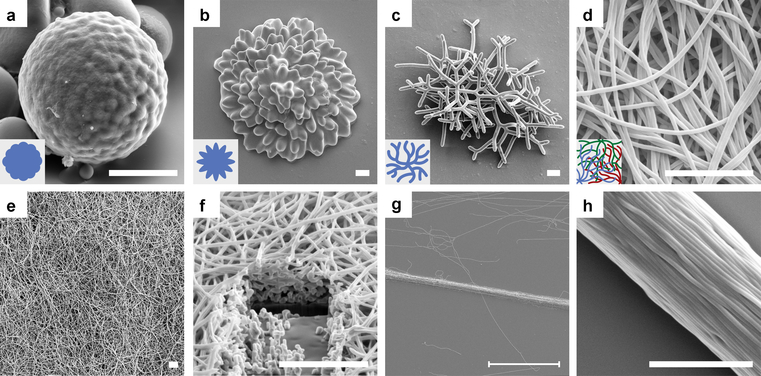
Fig. 4 |. SEM images of self-assembled NLCEs.
a-d, Crosslinked NLCEs: (a) roughened sphere, (b) flower, and (c-d) filamentous structures with decreasing diameter. See Fig. 2a–d for corresponding NLCO morphologies observed in bright-field microscopy. The objects shown in (a-c) evolved from a single drop and are depicted in inset schematics; thin filamentous structures grown from several drops are shown (intertwined) in (d) (Inset: filaments with different colors grow from different drops). e-f, Partial view of a centimeter-wide free-standing NLCE fibrous mat and its cross section (cut by a focused-Ga-ion-beam). See Extended Data Fig. 7 for macroscopic image of the mat. g-h, Oriented NLCE fiber yarn. The scale bar in (g) is 100 μm; all other scale bars are 5 μm.

