Abstract
Background:
Pain perception is critical for detection of noxious bodily insults. Gastrointestinal hypoalgesia in inflammatory bowel disease (IBD) is a poorly understood phenomenon previously linked to poor patient outcomes. We aimed to evaluate the risk factors associated with this condition and to discern characteristics that might differentiate these patients from pain-free quiescent counterparts.
Methods:
We performed a retrospective analysis using an IBD natural history registry based in a single tertiary care referral center. We compared demographic and clinical features in 3 patient cohorts defined using data from simultaneous pain surveys and ileocolonoscopy: a) active IBD without pain (hypoalgesic IBD); b) active IBD with pain; and c) inactive IBD without pain.
Results:
One hundred fifty-three IBD patients had active disease and 43 (28.1%) exhibited hypoalgesia. Hypoalgesic IBD patients were more likely to develop non-perianal fistulae (P=0.03). On logistic regression analysis, hypoalgesic IBD was independently associated with male sex, advancing age and mesalamine use, and inversely associated with anxious/depressed state and opiate use. Hypoalgesic IBD patients were demographically and clinically similar to the pain-free quiescent IBD cohort (n=59). Platelet count and C-reactive protein were more likely to be pathologically elevated in hypoalgesic IBD (P=0.03), though >25% did not exhibit elevated inflammatory markers.
Conclusions:
Hypoalgesia is common in IBD, particularly in male and older individuals, and is associated with an increased incidence of fistulae and corticosteroid use. Novel noninvasive diagnostic tools are needed to screen for this population, as inflammatory markers are not always elevated.
Keywords: Abdominal pain, hypoalgesia, hyposensitivity, inflammatory bowel disease, Crohn’s disease
Introduction
Abdominal pain is one of the most common symptoms associated with gastrointestinal disorders. It is a major driver behind the deterioration of patients’ quality of life and increased healthcare resource utilization in inflammatory bowel disease (IBD), including both Crohn’s disease (CD) and ulcerative colitis (UC) [1-4]. While there is little doubt that abdominal pain can have a detrimental impact on the lives of IBD patients, it also serves an indispensable physiological service. The ability to properly perceive pain is critical for the detection of noxious insults that have the potential to cause damage to the body. This is true even in the gastrointestinal tract, where perception of noxious exposures may be vaguer and poorly localized [5], and it is particularly relevant when considering IBD. If IBD patients with clinically significant disease activity do not simultaneously experience and/or report symptoms commensurate with the degree of inflammation, including abdominal pain, they are described as having silent IBD. Silent IBD is important because it has been associated with the development of more frequent and serious complications, including strictures, fistulae, and abscesses [6,7], as well as increased hospitalization [8]. As abdominal pain is one of the most frequently described and consequential symptoms in the setting of IBD [9], it is not difficult to imagine that a lack of or reduction in abdominal pain by itself during periods of significant disease activity, or “hypoalgesic IBD”, can impart a tremendous risk of poor outcomes over time.
Unfortunately, hypoalgesic IBD is a poorly understood condition and little is known about its pathophysiology or epidemiology. Investigators have gleaned some idea of the prevalence of this condition from the findings of prior studies evaluating silent IBD. Unfortunately, these studies were: a) not specifically designed to evaluate silent or hypoalgesic IBD; b) focused primarily on one form of IBD; and/or c) utilized less reliable symptom-based disease activity scores (e.g., CD activity index) [10] to make determinations about intestinal inflammatory status. Other clinical features, including disease location (e.g., upper gastrointestinal tract CD) [11], a variety of extraintestinal manifestations (EIMs) [12-16], and IBD-associated complications [17-20] have previously been implicated as potential signs of silent disease, but it is not clear how effective they are for screening.
Several endoscopic, radiologic and laboratory tests have been proposed as screening options for silent IBD [11,21-25], but their clinical practicality and cost-effectiveness are still uncertain, particularly when the scale of this issue remains incompletely understood. To this point, no studies have specifically evaluated for reduced abdominal pain perception in IBD patients while simultaneously evaluating disease activity using the most reliable assessment methods (e.g., direct endoscopic visualization). Thus, prior estimates as to the actual incidence of silent and/or hypoalgesic IBD may be inaccurate. Just as importantly, no prior investigation has been undertaken to identify risk factors for hypoalgesic IBD. To provide appropriate screening and care for these patients, it is imperative for IBD providers not only to recognize that this condition exists, but also to gain an improved understanding of the risk factors associated with it and how to screen for it.
Our primary aim in undertaking this study was to determine the incidence of abdominal hypoalgesia in both CD and UC, as well as to evaluate for demographic and clinical factors associated with the presence of this condition. Secondly, we sought to identify patient characteristics and test findings that could help differentiate hypoalgesic IBD patients from pain-free IBD patients with inactive disease.
Patients and methods
Study population
We performed a retrospective analysis using information derived from consecutive patients enrolled in the Intestinal Diseases Natural History Database at Pennsylvania State University Hershey Medical Center (PSHMC) between January 1, 2015, and August 31, 2018. This database includes clinical and research information related to the encounters of IBD patients undergoing clinical management at PSHMC, a tertiary care referral hospital with a dedicated IBD center that cares for over 5000 patients with these disorders. This work was performed in accordance with the rules and regulations set forth by the Pennsylvania State University College of Medicine Institutional Review Board and carried out under protocol PRAMSHY98-057.
To be included in this study, participants had to be adults (i.e., older than 17 years) and have an established diagnosis of CD, UC or IBD colitis of indeterminate nature (IC), based on standard clinical criteria routinely used to identify IBD. They also needed to have undergone an ileocolonoscopy and completed contemporaneous surveys on abdominal pain experience (including the Short Inflammatory Bowel Disease Questionnaire [SIBDQ] and Harvey-Bradshaw Index [HBI]). UC patients were excluded if they had undergone previous colonic surgery.
Definitions and data abstraction
Presence of “significant inflammation” was defined as moderate to severe activity based on findings during ileocolonoscopic evaluation, while quiescent disease was defined as the lack of any gross inflammation within the ileum or colon. Endoscopic activity in UC was determined using the Mayo endoscopy sub-score, which ranges from 0-3, with 0=no disease (“quiescent”) and 3=severe disease. Thus, “significant” UC-related inflammation was defined as a Mayo endoscopy subscore of 2-3. In CD, we used the Simple Endoscopic Score for CD (SES-CD), where scores of 7-15 indicated moderate disease and those >15 were representative of severe disease. Using this system, “significant” CD-related inflammation was defined as an SES-CD greater than or equal to 7. Pain ratings were obtained contemporaneously with each ileocolonoscopy and were based on responses to 2 separate items: 1) the fourth question in the SIBDQ (“How often over the past 2 weeks have you experienced abdominal pain?”, where patients respond using a frequency-based inverse Likert scale, with 1 representing pain “all of the time” and 7 representing pain “none of the time”); and 2) the second question from the HBI, which included potential responses of 0 (“no abdominal pain”), 1 (“mild”), 2 (“moderate”) and 3 (“severe”). For the purposes of this study, clinically relevant abdominal pain was defined as a numeric rating of <6 on the SIBDQ pain score and/or a score of 1 or greater on the HBI pain score. To qualify as having “hypoalgesic IBD” (see below), a patient could not have described clinically significant pain on either of these 2 measures. Age, sex, IBD duration, IBD extent/location (e.g., organ involvement, using the Montreal classification system), disease complications (including stricture, intra-abdominal fistula, abscess, and cancer development), EIMs (including inflammatory arthritides), IBD-associated dermatopathies (including pyoderma gangrenosum, erythema nodosum, uveitis, episcleritis, and primary sclerosing cholangitis), physician global assessment, endoscopic severity (defined by the Mayo Index endoscopy sub-score or the SES-CD as appropriate), medication use (including antidepressant/anxiolytic, corticosteroid, mesalamine, immunomodulator such as azathioprine, 6-mercaptopurine and/or methotrexate) and biologic therapy (infliximab, adalimumab, certolizumab, golimumab, vedolizumab and/or ustekinumab), surgical history, laboratory values (white blood cell count [WBC], erythrocyte sedimentation rate [ESR], C-reactive protein [CRP]), opiate, “other” pain medications (acetaminophen, nonsteroidal anti-inflammatory drugs [NSAIDs], dicyclomine and/or tricyclic agents), and tobacco use were obtained from the record. The presence of anxiety or depression symptoms was determined based upon responses to the Hospital Anxiety and Depression Scale completed at the time of the clinical encounter, using anxiety or depression subscores of 8 or greater to indicate the clinically significant presence of each.
Statistical analysis
Data were extracted and analyzed using GraphPad Prism version 8 (San Diego, CA) or SAS version 9.4 (Cary, NC). Initially, demographic and clinical variables were compared using univariate analysis (e.g., Student’s t-test, chi-square test or Fisher’s exact test, as appropriate) between 2 distinct cohorts: 1) IBD patients with active disease without abdominal pain (hereafter referred to as “hypoalgesic IBD”); and 2) IBD patients with active disease with abdominal pain (hereafter referred to as “active IBD with pain”). A multivariate logistic regression model was then created incorporating each significant variable identified during the univariate analysis to examine the odds of developing hypoalgesic IBD. A binary logistic regression was used for optimization. Univariate analysis was also used to compare demographic and clinical variables between: 1) hypoalgesic IBD patients; and 2) IBD patients with inactive disease without abdominal pain (hereafter referred to as “quiescent IBD without pain”) in order to evaluate for clinical factors that might be used to differentiate between these 2 groups. The primary endpoint for each of these analyses was hypoalgesic IBD (as defined above). Values listed represent means ± standard error of the mean (SEM) or odds ratio
Results
Prevalence and clinical characteristics of hypoalgesic IBD
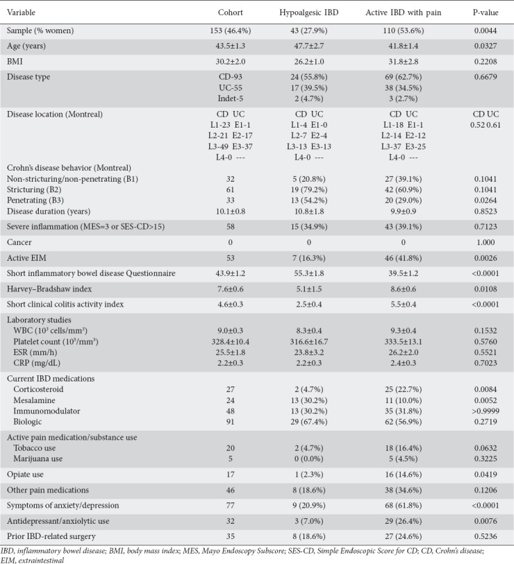
To assess the prevalence of hypoalgesic IBD, we evaluated consecutive patients who had undergone an ileocolonoscopy and completed concurrent validated pain-related surveys at our center. One hundred fifty-three individuals (71 female, 82 male) were found to have moderate-to-severe disease on gross endoscopic evaluation (i.e., Mayo endoscopy subscore of 2-3 or SES-CD of 7 or greater) during the study period (Table 1). Ninety-three individuals had CD (24.7% ileal CD [L1], 22.6% colonic CD [L2], 52.7% ileocolonic CD [L3]), 55 had UC (1.8% had proctitis, 30.9% L-sided UC, 67.3% pan-UC), and 5 individuals had IC. Of this cohort, 43 patients (28.1%) were found to have hypoalgesic IBD. Similar rates of hypoalgesic IBD (26.1% vs. 30.9% respectively, P=0.57) were exhibited in CD and UC. In view of the latter finding, we chose not to evaluate for other differences between IBD subtypes in this setting.
Table 1.
Demographic and clinical characteristics of the hypoalgesic and pain-perceiving active IBD cohorts

Though disease duration (10.8 vs. 9.9 years, P=0.63) and location (CD: L1 16.7% vs. 26.5%, L2 29.2% vs. 20.6%, L3 54.1% vs. 52.9%, P=0.52; UC: E1 0.0% vs. 2.5%, E2 23.5% vs. 32.5%, E3 76.5% vs. 65.0%, P=0.61) were similar between the hypoalgesic and pain-perceptive sub-cohorts respectively, hypoalgesic IBD patients were older (47.7 vs. 41.8 years, P=0.03) and were more likely to be male (72.1% vs. 46.4%, P=0.004). While hypoalgesic IBD patients had a statistically similar proportion of severe endoscopic scores (34.9% vs. 39.1%, P=0.71) and inflammatory laboratory values with regard to WBC (8300 vs. 9300 cells/mm3, P=0.15), CRP (1.7 vs. 2.4 mg/dL, P=0.55) and ESR (23.8 vs. 26.2 mm/hour, P=0.30), they were less likely to exhibit extraintestinal manifestations (16.3% vs. 41.8%, P=0.003) compared to pain-perceiving active IBD patients. Hypoalgesic IBD patients were also less likely to exhibit clinically significant symptoms of anxiety or depression (20.9% vs. 61.8%, P<0.0001). Notably, hypoalgesic CD patients had a significantly higher rate of non-perianal fistulae than their pain-reporting counterparts (54.2% vs. 29.4%, P=0.03).
Utilization of IBD-directed therapies and other medications in the active IBD cohorts
Hypoalgesic IBD patients used mesalamine products more frequently (30.2% vs. 10%, P=0.005) and corticosteroids less frequently (4.7% vs. 22.7%, P=0.008) compared to active IBD patients with pain. They exhibited very similar use of immunomodulator (30.2% vs. 31.8%, P=>0.99) and biologic therapies (67.4% vs. 56.7%, P=0.27) (Table 1). The two sub-cohorts also had statistically similar rates of having prior surgery (18.6% vs. 24.6%, P=0.52). Notably, hypoalgesic IBD patients were less likely to use opiates (2.3% vs. 14.6%, P=0.04) or to use antidepressants or anxiolytics (7.0% vs. 26.4%, P=0.008), and they exhibited a statistically insignificant difference in the use of other pain medications (including acetaminophen, NSAIDs, dicyclomine and tricyclic agents) at a similar frequency as pain-perceiving IBD patients (18.6% vs. 34.6%, P=0.12). Reported use of tobacco (4.7% vs. 16.4%, P=0.06) and marijuana (0% vs. 4.5%, P=0.32) was also not significantly different in hypoalgesic IBD. No other significant difference in demographics, disease characterization or medication use was found.
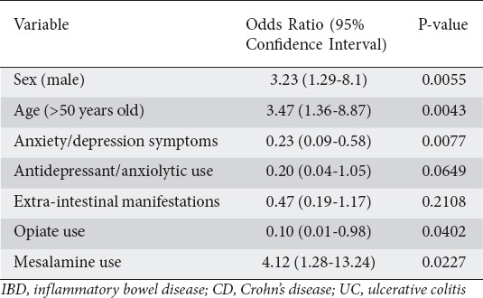
Employing a multivariate logistic regression analysis to evaluate the 153-patient study cohort described above, we found that hypoalgesic IBD was independently associated with male sex (OR 3.23, 95%CI 1.29-8.1; P=0.006), advancing age (>50 years of age) (OR 3.47, 95%CI 1.36-8.87; P=0.004) and mesalamine use (OR 4.12, 95%CI 1.28-13.24; P=0.02), but inversely associated with anxious and/or depressed state (OR 0.23, 95%CI 0.09-0.58; P=0.008), and opiate use (OR 0.10, 95%CI 0.01-0.98; P=0.04) (Table 2).
Table 2.
Multivariate analysis of factors associated with hypoalgesic IBD

Differentiating hypoalgesic IBD from quiescent IBD without abdominal pain
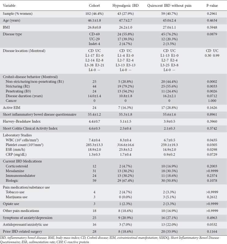
To find potential characteristics that might differentiate hypoalgesic IBD patients, we also used endoscopic and pain-related survey data to identify a cohort of asymptomatic quiescent IBD patients to compare to the hypoalgesic IBD group described above. Using this approach, we identified 59 individuals who qualified as having quiescent IBD without abdominal pain. The quiescent IBD without abdominal pain cohort was composed of mostly Crohn’s disease patients (45 CD: 13 ileal [L1], 7 colonic [L2], 25 ileocolonic [L3]; 12 UC: 4 left-sided [E2], 8 pan-colonic [E3]; 2 IC) and was predominantly male (24 female, 35 male), exhibiting disease distributions that were similar for both CD (P=0.30) and UC (P>0.99). It shared a similar demographic and clinical profile with the hypoalgesic IBD group, with the exception that individuals with quiescent IBD without pain trended toward being more likely to have CD (P=0.07) and have a longer mean disease duration (P=0.07). Hypoalgesic patients also trended toward utilizing antidepressants or anxiolytics (P=0.05) less frequently than the quiescent pain-free cohort (Table 3).
Table 3.
Demographic and clinical characteristics of the cohorts with hypoalgesic IBD and quiescent IBD without abdominal pain

However, several laboratory values differed significantly between these two sub-cohorts. Specifically, hypoalgesic IBD patients exhibited a higher mean WBC (8300 vs. 6700 cells/mm3, P=0.04), ESR (23.8 vs. 14.9 mm/h, P=0.03) and platelet count (316.6 × 10³ vs. 259.1 × 10³/mm3, P=0.03) and trended toward having a higher CRP (P=0.09) (Table 3). Using the laboratory-associated upper limit of normal (ULN) for each of these tests, we also found that hypoalgesic IBD patients more frequently had a significantly elevated ESR (50.0% vs. 25.7%, P=0.05), CRP (66.7% vs. 28.6%, P=0.02) and platelet count (40.0% vs. 12.5%, P=0.008). When we looked for a significant elevation in any one of these tests, we found that hypoalgesic IBD patients had exceeded the ULN for at least one of these tests at least 71.4% of the time, compared to 34.8% for the quiescent IBD group without pain (P=0.001).
Finally, we evaluated for gastrointestinal symptoms other than abdominal pain that are commonly experienced by IBD patients and might provide a clue to discriminating between these two cohorts. Recent or current symptoms of diarrhea (62.8% vs. 69.5%, P=0.52), nocturnal stooling (25.6% vs. 25.4%, P=0.99), blood in stools (32.6% vs. 17.0%, P=0.10), fecal urgency (55.8% vs. 49.2%, P=0.55), and sensation of an abdominal mass (4.7% vs. 3.4%, P=0.99) were statistically similar between the cohorts. Even when these symptoms were combined (86.1% vs. 79.7%, P=0.44), there was no significant difference.
Discussion
We demonstrated that hypoalgesic IBD is relatively common in both CD and UC, affecting over one-quarter of patients with moderate-to-severe disease activity in this study. We also found that hypoalgesic CD patients were at increased risk of developing IBD-associated complications (e.g., fistulae). Each of these findings was comparable to what has been reported in other studies that attempted to evaluate silent IBD [6-8,10]. These results reinforce the clinical relevance of hypoalgesic IBD and the importance of improving our understanding of this condition.
Our study is the first to characterize several key aspects of hypoalgesic IBD. We demonstrated that hypoalgesic IBD patients exhibit similar inflammatory severity and distribution of disease, and similar use of most IBD-associated therapies (with the exception of mesalamine), as well as pain-modifying prescription medications or illicit substances, compared to their pain-reporting counterparts. In fact, hypoalgesic patients use opiate medications less frequently and opiate use was inversely associated with this condition on multivariate analysis. These results suggest that our findings, and the differences in abdominal pain perception demonstrated by hypoalgesic IBD patients, are not related to disease type, severity or differences in analgesic use. Coupling this with the demonstration that male sex and advanced age are independently associated with hypoalgesic IBD and that symptoms of anxiety and/or depression and opiate use, were both inversely associated with hypoalgesic IBD, these findings each provide potential clues to the pathophysiological underpinnings of hypoalgesic IBD. For example, could differences in pain perception be explained by alterations in gonadal hormone signaling factors? Are there age-related effects on visceral sensory signaling pathways that diminish abdominal pain perception? These questions warrant further consideration in future studies of this condition.
Additionally, when comparing active and inactive IBD cohorts reporting little to no pain, we found that several inflammatory markers (including WBC, platelet count, ESR and CRP) may be elevated in hypoalgesic IBD, supporting the findings of previous studies [8,25]. However, a quarter or more of these patients will demonstrate normal inflammatory markers, demonstrating the potential limitation of this type of testing. Interestingly, when we compared the presence of EIMs between these cohorts, we found a potential trend but no significant difference in their incidence, suggesting that these clinical features may not be particularly sensitive for screening hypoalgesic IBD. Finally, when comparing the presence of other major gastrointestinal symptoms between these cohorts, there were no significant differences. Clearly endoscopy, radiological testing, laboratory testing and patient symptom history are critically important aspects of monitoring IBD disease activity. They all have limitations, however, in regard to cost, time, and/or efficacy. Our study highlights some of these potential deficiencies and reinforces the need to develop alternative approaches for identifying individuals at risk for this condition. To this end, our group has identified a genetic marker that could help risk-stratify patients for hypoalgesic IBD [7]. However, further investigation is required in this regard to clarify the potential utility of this approach for screening out hypoalgesic IBD patients.
Beyond the implications described above, there is also a great deal that could be learned from the hypoalgesic IBD patient. There are undoubtedly critical insights related to improving our understanding of human gastrointestinal pain perception and finding novel approaches to manage any condition associated with chronic visceral pain. There is still a lack of safe, efficacious analgesic options for abdominal pain in this setting. As evidenced above, hypoalgesic IBD patients demonstrate significantly better quality of life scores than their symptomatic counterparts with active disease, and exhibit symptom profiles that are similar to their quiescent counterparts. Although there are clearly consequences to the lack of appropriate symptomatology in the setting of IBD, conditions defined more by the presence of symptoms such as abdominal pain rather than an overt inflammatory process, including irritable bowel syndrome (IBS), might benefit from alternative approaches to pain management, particularly when we consider the lack of efficacy and/or toxicity of the available analgesics frequently utilized to manage disorders associated with chronic abdominal pain, including NSAIDs and opiates [26-29]. Notably, our study demonstrated an independent association between hypoalgesic IBD and mesalamine. This was somewhat surprising, considering that the bulk of mesalamine use was associated with the UC sub-cohort in this study. Although recent trials have suggested that mesalamine does not improve abdominal pain in IBS patients [30,31], perhaps there are benefits specifically for IBD populations, particularly those suffering from colitis. Additionally, there may be other novel diagnostic and therapeutic targets for pain management that could be identified with a more complete understanding of the underlying pathophysiology associated with hypoalgesic IBD.
This study had several limitations. First, it was undertaken in a single tertiary care center and evaluated a predominantly Caucasian population. Thus, these findings may not be relevant to every patient subtype. It is also a relatively small study, evaluating even smaller sub-cohorts of CD and UC, potentially limiting our ability to identify otherwise significant clinical and/or demographic associations, including sex differences within each disease subtype. There are several reasons that patients with CD and UC could exhibit gastrointestinal hypoalgesia in widely varying ways, including differing use of certain medications (e.g., mesalamine), anatomic disease distribution, disease complication impact (e.g., decompression related to fistula formation) and even variable impact on visceral sensory physiology. A large part of the data was also collected in a retrospective manner, so relevant clinical information may have been missed and there was the potential for selection bias. We also excluded UC patients who had undergone surgery, limiting our ability to compare surgical rates as well as potential complication rates (e.g., involving strictures and/or prior pre-cancerous or cancerous lesions) among UC-related cohorts. Although this was the most comprehensive study of hypoalgesic IBD to date, there are other potential factors worth investigating, including stool-based markers of intestinal inflammation (e.g., fecal calprotectin), concurrent radiological findings (e.g., computed tomography or magnetic resonance enterography), laboratory and clinical markers of nutritional status and lifestyle choices (e.g., exercise), that we were not able to evaluate consistently as part of this work. This study design also limited our ability to assess for potential cause and effect relationships. In order to verify the findings reported in this study and to determine if other relevant factors impact the development of hypoalgesic IBD, it will be important to undertake larger, prospectively designed investigations that evaluate each subtype separately.
However, the findings reported here are important, as they demonstrate how common hypoalgesic IBD is, reinforce the danger of reduced abdominal pain perception in IBD, and identify potential risk factors associated with this condition. This study emphasizes the importance of educating IBD providers to ensure that they are screening for this condition and the necessity of incorporating regular clinical evaluations into the care of IBD patients. The need for employing more objective determinants of disease activity, such as endoscopy, on an ongoing basis is reinforced. Given the time and expense associated with these interventions, however, our findings also highlight the need to develop quicker, cheaper and more tolerable methods for identifying patients at risk for this condition and its associated complications. This study also helps to answer questions about the nature of hypoalgesic IBD. Given the lack of evidence for any association with IBD disease type, activity, inflammatory or pain therapy, our data suggest that this condition is more likely related to other factors inherent to the patient. We demonstrated that age-related phenomena and sex-based differences probably play a role, but the exact mechanisms underlying these relationships are unclear. Given how common hypoalgesic IBD is, and the persistent questions that exist regarding its pathophysiology, as well as the great potential for learning more about human gastrointestinal pain perception and finding new methods for addressing conditions associated with chronic visceral pain, further larger scale natural history studies and more refined mechanistic investigations would be appropriate.
Summary Box.
What is already known:
Lack of symptomatology during active phases of inflammatory bowel disease (IBD), also known as silent IBD, has previously been associated with poor patient outcomes, including increased rates of complications and hospitalizations
Reduced or absent abdominal pain perception (or gastrointestinal hypoalgesia) is a major aspect of silent IBD but it is not clear how common or impactful this condition is
Further study on this topic is important to understand how this phenomenon develops and to identify more effective means of screening and identifying patients at risk for it
What the new findings are:
We demonstrated that gastrointestinal hypoalgesia is common in IBD and is associated with an increased frequency of complications, including non-perianal fistulae
Gastrointestinal hypoalgesia is also independently associated with male sex, advancing age and mesalamine use
While inflammatory markers may be helpful in differentiating some patients, at least a quarter of patients with gastrointestinal hypoalgesia and active IBD demonstrated no elevation in these tests
Acknowledgments
The authors would like to thank Anne Bobb, Sherry Booher and Melinda Kuhn (PSHMC IBD Center) along with the staff of the PSHMC Endoscopy Lab for providing assistance with the data collection associated with this study. This research was supported by the Peter and Marsha Carlino Early Career Professorship in Inflammatory Bowel Disease (MC) and the Margot E. Walrath Career Development Professorship in Gastroenterology (MC).
Biography
Pennsylvania State University College of Medicine, Hershey, PA, USA
Footnotes
Conflict of Interest: None
References
- 1.Peery AF, Crockett SD, Murphy CC, et al. Burden and cost of gastrointestinal, liver, and pancreatic diseases in the United States:Update 2018. Gastroenterology. 2019;156:254–272. doi: 10.1053/j.gastro.2018.08.063. [DOI] [PMC free article] [PubMed] [Google Scholar]
- 2.Bielefeldt K, Davis B, Binion DG. Pain and inflammatory bowel disease. Inflamm Bowel Dis. 2009;15:778–788. doi: 10.1002/ibd.20848. [DOI] [PMC free article] [PubMed] [Google Scholar]
- 3.Coates MD, Lahoti M, Binion DG, Szigethy EM, Regueiro MD, Bielefeldt K. Abdominal pain in ulcerative colitis. Inflamm Bowel Dis. 2013;19:2207–2214. doi: 10.1097/MIB.0b013e31829614c6. [DOI] [PMC free article] [PubMed] [Google Scholar]
- 4.Zeitz J, Ak M, Müller-Mottet S, et al. Swiss IBD Cohort Study Group. Pain in IBD patients:very frequent and frequently insufficiently taken into account. PLoS One. 2016;11:e0156666. doi: 10.1371/journal.pone.0156666. [DOI] [PMC free article] [PubMed] [Google Scholar]
- 5.Greenwood-Van Meerveld B, Prusator DK, Johnson AC. Animal models of gastrointestinal and liver diseases. Animal models of visceral pain:pathophysiology, translational relevance, and challenges. Am J Physiol Gastrointest Liver Physiol. 2015;308:G885–G903. doi: 10.1152/ajpgi.00463.2014. [DOI] [PubMed] [Google Scholar]
- 6.Bhattacharya A, Rao BB, Koutroubakis IE, et al. Silent Crohn's disease predicts increased bowel damage during multiyear follow-up:the consequences of under-reporting active inflammation. Inflamm Bowel Dis. 2016;22:2665–2671. doi: 10.1097/MIB.0000000000000935. [DOI] [PubMed] [Google Scholar]
- 7.Gonzalez-Lopez E, Imamura Kawasawa Y, Walter V, et al. Homozygosity for the SCN10A polymorphism rs6795970 is associated with hypoalgesic inflammatory bowel disease phenotype. Front Med (Lausanne) 2018;5:324. doi: 10.3389/fmed.2018.00324. [DOI] [PMC free article] [PubMed] [Google Scholar]
- 8.Click B, Vargas EJ, Anderson AM, et al. Silent Crohn's disease:asymptomatic patients with elevated C-reactive protein are at risk for subsequent hospitalization. Inflamm Bowel Dis. 2015;21:2254–2261. doi: 10.1097/MIB.0000000000000516. [DOI] [PubMed] [Google Scholar]
- 9.Perler B, Ungaro R, Baird G, et al. Presenting symptoms in inflammatory bowel disease:descriptive analysis of a community-based inception cohort. BMC Gastroenterol. 2019;19:47. doi: 10.1186/s12876-019-0963-7. [DOI] [PMC free article] [PubMed] [Google Scholar]
- 10.Peyrin-Biroulet L, Reinisch W, Colombel JF, et al. Clinical disease activity, C-reactive protein normalisation and mucosal healing in Crohn's disease in the SONIC trial. Gut. 2014;63:88–95. doi: 10.1136/gutjnl-2013-304984. [DOI] [PubMed] [Google Scholar]
- 11.Annunziata ML, Caviglia R, Papparella LG, Cicala M. Upper gastrointestinal involvement of Crohn's disease:a prospective study on the role of upper endoscopy in the diagnostic work-up. Dig Dis Sci. 2012;57:1618–1623. doi: 10.1007/s10620-012-2072-0. [DOI] [PubMed] [Google Scholar]
- 12.Tursi A. Concomitant hidradenitis suppurativa and pyostomatitis vegetans in silent ulcerative colitis successfully treated with golimumab. Dig Liver Dis. 2016;48:1511–1512. doi: 10.1016/j.dld.2016.09.010. [DOI] [PubMed] [Google Scholar]
- 13.Yilmaz B, Kahramanoglu Aksoy E, Efe C, Dayanan R. Pyoderma gangrenosum as an initial presenting symptom of silent ulcerative colitis. Gastroenterol Nurs. 2016;39:238–239. doi: 10.1097/SGA.0000000000000156. [DOI] [PubMed] [Google Scholar]
- 14.Markiewicz M, Suresh L, Margarone J, 3rd, Aguirre A, Brass C. Pyostomatitis vegetans:a clinical marker of silent ulcerative colitis. J Oral Maxillofac Surg. 2007;65:346–348. doi: 10.1016/j.joms.2005.07.020. [DOI] [PubMed] [Google Scholar]
- 15.Altomonte L, Zoli A, Veneziani A, et al. Clinically silent inflammatory gut lesions in undifferentiated spondyloarthropathies. Clin Rheumatol. 1994;13:565–570. doi: 10.1007/BF02242995. [DOI] [PubMed] [Google Scholar]
- 16.Atarbashi-Moghadam S, Lotfi A, Atarbashi-Moghadam F. Pyostomatitis vegetans:a clue for diagnosis of silent Crohn's disease. J Clin Diagn Res. 2016;10:ZD12–ZD13. doi: 10.7860/JCDR/2016/22573.9032. [DOI] [PMC free article] [PubMed] [Google Scholar]
- 17.Katsanos KH, Christodoulou D, Siozopoulou V, et al. Silent ulcerative colitis adjacent to a regular sigmoid adenocarcinoma. Eur J Gastroenterol Hepatol. 2011;23:957–960. doi: 10.1097/MEG.0b013e328348a605. [DOI] [PubMed] [Google Scholar]
- 18.Pichney LS, Fantry GT, Graham SM. Gastrocolic and duodenocolic fistulas in Crohn's disease. J Clin Gastroenterol. 1992;15:205–211. doi: 10.1097/00004836-199210000-00006. [DOI] [PubMed] [Google Scholar]
- 19.Slim R, Chemaly M, Yaghi C, Honein K, Moucari R, Sayegh R. Silent disease revealed by a fruit—ileal obstruction. Gut. 2006;55:181–190. doi: 10.1136/gut.2005.073270. [DOI] [PMC free article] [PubMed] [Google Scholar]
- 20.Magro F, Correia M, Moreira G, et al. Adenocarcinoma of the cecum as the first manifestation of ulcerative colitis complicated by primary sclerosing cholangitis and endomyocardial fibrosis. Inflamm Bowel Dis. 2002;8:287–290. doi: 10.1097/00054725-200207000-00008. [DOI] [PubMed] [Google Scholar]
- 21.Choi IY, Park SH, Park SH, et al. CT Enterography for surveillance of anastomotic recurrence within 12 months of bowel resection in patients with Crohn's disease:an observational study using an 8-year registry. Korean J Radiol. 2017;18:906–914. doi: 10.3348/kjr.2017.18.6.906. [DOI] [PMC free article] [PubMed] [Google Scholar]
- 22.Giaffer MH, Tindale WB, Holdsworth D. Value of technetium-99m HMPAO-labelled leucocyte scintigraphy as an initial screening test in patients suspected of having inflammatory bowel disease. Eur J Gastroenterol Hepatol. 1996;8:1195–1200. doi: 10.1097/00042737-199612000-00012. [DOI] [PubMed] [Google Scholar]
- 23.Hirche TO, Russler J, Schröder O, et al. The value of routinely performed ultrasonography in patients with Crohn disease. Scand J Gastroenterol. 2002;37:1178–1183. doi: 10.1080/003655202760373399. [DOI] [PubMed] [Google Scholar]
- 24.Magro F, Lopes S, Coelho R, et al. Portuguese IBD Study Group [GEDII] Accuracy of faecal calprotectin and neutrophil gelatinase b-associated lipocalin in evaluating subclinical inflammation in ulcerative colitis—the ACERTIVE study. J Crohns Colitis. 2017;11:435–444. doi: 10.1093/ecco-jcc/jjw170. [DOI] [PubMed] [Google Scholar]
- 25.Oh K, Oh EH, Baek S, et al. Elevated C-reactive protein level during clinical remission can predict poor outcomes in patients with Crohn's disease. PLoS One. 2017;12:e0179266. doi: 10.1371/journal.pone.0179266. [DOI] [PMC free article] [PubMed] [Google Scholar]
- 26.Grunkemeier DM, Cassara JE, Dalton CB, Drossman DA. The narcotic bowel syndrome:clinical features, pathophysiology, and management. Clin Gastroenterol Hepatol. 2007;5:1126–1139. doi: 10.1016/j.cgh.2007.06.013. [DOI] [PMC free article] [PubMed] [Google Scholar]
- 27.Long MD, Kappelman MD, Martin CF, Chen W, Anton K, Sandler RS. Role of nonsteroidal anti-inflammatory drugs in exacerbations of inflammatory bowel disease. J Clin Gastroenterol. 2016;50:152–156. doi: 10.1097/MCG.0000000000000421. [DOI] [PMC free article] [PubMed] [Google Scholar]
- 28.Burr NE, Smith C, West R, Hull MA, Subramanian V. Increasing prescription of opiates and mortality in patients with inflammatory bowel diseases in England. Clin Gastroenterol Hepatol. 2018;16:534–541. doi: 10.1016/j.cgh.2017.10.022. [DOI] [PubMed] [Google Scholar]
- 29.Russell RI. Non-steroidal anti-inflammatory drugs and gastrointestinal damage-problems and solutions. Postgrad Med J. 2001;77:82–88. doi: 10.1136/pmj.77.904.82. [DOI] [PMC free article] [PubMed] [Google Scholar]
- 30.Barbara G, Cremon C, Annese V, et al. Randomised controlled trial of mesalazine in IBS. Gut. 2016;65:82–90. doi: 10.1136/gutjnl-2014-308188. [DOI] [PMC free article] [PubMed] [Google Scholar]
- 31.Lam C, Tan W, Leighton M, et al. A mechanistic multicentre, parallel group, randomised placebo-controlled trial of mesalazine for the treatment of IBS with diarrhoea (IBS-D) Gut. 2016;65:91–99. doi: 10.1136/gutjnl-2015-309122. [DOI] [PMC free article] [PubMed] [Google Scholar]


