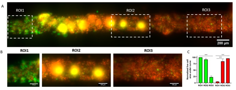
Figure 5.
Photodynamic therapy in the microfluidic array. (A) MDA-MB-231 green fluorescent protein (GFP) cells were injected in the lumen and incubated with 500 ng/mL verteporfin for 24 h. The right end of the lumen was exposed to 485/35 nm light for 45 s to photoactivate verteporfin. After another 24 h, cell viability was evaluated, showing a gradient of viability across the lumen. (B) Images showing the left, center, and right section of the lumen. (C) Graphs showing the normalized area under the curve of the luminescence plot for live cell (green) and dead cell (fluorescence) in the left, central, and right region of the lumen. *** p ≤ 0.001.

