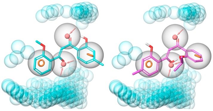
Figure 8.
Alignment of TD12 (cyan, left-hand side) and TD18 (magenta, right-hand side) to pharmacophore model 2. The pharmacophore features are shown as orange rings (aromatic ring), pink spheres (H-bond acceptor), and blue spheres (excluded volumes); feature tolerances are showed as grey spheres around the pharmacophores.

