

Abstract
In these short historical notes, we describe the early history of polymorphic ventricular tachycardia. Polymorphous ventricular tachycardia was probably first noted in 1918 by Wilson and Robinson. In a publication describing complete heart block and ventriculophasic arrhythmia, they noted a tachyarrhythmia characterized by multiple extrasystoles of different types at a rapid rate. Also, we briefly discuss the earliest recognized torsades de pointes by Dessertenes in 1966 and the first description of catecholaminergic polymorphic ventricular tachycardia, by Reid in 1977.
Keywords: polymorphic ventricular tachycardia, early history, torsades de pointes, catecholaminergic polymorphic ventricular tachycardia
Polymorphous ventricular tachycardia (PVT) is characterized by continuous change in the QRS morphology at a rate between 150 and 300 bpm. PVT is mostly seen in patients with congenital or acquired long QT syndrome and less frequently in ischemic heart disease, other structural cardiac abnormalities or in patients without organic heart disease. 1 Dessertenne in 1966 reported a specific form of PVT called “torsades de pointes” in a patient with atrioventricular block. 2 This short historical note discusses what we believe was the first reported PVT.
In 1918, Wilson and Robinson 3 published an article in Archives of Internal Medicine titled “Heart‐Block” with a subtitle “Two cases of complete Heart Block showing unusual features.” The main purpose of this article was the description of ventriculophasic sinus arrhythmia in complete AV block. According to Wilson and Robinson 3 this arrhythmia was rare with only one previously reported case in 1913 by Hecht. 4 The first patient was a 39‐year‐old housewife known to have slow pulse since the age of 19. The second patient was a cigarmaker who was hospitalized because of multiple syncopal episodes, vertigo, exertional shortness of breath and exercise induced PVT. His resting and postexercise ECGs are shown in Figures 1 and 2.
Figure 1.
Lead III shows complete AV block and slightly wider QRS complexes.
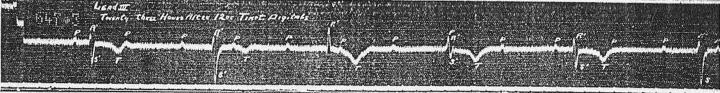
Figure 2.

Two runs of PVT in lead II. The upper one is initiated by the long‐short sequences and the lower strip which is the longer episode, is initiated by a late ventricular premature beat.
In Figure 1, lead 3 shows complete AV block with an atrial and ventricular rate of 67 and 33 bpm, respectively. The QRS complexes are slightly wider and there is left axis deviation. The P–P interval with and without QRS complex varied between 870 ms and 970 ms, respectively, consistent with ventriculophasic sinus arrhythmia. In Figure 2, there are two rhythm strips of lead II recorded shortly after exercise showing two paroxysms of PVT. The upper strip starts with an escape beat followed by a ventricular premature beat, a pause, another escape beat and a paroxysm of six beats PVT. Thus, the tachyarrhythmia is initiated by long–short sequence. The lower strip shows an eight‐beats PVT initiated by a late ventricular premature beat and more prominent changes in the QRS complex morphology. Both paroxysms of PVT were terminated spontaneously. The authors called this tachyarrhythmia “multiple extrasystoles after exercise” with the following comment, “the extrasystoles were not always of the same type and they occurred at a rapid rate. 3 ” Soon after Wilson and Robinson's article, 3 additional cases of ventricular tachycardia induced syncope were reported in patients taking quinidine, having AV block or both. 5 , 6
As mentioned earlier, the main purpose of Wilson and Robinson's article was not PVT, but ventriculophasic sinus arrhythmia and its mechanism. Normalization of the PP intervals by atropine and exercise supported the role of the parasympathetic tone in the pathogenesis ventriculophasic sinus arrhythmia. The authors also used atropine to localize the site of AV block. In the first patient, the ventricular rate increased from 43 to 60 bpm while in the second one there was no change in the ventricular rate after atropine. Wilson and Robinson 3 correctly assumed that improvement of AV conduction after parasympathetic blockade was consistent with AV nodal block, while no effect suggested conduction defect below the AV node.
TORSADES DE POINTES
It was not until 1966 that Dessertenne of France described the undulating nature of a type of PVT and coined the term “torsades de pointes.” 2 This too was in a patient with AV block. The patient's syncopal episodes were due to self‐terminating irregular ventricular arrhythmias resulting from intermittent 3rd degree AV block and bradycardia and not directly because of the AV block as was originally thought (Fig. 3).
Figure 3.

The first irregular ventricular rhythm termed “torsades de pointes” by F. Dessertenne.
Subsequent articles quickly reported that, prior the onset of torsades, QT lengthening was almost universally seen on ECG. 7 Soon after this, the characteristic ECG pattern of long–short cycle was recognized and it was postulated that early after depolarizations play a role in arrhythmia genesis. 8 , 9
CATECHOLAMINERGIC POLYMORPHIC VENTRICULAR TACHYCARDIA
It is now clear that not all polymorphic ventricular tachycardias require a long–short cycle or even a long QT interval for initiation. Catecholaminergic induced polymorphic VT is a rare arrhythmia seen primarily in children and adolescents characterized by a normal QT interval and the lack of a preceding pause. It is induced by stress or exercise and classic ECG findings include ventricular ectopy of RBBB morphology, bidirectional VT and polymorphic VT. 10 It is recognized as having a familial inheritance, in both autosomal dominant and recessive fashion. Mutations of either the ryanodine receptor (RyR2) or the calsequestrin gene (CASQ2) can result in abnormal calcium sequestration and concentrations within the sarcoplasmic reticulum and an increased vulnerability to ventricular arrhythmias. 10
The first description of ventricular tachycardia induced by catecholamines came in 1977 by Reid et al. 11 The patient, a 6‐year‐old girl, had symptoms precipitated by stress or effort. Reid was able to induce bidirectional ventricular tachycardia through rapid atrial pacing or isoproteronol infusion. He was careful to mention that the patient had no structural heart disease and had never received digitalis. The induced arrhythmia was proven to be ventricular in origin through the use of His electrocardiography.
CONCLUSIONS
The management and prevention of ventricular tachycardias have become a focus for much of cardiology research today. As we look to the future for advances in medications and device therapy, it is always interesting to reflect back on the origins of an arrhythmia or disease process.
This short historical note not only describes the first PVT but also shows the insightful thinking of Wilson and his coworkers almost 90 years ago. As we discussed in our two previous articles, 12 , 13 Wilson was one of the first who reported supraventricular tachycardia in preexcitation 14 and together with his coworkers first correctly classified right and left bundle branch block. 15
REFERENCES
- 1. Ho RT, Marchhlinkski FE. Approach to the patient with ventricular tachycardia or ventricular fibrillation In Management of Cardiac Arrhythmia ED. Totowa , NJ , Ganz LI Humana Press, 2002, pp. 233–264. [Google Scholar]
- 2. Dessertenne F. La tachycardia ventriculaire a deux foyers opposes variable. Arch Mal Coeur Vaiss 1966;59:263–272. [PubMed] [Google Scholar]
- 3. Wilson FN, Robinson CC. Heart‐Block I. Two cases of complete heart block showing unusual features. Arch Intern Med 1918;21:166–175. [Google Scholar]
- 4. Hecht AF. quoted by Wilson and Robinson.
- 5. Kerr WMJ, Bender WML. Paroxysmal ventricular fibrillation with cardiac recovery in a case of auricular fibrillation and complete heart‐block while under quinidine sulphate therapy. Heart 1922;9:269–281. [Google Scholar]
- 6. Davis D, Sprague HB. Ventricular fibrillation: Its relation to heart block. Report of a case in which syncopal attacks and death occurred in the course of quinidine therapy. Am Heart J 1929;4:559–572. [Google Scholar]
- 7. Cokkinos DV, Mallios C, Philias N, et al “Torsades de pointe”: A distinct entity of ventricular arrhythmia? Acta Cardiol 1978;33(3):167–184. [PubMed] [Google Scholar]
- 8. Brachmann J, Scherlag BJ, Rosenshtraukh LV, et al Bradycardia‐dependent triggered activity: Relevance to drug‐induced multiform ventricular tachycardia. Circulation 1983;68(4):846–856. [DOI] [PubMed] [Google Scholar]
- 9. El‐Sherif N. Electrophysiologic mechanisms of ventricular arrhythmias. Int J Card Imaging 1991;7(3–4):141–150. [DOI] [PubMed] [Google Scholar]
- 10. Francis J, Sankar V, Nair VK, et al Catecholaminergic polymorphic ventricular tachycardia. [Review][30 refs][Journal Article. Review] Heart Rhythm 2005;2(5):550–554. [DOI] [PubMed] [Google Scholar]
- 11. Reid DS, Tynan M, Braidwood L, et al Bidirectional tachycardia in a child. A study using His bundle electrography. Br Heart J 1975;37:339–344. [DOI] [PMC free article] [PubMed] [Google Scholar]
- 12. Hanon S, Shapiro M, Schweitzer P. Early history of the pre‐excitation syndrome. Europace 2005;7:28–33. [DOI] [PubMed] [Google Scholar]
- 13. Schweitzer P, Couturier G. The history of bundle branch block. Ann Noninv Electrocardiol 2001;6:243–245. [DOI] [PMC free article] [PubMed] [Google Scholar]
- 14. Wilson FN. A case in which the vagus influenced the form of the ventricular complex of the electrocardiogram. Arch Intern Med 1915;16:1008–1027. [DOI] [PMC free article] [PubMed] [Google Scholar]
- 15. Wilson FN, Macleod AG, Baker PS. The interpretation of the initial deflection of ventricular complex of the electrocardiogram. Am Heart J 1931;6:637–664. [Google Scholar]