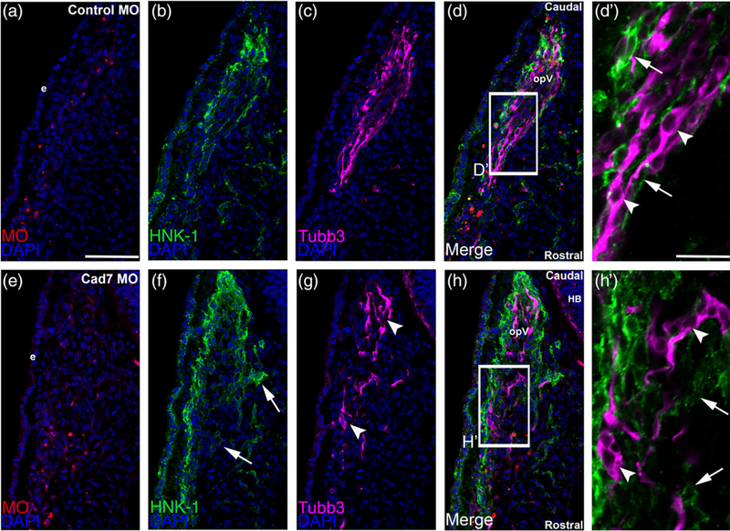
FIGURE 4.
Morpholino-mediated depletion of Cadherin-7 from migratory cranial neural crest cells alters the distribution of neural crest cells and placodal neurons within the forming trigeminal ganglion. Representative transverse sections taken at the axial level of the forming ophthalmic lobe of the trigeminal ganglion after electroporation of a 5 bp mismatch control Cadherin-7 morpholino (control MO, a–d′) or Cadherin-7 MO (Cad7 MO, e–h′) into premigratory neural crest cells at the 3ss followed by immunohistochemistry for HNK-1 (green) and Tubb3 (purple) at HH15. (d′, h′) Higher magnification images of the boxed regions in (d, h). (a–d′) Control MO (a)-containing neural crest cells (b) coalesce with placodal neurons (c, d). At higher magnification (d′), HNK-1-positive neural crest cells (arrows) surround Tubb3-positive placodal neurons (arrowheads), many of which are already forming neurites or adopting the bipolar morphology associated with neuronal maturation. Conversely, a trigeminal ganglion containing the Cadherin-7 MO (e) in the neural crest (f) reveals differences in neural crest cell (f, h, arrows) and placodal neurons (g, h, arrowheads) distribution within the anlage, with neural crest cells sometimes appearing more dispersed compared to control. At higher magnification (h′), it is apparent that neural crest cells still surround the placodal neurons (arrows), but the shape adopted by the placodal neurons is aberrant, with neurons appearing round (arrowheads). Rostral and caudal orientation, the hindbrain (HB), and the ophthalmic lobe of the trigeminal ganglion (opV) are indicated in (d) and (h). DAPI (blue) labels cell nuclei. e = ectoderm. Scale bar in (a) is 67 μm and applicable to (b–h), while scale bar in (d′) is 20 μm and applicable to (h′)

