Abstract
Objective
Acromegaly is characterized by an excess of growth hormone (GH) and insulin-like growth factor 1 (IGF1). Cardiovascular disease (CVD) risk factors are common in acromegaly and often persist after treatment. Both acute and long-lasting pro-inflammatory effects have been attributed to IGF1. Therefore, we hypothesized that inflammation persists in treated acromegaly and may contribute to CVD risk.
Methods
In this cross-sectional study, we assessed cardiovascular structure and function, and inflammatory parameters in treated acromegaly patients. Immune cell populations and inflammatory markers were assessed in peripheral blood from 71 treated acromegaly patients (with controlled or uncontrolled disease) and 41 matched controls. Whole blood (WB) was stimulated with Toll-like receptor ligands. In a subgroup of 21 controls and 33 patients with controlled disease, vascular ultrasound measurements were performed.
Results
Leukocyte counts were lower in patients with controlled acromegaly compared to patients with uncontrolled acromegaly and controls. Circulating IL18 concentrations were lower in patients; concentrations of other inflammatory mediators were comparable with controls. In stimulated WB, cytokine production was skewed toward inflammation in patients, most pronounced in those with uncontrolled disease. Vascular measurements in controlled patients showed endothelial dysfunction as indicated by a lower flow-mediated dilatation/nitroglycerine-mediated dilatation ratio. Surprisingly, pulse wave analysis and pulse wave velocity, both markers of endothelial dysfunction, were lower in patients, whereas intima-media thickness did not differ.
Conclusions
Despite treatment, acromegaly patients display persistent inflammatory changes and endothelial dysfunction, which may contribute to CVD risk and development of CVD.
Keywords: inflammation, cardiovascular disease, IGF1, endothelial dysfunction, acromegaly
Introduction
Acromegaly is caused by overproduction of growth hormone (GH), in most cases by a pituitary adenoma. GH in turn induces production of insulin-like growth factor 1 (IGF1) (1). Both GH and IGF1 have numerous metabolic and trophic effects (2). Apart from disease-specific complications, patients with active acromegaly suffer from an increased morbidity and mortality due to cardiovascular disease (CVD) (3, 4). With disease control (i.e. normalized circulating GH and IGF1 concentrations), the increased prevalence of CVD normalizes to a great extent (5). However, the prevalence of CVD risk factors as hypertension and diabetes mellitus (DM) remains elevated (6, 7, 8), which implies persistence of the elevated CVD risk in controlled acromegaly patients. The cause of this phenomenon is incompletely understood, and it is debated whether it could be attributed to direct deleterious effects of GH and IGF1 on the cardiovascular system or is also caused by concomitant cardiovascular and metabolic disturbances that cause hypertension, insulin resistance and dyslipidemia in acromegaly patients (6).
Interestingly, CVD is strongly associated with subclinical systemic inflammation (9, 10). Vascular wall inflammation is an important driver of the initiation and progression of atherosclerosis, which is the main pathophysiological process driving CVD. Circulating immune cells invade the vasculature and induce expression of adhesion molecules and subsequent leukocyte adherence, which promotes a pro-inflammatory and pro-atherogenic environment. Although the importance of innate immune cells in the development and progression of atherosclerosis is widely accepted, the unresolving character of the low-grade inflammation that drives it remains poorly understood. Recently, our group described that innate immune cells can develop long-term functional reprogramming characterized by hyperresponsiveness, termed ‘trained immunity’ (11). Short-term exposure to stimuli can induce a long-term pro-inflammatory phenotype of monocyte-derived macrophages (12, 13), and circulating monocytes obtained from patients with risk factors for atherosclerosis or established atherosclerosis display a pro-inflammatory phenotype (14, 15).
Intriguingly, immune cells express GH and IGF1 receptors (16, 17). Previously, we found that IGF1 can impact on monocyte inflammatory function in vitro (18). Moreover, exposure to IGF1 induces trained immunity (19). However, studies on the inflammatory profile of acromegaly patients rendered conflicting results: both unaltered as well as pro-inflammatory phenotypes have been reported (20, 21, 22). On the other hand, previous studies on the risk of CVD in (treated) acromegaly imply that the arterial structure and function of patients with acromegaly is impaired, which might contribute to the development of CVD (6). We therefore hypothesized that treated acromegaly patients are characterized by prolonged inflammatory changes, which might contribute to the persistence of CVD risk factors and development of CVD. To test this hypothesis, we comprehensively assessed vascular structure and function, circulating inflammatory markers and ex vivo cytokine production capacity in acromegaly patients and healthy controls. By including both patients with active disease under treatment and controlled disease, we aimed to elucidate whether these properties are reversible after disease control.
Materials and methods
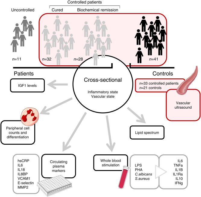
This cross-sectional case–control study was conducted in an academic referral center (Radboudumc Nijmegen, the Netherlands). The study structure is displayed in Fig. 1.
Figure 1.
Study structure and assessments. IGF1, insulin-like growth factor 1; hsCRP, high sensitivity C-reactive protein; IL, interleukin; IL18BP, IL18 binding protein; VCAM1, vascular cell adhesion molecule 1; MMP2, matrix metalloproteinase 2; LPS, lipopolysaccharide; PHA, phytohemagglutinin; IL, interleukin; TNFa, tumor necrosis factor alpha; Ra, receptor antagonist; IFNg, interferon gamma.
Subjects
Seventy-one adult patients with acromegaly and forty-one healthy controls (Table 1) were included between February 2016 and April 2017. All patients that were currently treated at our center or were treated within the last 5 years were selected. Subjects with inflammatory comorbidities, active malignancies or those using statins or systemic immunosuppressive medication were excluded. In addition, we excluded patients with inadequately treated hypertension (systolic blood pressure ≥160 mmHg or diastolic blood pressure ≥100 mmHg), poorly controlled DM (HbA1c >69 mmol/mol for >1 year) or ischemic cardiovascular diseases, or with an alcohol intake of >21 IU per week. Non-pregnant adults with established and treated acromegaly were asked to participate (n = 101), and 29 patients declined. Reasons for decline were not being able to travel to the hospital, lack of time, and difficulties with being fasted. The remaining 72 patients were enrolled in the study. One patient was excluded because a previously unknown inflammatory disorder manifested during the study.
Table 1.
Clinical characteristics in patients and controls.
| Clinical characteristics | Controls (n = 41) | Patients (n = 71) | P |
|---|---|---|---|
| Sex: male | 20 | 36 | 1.0 |
| Age (years) | 51.9 (14.3) | 54.5 (12.1) | 0.33 |
| Height (m) | 1.75 (0.09) | 1.76 (0.11) | 0.73 |
| Weight (kg) | 81.2 (54.8–129.8) | 83.7 (51.4–150.8) | 0.06 |
| BMI (kg/m2) | 27.07 (18.3–46) | 27.7 (20–49.1) | 0.13 |
| Waist-to-hip ratio | 0.93 (0.08) | 0.94 (0.08) | 0.82 |
| Systolic BP (mmHg) | 125 (15) | 129 (16) | 0.9 |
| Diastolic BP (mmHg) | 75 (9) | 80 (10) | 0.52 |
| Heart rate (/min) | 64 (44–80) | 60 (44–78) | 0.87 |
| Anti-hypertensives | 3 | 18 | 0.02 |
| Diabetes mellitus | 0 | 8 | 0.03 |
| Smoker; current/past | 10/10 | 8/32 | 0.05 |
| Alcohol use (units/week) | 3 (0–20) | 2 (0–21) | 0.54 |
| IGF1 (nmol/L) | 17.5 (7.9–35.8) | 18.2 (8.3–46.7) | 0.08 |
| Hormonal deficiency | 2 | 30 | <0.001 |
| Estrogen depletion | 13 | 25 | 0.56 |
| Hypothyroidism | 2 | 18 | 0.01 |
| Hypogonadism | 0 | 20 | <0.001 |
| Hypocortisolism | 0 | 15 | <0.001 |
| GH deficiency | 0 | 2 | 0.53 |
| Diabetes insipidus | 0 | 6 | 0.08 |
| Hyperprolactinemia | 0 | 1 | 0.63 |
| Medical treatment | 0 | 35 | <0.001 |
| SSA | 0 | 30 | <0.001 |
| Dopamine agonist | 0 | 6 | 0.08 |
| Pegvisomant | 0 | 8 | 0.03 |
| Surgery | 0 | 65 | <0.001 |
| Radiotherapy | 0 | 10 | 0.01 |
| Total cholesterol (mmol/L) | 5.51 (1.24) | 5.23 (1.1) | 0.13 |
| HDL cholesterol (mmol/L) | 1.47 (0.51–2.84) | 1.43 (0.57–2.88) | 0.55 |
| LDL cholesterol (mmol/L) | 3.27 (1.17) | 3.1 (0.93) | 0.23 |
| Triglycerides (mmol/L) | 1.35 (0.58–3.55) | 1.11 (0.53–5.46) | 0.2 |
| Non-HDL cholesterol (mmol/L) | 3.96 (1.26) | 3.72 (1.02) | 0.22 |
Values are displayed as mean with s.d. or as median with minimum and maximum, depending on the normality of the distribution. Categorical variables are displayed as numbers.
BMI, body mass index in kg/m2; BP, blood pressure; estrogen depletion (in women), postmenopausal women not using estrogen substitution; GH, growth hormone; HDL, high-density lipoprotein; IGF1, insulin-like growth factor 1; LDL, low-density lipoprotein; SSA, somatostatin analogue.
In order to provide a sex-, age- and body composition-matched control group, patients were asked to provide a healthy volunteer from their own living environment with, preferably, a similar age, sex, and physique. The above-mentioned criteria, except for the presence of acromegaly, were also applied to controls. In addition, controls with hormonal disturbances, except for adequately supplemented primary hypothyroidism (based on normal TSH-levels after suppletion for >3 months), were excluded. Forty-four controls were willing to participate; three candidates were excluded based on above-mentioned exclusion criteria.
All patients had a history of biochemically and radiologically confirmed acromegaly, defined as an increased serum IGF1 level (>2 s.d. above the mean corrected for sex and age) and insufficient suppression of serum GH levels (≥0.4 µg/L) during an oral glucose tolerance test (OGTT) (1), combined with the presence of a pituitary adenoma on a MRI or CT scan. All patients had received treatment (i.e. surgery, radiotherapy and/or medication; Table 2). Disease duration was based on patients’ reports that were obtained during a thorough medical history. Patients were considered cured if their serum IGF1 level fell within the reference range for sex and age, and when patients had a sufficient suppression of serum GH levels during an OGTT (GH <0.4 µg/L), that was performed after surgery and/or radiation therapy (23). Biochemical control was defined as IGF1 levels in the reference range for sex and age with use of GH or IGF1 lowering therapy. Both cured and biochemically controlled patients were considered controlled patients. Uncontrolled patients had elevated IGF1 levels despite medical, surgical and/or radiation treatment.
Table 2.
Clinical characteristics of patient subgroups.
| Clinical characteristics | Controlled (n = 60) | Uncontrolled (n = 11) | P | P* |
|---|---|---|---|---|
| Sex: male | 31 | 5 | 0.75 | 0.93 |
| Age (years) | 56.3 (11.1) | 44.6 (13.1) | 0.01 | 0.012 |
| Height (m) | 1.76 (0.11) | 1.79 (0.11) | 0.61 | 0.66 |
| Weight (kg) | 83.2 (51.4–150.8) | 111 (64.9–147.2) | 0.052 | 0.032 |
| BMI (kg/m2) | 27 (20–49.1) | 32.3 (24.1–41.4) | 0.029 | 0.029 |
| Waist-to-hip ratio | 0.93 (0.77–1.16) | 0.91 (0.82–1.04) | 0.86 | 0.98 |
| Systolic BP (mmHg) | 129 (17) | 125 (13) | 0.25 | 0.31 |
| Diastolic BP (mmHg) | 80 (10) | 76 (10) | 0.72 | 0.05 |
| Heart rate (/min) | 61 (44–78) | 60 (56–72) | 0.81 | 0.95 |
| Anti-hypertensives | 17 | 1 | 0.27 | 0.018 |
| Diabetes mellitus | 5 | 3 | 0.1 | 0.006 |
| Smoker; current/past | 7/27 | 1/5 | 0.67 | 0.97 |
| Alcohol use (units/week) | 2 (0–21) | 2 (0–21) | 0.56 | 0.69 |
| IGF1 (nmol/L) | 17.6 (4.1) | 32.6 (6.9) | 0.014 | <0.001 |
| Disease duration (years) | 9 (1–40) | 3 (1–22) | 0.05 | NA |
| Hormonal deficiency | 24 | 6 | 0.51 | <0.001 |
| Estrogen depletion | 22 | 3 | 0.39 | 0.66 |
| Hypothyroidism | 16 | 2 | 0.72 | 0.012 |
| Hypogonadism | 15 | 5 | 0.27 | <0.001 |
| Hypocortisolism | 11 | 4 | 0.23 | <0.001 |
| GH deficiency | 2 | 0 | 1 | 0.60 |
| Diabetes insipidus | 5 | 1 | 1 | 0.11 |
| Hyperprolactinemia | 0 | 1 | 0.16 | 0.1 |
| Medical treatment | 28 | 7 | 0.34 | NA |
| SSA | 24 | 6 | 0.51 | NA |
| Dopamine agonist | 4 | 2 | 0.23 | NA |
| Pegvisomant | 5 | 3 | 0.1 | NA |
| Surgery | 55 | 10 | 1 | NA |
| Radiotherapy | 7 | 3 | 0.18 | NA |
| Total cholesterol (mmol/L) | 5.27 (1.13) | 5.05 (0.94) | 0.5 | 0.39 |
| HDL cholesterol (mmol/L) | 1.47 (0.77–2.88) | 1.16 (0.57–2.26) | 0.003 | 0.067 |
| LDL cholesterol (mmol/L) | 3.14 (0.95) | 2.88 (0.73) | 0.33 | 0.54 |
| Triglycerides (mmol/L) | 1 (0.53–3.34) | 1.71 (1.06–5.46) | 0.036 | 0.003 |
| Non-HDL cholesterol (mmol/L) | 3.70 (1.05) | 3.83 (0.84) | 0.33 | 0.5 |
Values are displayed as mean with s.d. or as median with minimum and maximum, depending on the normality of the distribution. Categorical variables are displayed as numbers.
BMI, body mass index in kg/m2; BP, blood pressure; estrogen depletion, postmenopausal women not using estrogen substitution; GH, growth hormone; HDL, high-density lipoprotein; IGF1, insulin-like growth factor 1; LDL, low-density lipoprotein; NA, not applicable; P, P values when comparing subgroups of controlled and uncontrolled patients; P*, P values when controls are included as third subgroup in the analysis; SSA, somatostatin analogue.
Adrenal insufficiency was defined as an insufficient response (serum cortisol levels <0.55 µmol/L) during an insulin tolerance test or a 250 μg ACTH (Synacthen) stimulation test (24), which has been performed in each patient prior to study participation following the standard of care in our hospital. Hypogonadism in premenopausal women was defined as the presence of secondary amenorrhea combined with estrogen values below the reference range. The physiological postmenopausal state, defined as gonadotrophin levels that fall in the postmenopausal range, was not considered as hypogonadism. In men, hypogonadism was defined as a testosterone level below the reference range (<11 nmol/L). Patients with hormonal deficiencies were all on stable substitution therapy, except for postmenopausal women. Testosterone respectively thyroid hormone substitution therapy was monitored with serum testosterone respectively fT4 levels.
In a subgroup of 21 controls and 33 patients, vascular measurements were performed. Because our study focused on the persistent long-term risk of CVD in patients with acromegaly, only controlled patients were included in the vascular analysis. The group of controlled patients was divided intro cured and biochemically controlled patients, given the potential beneficial effects of SSA on vascular function (25). Furthermore, to avoid potential interference with our results, only patients without hormonal deficiencies (except for diabetes insipidus) were selected.
This study was conducted in accordance with the Declaration of Helsinki and approved by our local ethical committee (CMO regio Arnhem-Nijmegen; 2015-2023). All subjects signed informed consent prior to participation.
Anthropometric measurements
Blood pressure and heart rate were measured in supine position on both arms after at least 10 min of rest. Height, weight, waist and hip circumference were determined between 08:30 and 10:30 h in a fasted state. All measurements were performed by a single non-blinded investigator.
Circulating inflammatory and cardiovascular markers
Venous blood was drawn from the brachial vein in a fasted state, between 08:00 and 10:00 h, in 10 ml EDTA tubes (Vacutainer, BD; Franklin Lakes, NJ, USA). Within 3 h, tubes were centrifuged (Hettich Rotina 420R, radius 183 mm; 3800 rpm (2954 g), 10 min, room temperature), and plasma was collected and stored at −80°C until assayed. Plasma IGF1 levels were determined by a chemiluminescent immunometric assay (Liaison, DiaSorin, Saluggia, Italy) according to the 1st WHO International Standard for insulin-like growth factor 1 (NIBSC code: 02/25). Levels of total cholesterol, LDL cholesterol, HDL cholesterol, non-HDL cholesterol and triglycerides were determined using an in-house analyzer (Cobas 8000; Roche Diagnostics).
Plasma E-selectin, matrix metalloproteinase (MMP)2, vascular cell adhesion molecule (VCAM)1, high-sensitivity C-reactive protein (hsCRP), interleukin (IL)18, IL18-binding protein (IL18BP) and IL6 levels were determined with ELISA. For E-Selectin, MMP2, VCAM1, hsCRP, and IL18, DuoSet ELISA (R&D Systems, Abingdon, United Kingdom) was used with a sensitivity of 93.8 pg/mL for E-Selectin, 625 pg/mL for MMP2, 15.6 pg/mL for VCAM1 and hsCRP, and 11.7 pg/mL for IL18. For measurement of IL6 and IL18BP, high-sensitivity Quantikine ELISA assays (R&D) were used with a sensitivity of 0.11 pg/mL for IL6 and 7.52 pg/mL for IL18BP.
Cell counts
Cell counts were obtained in fresh EDTA blood with a Sysmex automated hematology analyzer (XN-450; Sysmex Corporation, Kobe, Japan).
Ex vivo stimulation of whole blood (WB)
E. coli lipopolysaccharide (LPS; serotype 055:B5) was purchased from Sigma-Aldrich, repurified as previously described, and used as an ultrapure Toll-like receptor 4 ligand (26). Phytohemagglutinin (PHA) was purchased from Sigma-Aldrich (PHA-P; L1668). Candida albicans (C. albicans) ATCC MYA-3573 (UC 820) and Staphylococcus aureus (S. aureus) Rosenbach ATCC 25923 were used. C. albicans and S. aureus were grown overnight at 37°C in Sabouraud and Brain Heart Infusion broth, respectively. Microorganisms were harvested by centrifugation, washed twice, and resuspended in Roswell Park Memorial Institute (RPMI) 1640 culture medium (Dutch Modification, Gibco, Thermo Scientific) (27). C. albicans yeasts were heat-killed for 30 min at 95°C.
Venous blood was drawn from the brachial vein in a fasted state, between 08:00 and 10:00 h, in 4 mL lithium-heparin tubes (Vacutainer). Within 3 h, 100 μL of WB was incubated at 37°C in round-bottom 48-well plates (Greiner; Kremsmünster, Austria) with 400 μL of stimulus (LPS 100 ng/mL, PHA 10 µg/mL, C. albicans 1 × 10⁶/mL, S. aureus 1 × 10⁶/mL) or RPMI (basal unstimulated condition) per well. After 48 h of incubation, supernatants were collected and stored at −20°C until assayed.
Cytokine concentrations were measured in supernatants by commercial ELISA kits according to the manufacturer’s instructions: tumor necrosis factor alpha (TNFa), IL1B, IL1 receptor antagonist (IL1Ra), IL6 (DuoSet ELISA, R&D), IL10, interferon gamma (IFNg) (PeliKine Compact, Sanquin; Amsterdam). The sensitivity of the assays was 2.34 pg/mL for IL10, 3.9 pg/mL for IL1B and IFNg, 4.7 pg/mL for IL6, 7.8 pg/mL for TNFa, and 39.0 pg/mL for IL1Ra. The inter-assay coefficients of variability were 9.5% for IL10, 5.6% for IL1B, 12.8% for IFNg, 8.9% for IL6, 6.9% for TNFa, and 8.4% for IL1Ra.
All samples were analyzed in the same batch without previous freeze-thaw cycles.
Vascular measurements
Subjects that underwent vascular measurements refrained from exercise and consumption of caffeine, alcohol, dark chocolate, vitamin C-rich products and vitamin supplements for 24 h and fasted for at least 6 h. All vascular measurements were performed between 09:00 and 12:00 h in a supine position after at least 15 min of rest under standardized conditions in a temperature-controlled room (28, 29).
Pulse wave velocity and pulse wave analysis
Pulse wave velocity (PWV) and pulse wave analysis (PWA) measurements were performed with a SphygmoCor EM3 tonometry device (AtCor Medical, Sydney, Australia) by a single investigator according to the manufacturer’s instructions.
Heart Rate Corrected Central Augmented Pressure (C_AP_HR75) was calculated based on PWA of the right radial artery, the median of three measurements was used for data analysis. For calculation of PWV, 80% of the direct distance between the palpation site of the right common carotid and the right femoral artery was divided by the pulse transit time (in seconds) (30).
Ultrasound measurements
All ultrasound measurements were performed by a single technician on a Terason t3000 ultrasound device (Aloka, UK). All ultrasound images were analyzed by a single observer using computer-assisted analysis with edge-detection and wall-tracking software (DICOM Encoder Analysis Combo) (28).
Flow-mediated dilation (FMD)
FMD (% diameter change: (peak diameter − baseline diameter)/baseline diameter) and shear rate (Arbitrary Units; AU) were measured in the distal third of the brachial artery of the right arm using high-resolution B-mode 10 MHz ultrasonography and simultaneous acquisition of pulsed-wave Doppler velocity signals according to a validated protocol (28, 29).
Nitroglycerine-mediated dilation (NMD)
One minute prior and 10 min after 0.4 mg nitroglycerine sublingually, brachial artery diameter and blood flow velocity were measured and analyzed following the same protocol as was used for FMD analysis.
Intima-media thickness (IMT)
IMT was measured using high-resolution B-mode 10 MHz ultrasonography in the common carotid artery on the far wall, at three different angles (31). IMT was identified as the region between the lumen-intima border and the media-adventitia border. Regions of interest were manually marked and at least 50 frames per scan were analyzed to gain a representative mean of lumen diameter and IMT. These analyses were randomly repeated in order to retain accuracy. Mean IMT was calculated from at least 40 useful frames at three different angles.
Statistical analysis
Data were analyzed with SPSS 25.0. Data are presented as unadjusted means with s.d. or medians with minimum and maximum values for continuous variables, depending on the normality of the distribution, which was tested by the Shapiro–Wilk test. Differences between patients and controls were tested with an independent samples t-test or a Mann–Whitney U test (depending on the normality of the distribution) for continuous parameters and with the Fisher Exact test in case of categorical data. Differences between subgroups were tested using ANOVA. Group matching of patients and controls was performed by testing for differences in age, gender, and BMI.
Data on cytokines and circulating parameters were log-transformed using the natural logarithm prior to analysis with ANCOVA; BMI, age, and leukocyte count were associated with cytokine production and circulating parameters and were therefore included as covariates. For leukocyte counts, IGF1 levels, BMI and age were used. We performed a sensitivity analysis using forward selection, by alternately adding estrogen depletion, use of antihypertensives and presence of diabetes mellitus as covariates to our ANCOVA model, which did not improve goodness of fit of the model.
For vascular ultrasound and PWV, age and systolic blood pressure were used as covariates (32, 33) and for C_AP_HR75, sex and systolic blood pressure were used. Correlations were determined on non-transformed data using Spearman rank correlation. All tests were two-tailed. P values of <0.05 were considered statistically significant. When comparing three groups of subjects, the Bonferroni correction for multiple testing was applied, which rendered an adjusted P value of 0.0167; corrected P values were displayed.
Results
Subject characteristics: total group (inflammatory parameters)
Of the 71 patients, 60 were controlled (of whom 32 were cured and 28 biochemically controlled), and 11 uncontrolled. The prevalence of hormonal deficiencies differed between patients and controls, but not between the subgroups. Sex, age and anthropometrical measurements were not statistically different between the total patient group and controls, which indicates adequate group matching regarding these parameters. Use of antihypertensive medication was more prevalent in patients, but blood pressure did not differ between patients and controls nor between patient subgroups. DM was not present in controls, but was present in eight patients; all but one had a HbA1c <58 mmol/mol (median 53 (40–69)). None of the subjects had established coronary artery disease. The control group contained more current smokers and the patient group more former smokers (Tables 1 and 2).
In the subgroup analysis, we observed that uncontrolled patients were younger and had a higher weight and BMI. Disease duration tended to be shorter in uncontrolled patients, although this difference was not statistically significant. All other features were similar in both patient subgroups (Table 2).
Subject characteristics: subgroup selected for vascular measurements
Thirty-three controlled patients without hormonal deficiencies and 21 healthy controls underwent additional vascular measurements. They were comparable to the subjects of the total group, except that the patients in this subgroup used slightly more antihypertensive drugs. However, including use of antihypertensive medication in our model did not change our results.
IGF1 levels
There was no difference between the plasma IGF1 levels of controlled patients (17.6 ± 4.1 nmol/L) and controls (17.3 ± 5.4 nmol/L; P = 0.7). Uncontrolled patients had higher IGF1 levels (32.6 ± 6.9 nmol/L) than the other two groups (P < 0.001).
Cell counts
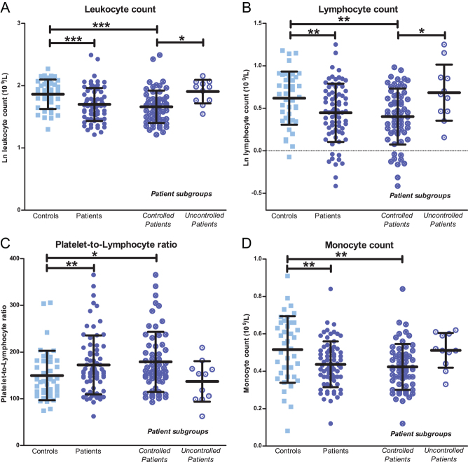
Total leukocyte count was lower in patients (5.38 (3.36–12.06) × 109/L) compared to controls (6.81 (3.66–11.62) × 109/L; P < 0.001), as were monocyte, lymphocyte and neutrophil counts. However, the lower leukocyte count was triggered only by the controlled patients, while the leukocyte count in uncontrolled patients (7.24 (4.68–8.63) × 109/L) was not different compared to controls. Relative leukocyte counts did not differ between patients and controls. The inflammatory marker neutrophil-to-lymphocyte (NtL) ratio did not differ between groups, whereas its analogue, the platelet-to-lymphocyte (PtL) ratio was higher in patients compared to controls (158.2 (62.5–365.2) vs 137.6 (74.4–305.4); P = 0.007). In patients, we observed a positive correlation between IGF1 levels and leukocyte counts (R0.28; P = 0.02), whereas in controls, a negative correlation was present (R−0.32; P = 0.04). IGF1 levels were also positively correlated with monocyte counts in patients (R0.30; P = 0.01), but not in controls (Fig. 2).
Figure 2.
Leukocyte (A), lymphocyte counts (B), platelet-to-lymphocyte ratio (PtL) (C) and monocyte counts (D). Leukocyte and lymphocyte counts were log-transformed using the natural logarithm; mean with s.d. is displayed for all parameters. *P <0.05; **P <0.01; ***P <0.001.
Circulating markers of inflammation and endothelial dysfunction
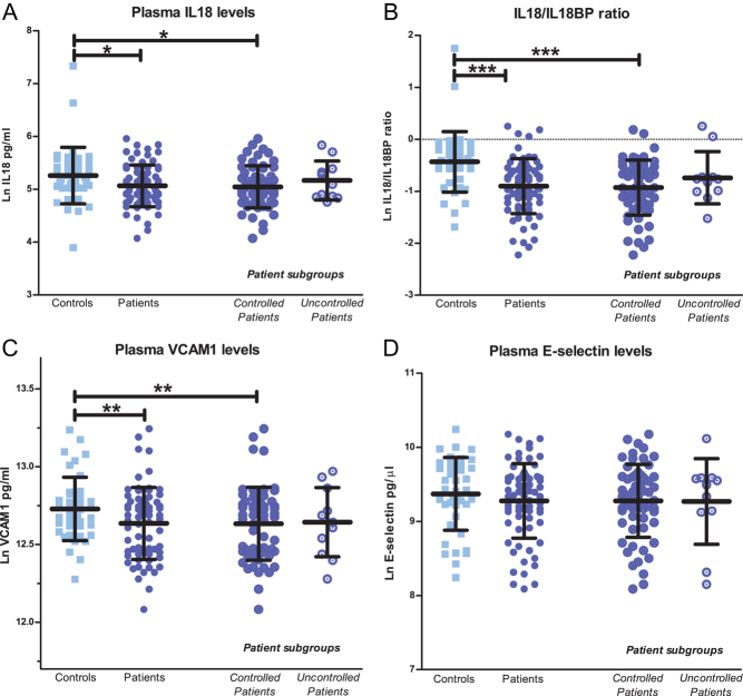
In the total patient group, plasma IL18 concentrations were significantly lower than in controls (151.9 (58.6–387.4) vs 178.5 (49.2–1528.3) pg/mL; P = 0.01). IL18BP concentrations were higher in patients compared to controls (356.1 (265.6–1341.2) vs 265.6 (265.6-601.1) pg/mL; P < 0.001). Consequently, the IL18/IL18BP ratio was significantly lower in patients than in controls (0.44 (0.11–1.29) vs 0.65 (0.19–5.75); P < 0.001); these differences were triggered by controlled patients, since uncontrolled patients did not differ from controls. VCAM1 levels were lower in patients compared to controls (320 (177–565) vs 326 (215–561) pg/µL; P = 0.008), which was caused by lower levels in controlled patients compared to controls (322 (177–565) pg/µL; P = 0.003). IL18 levels correlated with VCAM1 levels (R0.514; P < 0.001). The other circulating factors investigated did not differ significantly between patients and controls nor between patient subgroups (Fig. 3).
Figure 3.
Circulating inflammatory markers. Anti-inflammatory IL18 (A), IL18/IL18BP ratio (B), proinflammatory VCAM1 (C) and pro-inflammatory E-selectin levels (D). Cytokine concentrations were logtransformed using the natural logarithm, and mean with s.d. is displayed. IL18, interleukin 18; IL18BP, IL18-binding protein; VCAM1, vascular cell adhesion molecule 1. *P <0.05; **P <0.01; ***P <0.001.
Ex vivo cytokine production in whole blood
Monocyte-derived cytokine production
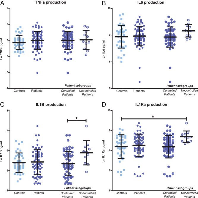
Uncontrolled patients had higher S.aureus-stimulated IL1B production compared to controlled patients (381.6 (140–1387.9) vs 194.8 (87.1–653.4) pg/mL; P = 0.02) and higher IL1Ra production compared to controls (5847.7 (4197–11760.2) vs 3375.9 (874.1–8797.5) pg/mL; P = 0.03). Controlled patients showed a IL1B and IL1Ra production that was comparable to controls. A similar pattern was seen for IL1B and IL1Ra production in response to other WB stimuli, although these differences were not statistically significant (Fig. 4).
Figure 4.
Monocyte-derived pro-inflammatory cytokine production. LPS-stimulated TNFa (A) and IL6 production (B), and S. aureus-stimulated IL1B (C) and IL1Ra production (D). Cytokine concentrations were log-transformed using the natural logarithm, and mean with s.d. is displayed. LPS, lipopolysaccharide; TNFa, tumor necrosis factor alpha; IL, interleukin; Ra, receptor antagonist. *P <0.05.
No differences were observed in the production of monocyte-derived pro-inflammatory cytokines IL6 and TNFa between patients and controls, nor between patient subgroups.
Th-derived cytokine production
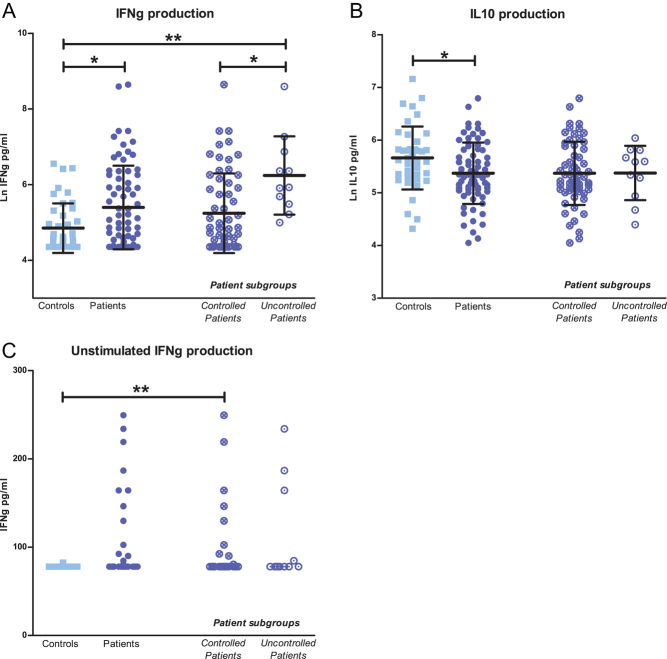
We found unstimulated IFNg production in patients, but not in controls (Fig. 5C). In addition, the S. aureus-stimulated IFNg production was significantly higher in patients compared to controls (148.1 (78–5672.7) vs 92.7 (78–701.7) pg/mL; P = 0.02); the highest IFNg production was observed in uncontrolled patients (374.4 (148.1–5389.5) pg/mL; P = 0.001 vs controls, and P = 0.012 vs controlled patients ((118.8 (78–5672.7) pg/mL). Again, a similar pattern was seen for the other WB stimuli. LPS-stimulated anti-inflammatory IL10 production was lower in patients compared to controls (208.1 (57.2–890.4) vs 275.5 (74.9–1285.8) pg/mL; P = 0.04). IGF1 levels positively correlated with IL6 (R0.31; P = 0.008), IL1B (R0.42; P < 0.001), IL1Ra (R0.51; P < 0.001), and IFNg production (R0.34; P = 0.004) in patients (Fig. 5).
Figure 5.
Lymphocyte-derived cytokine production. S. aureus-stimulated pro-inflammatory IFNg production (A), LPS-stimulated anti-inflammatory IL10 production (B) and unstimulated IFNg production (C). S. aureus-stimulated IFNg production and LPS-stimulated IL10 production were log-transformed using the natural logarithm. For S. aureus-stimulated IFNg production and LPS-stimulated IL10 production mean with s.d. is displayed. IFNg, interferon gamma; LPS, lipopolysaccharide; IL, interleukin. *P <0.05; **P <0.01.
Subgroup: vascular measurements
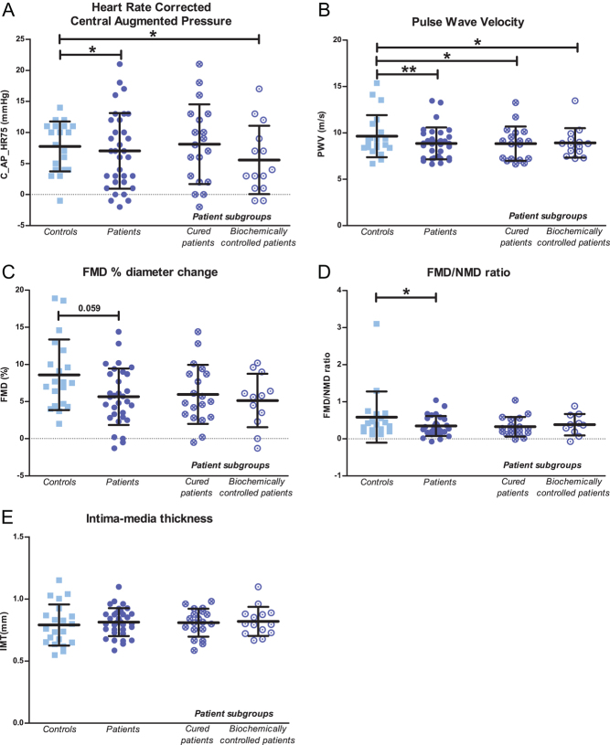
Serum lipid and IGF1 levels were not significantly different between the groups. All subjects had IGF1 levels that were in the normal reference range for age and sex (Fig. 6).
Figure 6.
Vascular measurements. Heart rate-corrected central augmented pressure (C_AP_HR75; A), PWV (B), FMD (C), FMD/NMD ratio (D) and IMT (E). Values were log-transformed using the natural logarithm prior to analysis and mean with s.d. is displayed. PWV, pulse wave velocity; IMT, intima-media thickness; FMD, flow-mediated dilatation; NMD, nitroglycerine-mediated dilatation. *P <0.05; **P <0.01.
FMD was lower in patients than in controls (5.22 ± 3.58% vs 8.68 ± 4.87%; P = 0.06), but did not differ significantly between the patient groups. The FMD/NMD ratio was lower in patients compared to controls (0.27 (−0.08; 0.15) vs 0.42 (0.12–5.95); P = 0.04). Shear rate was lower in patients compared to controls (15,997 (4676–39,954) vs 26,245 (14,287–53,297) AU; P = 0.002).
Compared to controls, patients had both a lower C_AP_HR75 (7.75 ± 4.03 vs 6.68 ± 6.12 mmHg; P = 0.04) and PWV (9.14 (7.1–15.36) vs 8.83 (6.63–13.46) m/s; P = 0.002). When comparing patient subgroups to controls, the lower C_AP_HR75 was only present in biochemically controlled patients (5.57 ± 5.5 mmHg; P = 0.02), whereas PWV was lower in both cured (9.09 (6.63–13.27) m/s; P = 0.02) and biochemically controlled patients (8.74 (7.24–13.46) m/s; P = 0.03). Patients using somatostatin analogues (SSA) had a lower C_AP_HR75 (8.8 ± 6.3 vs 3.6 ± 3.9 mmHg; P = 0.006). IMT did not differ between controls and patients.
Discussion
To our best knowledge, this is the first study that comprehensively examined the multifaceted aspects of inflammation in patients with treated acromegaly and relates them to structural and functional vascular characteristics. We hypothesized that persistent inflammation contributes to the persistence of CVD risk and the development of CVD in acromegaly patients despite adequate treatment. Indeed, we observed pro-inflammatory changes in the function of the immune system in patients with acromegaly, most pronounced in those having active disease, but partly persisting in those with controlled disease. This was paralleled by persistent endothelial dysfunction in controlled patients. These findings suggest that chronic inflammation could contribute to the high prevalence of CVD risk factors in acromegaly both during active disease as well as in adequately treated patients.
Recent evidence suggests that IGF1 levels are related to CVD in an U-shaped fashion, with both low and high circulating IGF1 levels being associated with an increased CVD risk (34, 35). Since the importance of inflammation in the development of CVD is well established (9, 10), we previously investigated the effects of IGF1 in vitro. We showed direct pro-inflammatory effects of IGF1 on human white blood cells in supraphysiological concentrations that reflect IGF1 levels in patients with acromegaly (18). Our group has shown long-lasting pro-inflammatory effects of IGF1 on human monocytes, a phenomenon termed ‘trained immunity’ (11). In this study we observed that acromegaly patients with active disease under treatment are characterized by high IL1B and IL1Ra as well as IFNg production capacity ex vivo in stimulated whole blood (WB). The finding that these changes in cytokine production were more pronounced in reaction to certain stimuli (i.e. all stimuli gave a similar pattern of cytokine production, but not all differences between groups were significant), suggests that the functional reprogramming of monocytes in acromegaly is selective. These pro-inflammatory changes appeared reversible after normalization of IGF1 levels, since these findings were not observed in patients with controlled disease. However, we did observe a lower production capacity of the anti-inflammatory IL10 in the total patient group, suggestive of a pro-inflammatory change in immune cell function that persisted after normalization of IGF1 levels. In addition, WB cells produced IFNg in the absence of a stimulus in patients but not in controls, indicating a more inflammatory tendency in patients. Intriguingly, our data furthermore suggest an altered interaction between IGF1 and the immune system in acromegaly, as was reported earlier (36, 37): IGF1 levels were positively correlated with IL6, IFNg, IL1B and IL1Ra production in stimulated WB of patients, but not in WB of controls. Moreover, whereas IGF1 levels and leukocyte counts were negatively correlated in controls, we observed a positive correlation in patients. Also, the platelet-to-lymphocyte ratio was significantly higher in patients compared to controls. This inflammatory biomarker was recently shown to predict inflammatory and cardiovascular events (38). These findings are in concordance with previous reports that CVD risk decreases, but not normalizes with treatment of acromegaly (8, 39), although the prevalence of evident CVD as myocardial infarction and stroke was comparable in acromegaly patients treated in specialized centers compared to the general population (5).
An additional argument suggesting that acromegaly leaves a long-lasting immunological imprint is the observation that patients have lower circulating IL18 levels, paralleling higher IL18 binding protein (BP) levels. This is the first study to report on IL18 homeostasis in acromegaly. The effects of IL18 on CVD are controversial: some report IL18 to be associated with atherosclerosis, while others have shown that IL18 improves insulin sensitivity and attenuates the metabolic syndrome (40, 41, 42). These effects of IL18 are counteracted by IL18BP, which binds to IL18 and reduces the amount of free (active) IL18. The low IL18 biological activity in the circulation of acromegaly patients could therefore have deleterious metabolic effects. Interestingly, IL18 induces VCAM1 expression (43), and IL18 levels strongly correlated with VCAM1 levels in our patient cohort. We found lower VCAM1 levels in controlled patients compared to controls, which raises the question whether the lower VCAM1 levels in our cohort are a consequence of lower IL18 levels. In previous studies both similar and higher levels of VCAM1 have been reported in active acromegaly patients compared to controls (20, 44). Differences in disease activity, metabolic profiles and treatment between study populations could explain these discrepant results, since previous studies reported on untreated patients. In addition, we applied strict correction for potential confounders (age, BMI and leukocyte counts).
Given the fact that the vast majority of our patients had controlled disease with normal IGF1 levels, it was expected that the levels of hsCRP, which is a surrogate marker of low-grade inflammation, were not different between patients and controls. Previously, levels of hsCRP were reported to be similar in patients with controlled disease and healthy controls (3, 45).
To investigate if persistent structural and functional vascular changes could be observed after successful acromegaly treatment, and to assess the possible link between inflammation and CVD, we performed vascular measurements in a subgroup of controlled patients without hormonal deficiencies and their matched controls, a comparison that has not been previously reported. We observed impaired endothelium-dependent vascular dilatation in patients compared to controls as measured by the FMD/NMD ratio, reflecting an impairment of arterial vasoprotective functioning (46, 47). Endothelial dysfunction is considered the earliest stage of atherosclerotic disease (48), and has been reported to be present in acromegaly patients (3, 6, 33).
Findings suggesting more advanced atherosclerosis, such as structural changes (IMT) or arterial stiffening (PWV, PWA) (47), were not observed in controlled patients. Surprisingly, our data suggested less arterial stiffness in these patients than in matched controls. A lower C_AP_HR75 was observed in biochemically controlled patients, as well as a lower PWV in both biochemically controlled and cured patients. In contrast to our results, previous studies reported higher (49, 50) or similar PWV in patients compared to controls (32, 33, 51) and a similar (32, 33, 52, 53) or higher IMT (3, 6). These difference may be due to differences in study populations, since the aforementioned studies also included patients with active acromegaly, used non-cardiovascular matched controls or included patients using hormonal replacement therapy (3, 32, 33): all factors known to affect vascular measurements (54, 55, 56). Also, SSA – which were used by 11 out of the 14 biochemically controlled patients that underwent vascular measurements in our series – are reported to lower arterial stiffness (25). Indeed, we observed lower C_AP_HR75 values in patients using SSA.
Last, we excluded patients using statins, thereby indirectly excluding those with established CVD. This has likely resulted in the inclusion of patients who are less cardiovascular and metabolic compromised, which could explain the discrepancy between our results and earlier reports.
This study has some limitations. Our cross-sectional study showed associations between IGF1 and cytokine levels and suggested that certain inflammatory and vascular changes are reversible after disease control. However, a causal relation between IGF1 excess, inflammatory and vascular changes, and the reversibility of these changes can only be assessed in a prospective manner. Second, due to the relatively small number of subjects, the statistical power of the subgroup analysis is limited and the presence of confounding factors (e.g. DM, effects of antihypertensive drugs, and hormonal deficiencies) that may influence inflammatory status cannot be ruled out. For example, serum triglyceride levels and the prevalence of DM were higher in uncontrolled patients, patients used more antihypertensive drugs and no controls had DM. When using use of antihypertensives or presence of DM as covariates in our model, no significant influence or pro-inflammatory effect of these covariates was found, which argues against the presence of important effects of these potential confounders. However, a confounding effect cannot be completely ruled out. In addition, we observed multiple trends that need validation in larger and better matched cohorts of patients and controls. So, despite the finding that group differences were not significant, they might be clinically relevant. Third, since acromegaly has an insidious onset and is often diagnosed with a significant delay, it is usually impossible to define the exact duration of the disease or the time that patients are exposed to high IGF1 levels; these factors and previous treatments may have impacted on our outcome. Fourth, in this series we did not have information on the circulating GH levels at the time of the experiments. Although most studies have focused on the effect of IGF1 on CVD and inflammation, we cannot exclude independent effects of GH (34, 57). Finally, a significant proportion of patients used SSA, which can exert anti-inflammatory effects (58), and therefore possibly affected arterial stiffness and cytokine production in peripheral blood cells (59, 60). If this is the case, use of SSA may have alleviated some of the effects of the GH and IGF1 on systemic inflammation and vascular impairment in patients under pharmacological treatment.
Although our study did not provide definitive evidence of the presence of chronic inflammation in patients with treated acromegaly, we have found several clues that point toward persistent pro-inflammatory changes in treated acromegaly patients. Further research is needed to validate our results, especially prospective studies in larger, homogeneous cohorts of patients, and research on the underlying inflammatory mechanisms that may link GH/IGF1 excess to cardiovascular disturbances.
In conclusion, the immune profile and the interplay between IGF1 and the immune system are skewed toward inflammation in acromegaly patients who are controlled or uncontrolled under treatment. The most profound changes in the inflammatory state were found in patients that were uncontrolled despite treatment. However, even after normalization of IGF1 levels, acromegaly appears to leave an immunological footprint. These persistent inflammatory changes could contribute to the sustained endothelial dysfunction that we observed in patients who are successfully treated and add to the development and persistence of cardiovascular risk in patients with controlled acromegaly.
Declaration of interest
The authors declare that there is no conflict of interest that could be perceived as prejudicing the impartiality of the research reported.
Funding
This investigator-initiated study was supported by an unrestricted research grant from Ipsen Pharmaceuticals. N P R and M G N received funding from the European Union’s Horizon 2020 research and innovation program (grant agreement No 667837). M G N was supported by an ERC Consolidator Grant (#310372) and a Spinoza grant of the Netherlands Organization for Scientific Research (NWO).
Acknowledgements
The authors sincerely thank R B T M Sterenborg, L C A Drenthen, I F Mustafajev, I Velthuis, H I Dijkstra, and H L M Lemmers for their support. Figure 1 in this paper uses elements derived and adapted from Servier Medical Art by Servier (https://smart.servier.com/), licensed under a Creative Commons Attribution 3.0 Unported License (https://creativecommons.org/licenses/by/3.0/).
References
- 1.Katznelson L, Laws ER, Jr, Melmed S, Molitch ME, Murad MH, Utz A, Wass JA. & Endocrine Society. Acromegaly: an Endocrine Society Clinical Practice guideline. Journal of Clinical Endocrinology and Metabolism 2014. 3933–3951. ( 10.1210/jc.2014-2700) [DOI] [PubMed] [Google Scholar]
- 2.Vijayakumar A, Novosyadlyy R, Wu Y, Yakar S, LeRoith D. Biological effects of growth hormone on carbohydrate and lipid metabolism. Growth Hormone and IGF Research 2010. 1–7. ( 10.1016/j.ghir.2009.09.002) [DOI] [PMC free article] [PubMed] [Google Scholar]
- 3.Ozkan C, Altinova AE, Cerit ET, Yayla C, Sahinarslan A, Sahin D, Dincel AS, Toruner FB, Akturk M, Arslan M. Markers of early atherosclerosis, oxidative stress and inflammation in patients with acromegaly. Pituitary 2015. 621–629. ( 10.1007/s11102-014-0621-6) [DOI] [PubMed] [Google Scholar]
- 4.Akgul E, Tokgozoglu SL, Erbas T, Kabakci G, Aytemir K, Haznedaroglu I, Oto A, Kes SS. Evaluation of the impact of treatment on endothelial function and cardiac performance in acromegaly. Echocardiography 2010. 990–996. ( 10.1111/j.1540-8175.2010.01179.x) [DOI] [PubMed] [Google Scholar]
- 5.Schofl C, Petroff D, Tonjes A, Grussendorf M, Droste M, Stalla G, Jaursch-Hancke C, Stormann S, Schopohl J. Incidence of myocardial infarction and stroke in acromegaly patients: results from the German Acromegaly Registry. Pituitary 2017. 635–642. ( 10.1007/s11102-017-0827-5) [DOI] [PubMed] [Google Scholar]
- 6.Parolin M, Dassie F, Martini C, Mioni R, Russo L, Fallo F, Rossato M, Vettor R, Maffei P, Pagano C. Preclinical markers of atherosclerosis in acromegaly: a systematic review and meta-analysis. Pituitary 2018. 653–662. ( 10.1007/s11102-018-0911-5) [DOI] [PubMed] [Google Scholar]
- 7.Ramos-Levi AM, Marazuela M. Bringing cardiovascular comorbidities in acromegaly to an update. How should we diagnose and manage them? Frontiers in Endocrinology 2019. 120 ( 10.3389/fendo.2019.00120) [DOI] [PMC free article] [PubMed] [Google Scholar]
- 8.Amado A, Araujo F, Carvalho D. Cardiovascular risk factors in acromegaly: what’s the impact of disease control? Experimental and Clinical Endocrinology and Diabetes 2018. 505–512. ( 10.1055/s-0043-124668) [DOI] [PubMed] [Google Scholar]
- 9.Daiber A, Steven S, Weber A, Shuvaev VV, Muzykantov VR, Laher I, Li H, Lamas S, Munzel T. Targeting vascular (endothelial) dysfunction. British Journal of Pharmacology 2017. 1591–1619. ( 10.1111/bph.13517) [DOI] [PMC free article] [PubMed] [Google Scholar]
- 10.Ridker PM, Everett BM, Thuren T, MacFadyen JG, Chang WH, Ballantyne C, Fonseca F, Nicolau J, Koenig W, Anker SD, et al. Antiinflammatory therapy with canakinumab for atherosclerotic disease. New England Journal of Medicine 2017. 1119–1131. ( 10.1056/NEJMoa1707914) [DOI] [PubMed] [Google Scholar]
- 11.Netea MG, Joosten LAB, Latz E, Mills KHG, Natoli G, Stunnenberg HG, O’Neill LAJ, Xavier RJ. Trained immunity: a program of innate immune memory in health and disease. Science 2016. aaf1098 ( 10.1126/science.aaf1098) [DOI] [PMC free article] [PubMed] [Google Scholar]
- 12.Arts RJW, Carvalho A, La Rocca C, Palma C, Rodrigues F, Silvestre R, Kleinnijenhuis J, Lachmandas E, Goncalves LG, Belinha A, et al. Immunometabolic pathways in BCG-induced trained immunity. Cell Reports 2016. 2562–2571. ( 10.1016/j.celrep.2016.11.011) [DOI] [PMC free article] [PubMed] [Google Scholar]
- 13.Christ A, Gunther P, Lauterbach MAR, Duewell P, Biswas D, Pelka K, Scholz CJ, Oosting M, Haendler K, Bassler K, et al. Western diet triggers NLRP3-dependent innate immune reprogramming. Cell 2018. 162.e14–175.e14. ( 10.1016/j.cell.2017.12.013) [DOI] [PMC free article] [PubMed] [Google Scholar]
- 14.van der Valk FM, Bekkering S, Kroon J, Yeang C, Van den Bossche J, van Buul JD, Ravandi A, Nederveen AJ, Verberne HJ, Scipione C, et al. Oxidized phospholipids on lipoprotein(a) elicit arterial wall inflammation and an inflammatory monocyte response in humans. Circulation 2016. 611–624. ( 10.1161/CIRCULATIONAHA.116.020838) [DOI] [PMC free article] [PubMed] [Google Scholar]
- 15.Bekkering S, van den Munckhof I, Nielen T, Lamfers E, Dinarello C, Rutten J, de Graaf J, Joosten LA, Netea MG, Gomes ME, et al. Innate immune cell activation and epigenetic remodeling in symptomatic and asymptomatic atherosclerosis in humans in vivo. Atherosclerosis 2016. 228–236. ( 10.1016/j.atherosclerosis.2016.10.019) [DOI] [PubMed] [Google Scholar]
- 16.Wit JM, Kooijman R, Rijkers GT, Zegers BJ. Immunological findings in growth hormone-treated patients. Hormone Research 1993. 107–110. ( 10.1159/000182708) [DOI] [PubMed] [Google Scholar]
- 17.Stuart CA, Meehan RT, Neale LS, Cintron NM, Furlanetto RW. Insulin-like growth factor-I binds selectively to human peripheral blood monocytes and B-lymphocytes. Journal of Clinical Endocrinology and Metabolism 1991. 1117–1122. ( 10.1210/jcem-72-5-1117) [DOI] [PubMed] [Google Scholar]
- 18.Wolters TLC, Netea MG, Hermus ARMM, Smit JWA, Netea-Maier RT. IGF1 potentiates the pro-inflammatory response in human peripheral blood mononuclear cells via MAPK. Journal of Molecular Endocrinology 2017. 129–139. ( 10.1530/JME-17-0062) [DOI] [PubMed] [Google Scholar]
- 19.Bekkering S, Arts RJW, Novakovic B, Kourtzelis I, van der Heijden CDCC, Li Y, Popa CD, Ter Horst R, van Tuijl J, Netea-Maier RT, et al. Metabolic induction of trained immunity through the mevalonate pathway. Cell 2018. 135.e9–146.e9. ( 10.1016/j.cell.2017.11.025) [DOI] [PubMed] [Google Scholar]
- 20.Boero L, Manavela M, Merono T, Maidana P, Gomez Rosso L, Brites F. GH levels and insulin sensitivity are differently associated with biomarkers of cardiovascular disease in active acromegaly. Clinical Endocrinology 2012. 579–585. ( 10.1111/j.1365-2265.2012.04414.x) [DOI] [PubMed] [Google Scholar]
- 21.Ucler R, Aslan M, Atmaca M, Alay M, Ademoglu EN, Gulsen I. Evaluation of blood neutrophil to lymphocyte and platelet to lymphocyte ratios according to plasma glucose status and serum insulin-like growth factor 1 levels in patients with acromegaly. Human and Experimental Toxicology 2016. 608–612. ( 10.1177/0960327115597313) [DOI] [PubMed] [Google Scholar]
- 22.Arikan S, Bahceci M, Tuzcu A, Gokalp D. Serum tumour necrosis factor-alpha and interleukin-8 levels in acromegalic patients: acromegaly may be associated with moderate inflammation. Clinical Endocrinology 2009. 498–499. ( 10.1111/j.1365-2265.2008.03362.x) [DOI] [PubMed] [Google Scholar]
- 23.Giustina A, Chanson P, Bronstein MD, Klibanski A, Lamberts S, Casanueva FF, Trainer P, Ghigo E, Ho K, Melmed S, et al. A consensus on criteria for cure of acromegaly. Journal of Clinical Endocrinology and Metabolism 2010. 3141–3148. ( 10.1210/jc.2009-2670) [DOI] [PubMed] [Google Scholar]
- 24.Arlt W, Allolio B. Adrenal insufficiency. Lancet 2003. 1881–1893. ( 10.1016/S0140-6736(03)13492-7) [DOI] [PubMed] [Google Scholar]
- 25.Smith JC, Lane H, Davies N, Evans LM, Cockcroft J, Scanlon MF, Davies JS. The effects of depot long-acting somatostatin analog on central aortic pressure and arterial stiffness in acromegaly. Journal of Clinical Endocrinology and Metabolism 2003. 2556–2561. ( 10.1210/jc.2002-021746) [DOI] [PubMed] [Google Scholar]
- 26.Hirschfeld M, Weis JJ, Toshchakov V, Salkowski CA, Cody MJ, Ward DC, Qureshi N, Michalek SM, Vogel SN. Signaling by toll-like receptor 2 and 4 agonists results in differential gene expression in murine macrophages. Infection and Immunity 2001. 1477–1482. ( 10.1128/IAI.69.3.1477-1482.2001) [DOI] [PMC free article] [PubMed] [Google Scholar]
- 27.van der Graaf CA, Netea MG, Verschueren I, van der Meer JW, Kullberg BJ. Differential cytokine production and toll-like receptor signaling pathways by Candida albicans blastoconidia and hyphae. Infection and Immunity 2005. 7458–7464. ( 10.1128/IAI.73.11.7458-7464.2005) [DOI] [PMC free article] [PubMed] [Google Scholar]
- 28.Thijssen DHJ, Black MA, Pyke KE, Padilla J, Atkinson G, Harris RA, Parker B, Widlansky ME, Tschakovsky ME, Green DJ. Assessment of flow-mediated dilation in humans: a methodological and physiological guideline. American Journal of Physiology: Heart and Circulatory Physiology 2011. H2–H12. ( 10.1152/ajpheart.00471.2010) [DOI] [PMC free article] [PubMed] [Google Scholar]
- 29.Corretti MC. Guidelines for the ultrasound assessment of endothelial-dependent flow-mediated vasodilation of the brachial artery: a report of the international brachial artery reactvity task force. Journal of American College of Cardiology 2002. 257–265. [DOI] [PubMed] [Google Scholar]
- 30.Van Bortel LM, Laurent S, Boutouyrie P, Chowienczyk P, Cruickshank JK, De Backer T, Filipovsky J, Huybrechts S, Mattace-Raso FU, Protogerou AD, et al. Expert consensus document on the measurement of aortic stiffness in daily practice using carotid-femoral pulse wave velocity. Journal of Hypertension 2012. 445–448. ( 10.1097/HJH.0b013e32834fa8b0) [DOI] [PubMed] [Google Scholar]
- 31.Bruno RM, Bianchini E, Faita F, Taddei S, Ghiadoni L. Intima media thickness, pulse wave velocity, and flow mediated dilation. Cardiovascular Ultrasound 2014. 34 ( 10.1186/1476-7120-12-34) [DOI] [PMC free article] [PubMed] [Google Scholar]
- 32.Paisley AN, Banerjee M, Rezai M, Schofield RE, Balakrishnannair S, Herbert A, Lawrance JA, Trainer PJ, Cruickshank JK. Changes in arterial stiffness but not carotid intimal thickness in acromegaly. Journal of Clinical Endocrinology and Metabolism 2011. 1486–1492. ( 10.1210/jc.2010-2225) [DOI] [PubMed] [Google Scholar]
- 33.Yaron M, Izkhakov E, Sack J, Azzam I, Osher E, Tordjman K, Stern N, Greenman Y. Arterial properties in acromegaly: relation to disease activity and associated cardiovascular risk factors. Pituitary 2016. 322–331. ( 10.1007/s11102-016-0710-9) [DOI] [PubMed] [Google Scholar]
- 34.Higashi Y, Gautam S, Delafontaine P, Sukhanov S. IGF-1 and cardiovascular disease. Growth Hormone and IGF Research 2019. 6–16. ( 10.1016/j.ghir.2019.01.002) [DOI] [PMC free article] [PubMed] [Google Scholar]
- 35.Jing Z, Hou X, Wang Y, Yang G, Wang B, Tian X, Zhao S, Wang Y. Association between insulin-like growth factor-1 and cardiovascular disease risk: evidence from a meta-analysis. International Journal of Cardiology 2015. 1–5. ( 10.1016/j.ijcard.2015.06.114) [DOI] [PubMed] [Google Scholar]
- 36.Colao A, Ferone D, Marzullo P, Panza N, Pivonello R, Orio F, Jr, Grande G, Bevilacqua N, Lombardi G. Lymphocyte subset pattern in acromegaly. Journal of Endocrinological Investigation 2002. 125–128. ( 10.1007/BF03343975) [DOI] [PubMed] [Google Scholar]
- 37.Hettmer S, Dannecker L, Foell J, Elmlinger MW, Dannecker GE. Effects of insulin-like growth factors and insulin-like growth factor binding protein-2 on the in vitro proliferation of peripheral blood mononuclear cells. Human Immunology 2005. 95–103. ( 10.1016/j.humimm.2004.10.014) [DOI] [PubMed] [Google Scholar]
- 38.Budzianowski J, Pieszko K, Burchardt P, Rzezniczak J, Hiczkiewicz J. The role of hematological indices in patients with acute coronary syndrome. Disease Markers 2017. 3041565 ( 10.1155/2017/3041565) [DOI] [PMC free article] [PubMed] [Google Scholar]
- 39.Dekkers OM, Biermasz NR, Pereira AM, Romijn JA, Vandenbroucke JP. Mortality in acromegaly: a metaanalysis. Journal of Clinical Endocrinology and Metabolism 2008. 61–67. ( 10.1210/jc.2007-1191) [DOI] [PubMed] [Google Scholar]
- 40.Dinarello CA, Novick D, Kim S, Kaplanski G. Interleukin-18 and IL-18 binding protein. Frontiers in Immunology 2013. 289 ( 10.3389/fimmu.2013.00289) [DOI] [PMC free article] [PubMed] [Google Scholar]
- 41.Murphy AJ, Kraakman MJ, Kammoun HL, Dragoljevic D, Lee MK, Lawlor KE, Wentworth JM, Vasanthakumar A, Gerlic M, Whitehead LW, et al. IL-18 production from the NLRP1 inflammasome prevents obesity and metabolic syndrome. Cell Metabolism 2016. 155–164. ( 10.1016/j.cmet.2015.09.024) [DOI] [PubMed] [Google Scholar]
- 42.Ballak DB, Stienstra R, Tack CJ, Dinarello CA, van Diepen JA. IL-1 family members in the pathogenesis and treatment of metabolic disease: focus on adipose tissue inflammation and insulin resistance. Cytokine 2015. 280–290. ( 10.1016/j.cyto.2015.05.005) [DOI] [PMC free article] [PubMed] [Google Scholar]
- 43.Durpes MC, Morin C, Paquin-Veillet J, Beland R, Pare M, Guimond MO, Rekhter M, King GL, Geraldes P. PKC-beta activation inhibits IL-18-binding protein causing endothelial dysfunction and diabetic atherosclerosis. Cardiovascular Research 2015. 303–313. ( 10.1093/cvr/cvv107) [DOI] [PMC free article] [PubMed] [Google Scholar]
- 44.Topaloglu O, Sayki Arslan M, Turak O, Ginis Z, Sahin M, Cebeci M, Ucan B, Cakir E, Karbek B, Ozbek M, et al. Three noninvasive methods in the evaluation of subclinical cardiovascular disease in patients with acromegaly: epicardial fat thickness, aortic stiffness and serum cell adhesion molecules. Clinical Endocrinology 2014. 726–734. ( 10.1111/cen.12356) [DOI] [PubMed] [Google Scholar]
- 45.Sesmilo G, Fairfield WP, Katznelson L, Pulaski K, Freda PU, Bonert V, Dimaraki E, Stavrou S, Vance ML, Hayden D, et al. Cardiovascular risk factors in acromegaly before and after normalization of serum IGF-I levels with the GH antagonist pegvisomant. Journal of Clinical Endocrinology and Metabolism 2002. 1692–1699. ( 10.1210/jcem.87.4.8364) [DOI] [PubMed] [Google Scholar]
- 46.Thijssen DH, Black MA, Pyke KE, Padilla J, Atkinson G, Harris RA, Parker B, Widlansky ME, Tschakovsky ME, Green DJ. Assessment of flow-mediated dilation in humans: a methodological and physiological guideline. American Journal of Physiology: Heart and Circulatory Physiology 2011. H2–H12. ( 10.1152/ajpheart.00471.2010) [DOI] [PMC free article] [PubMed] [Google Scholar]
- 47.Bruno RM, Bianchini E, Faita F, Taddei S, Ghiadoni L. Intima media thickness, pulse wave velocity, and flow mediated dilation. Cardiovascular Ultrasound 2014. 34 ( 10.1186/1476-7120-12-34) [DOI] [PMC free article] [PubMed] [Google Scholar]
- 48.Kobayashi K, Akishita M, Yu W, Hashimoto M, Ohni M, Toba K. Interrelationship between non-invasive measurements of atherosclerosis: flow-mediated dilation of brachial artery, carotid intima-media thickness and pulse wave velocity. Atherosclerosis 2004. 13–18. ( 10.1016/j.atherosclerosis.2003.10.013) [DOI] [PubMed] [Google Scholar]
- 49.Annamalai AK, Webb A, Kandasamy N, Elkhawad M, Moir S, Khan F, Maki-Petaja K, Gayton EL, Strey CH, O’Toole S, et al. A comprehensive study of clinical, biochemical, radiological, vascular, cardiac, and sleep parameters in an unselected cohort of patients with acromegaly undergoing presurgical somatostatin receptor ligand therapy. Journal of Clinical Endocrinology and Metabolism 2013. 1040–1050. ( 10.1210/jc.2012-3072) [DOI] [PubMed] [Google Scholar]
- 50.Cansu GB, Yilmaz N, Yanikoglu A, Ozdem S, Yildirim AB, Suleymanlar G, Altunbas HA. Assessment of diastolic dysfunction, arterial stiffness and carotid intima-media thickness in patients with acromegaly. Endocrine Practice 2017. 536–545. ( 10.4158/EP161637.OR) [DOI] [PubMed] [Google Scholar]
- 51.Sakai H, Tsuchiya K, Nakayama C, Iwashima F, Izumiyama H, Doi M, Yoshimoto T, Tsujino M, Yamada S, Hirata Y. Improvement of endothelial dysfunction in acromegaly after transsphenoidal surgery. Endocrine Journal 2008. 853–859. ( 10.1507/endocrj.k07e-125) [DOI] [PubMed] [Google Scholar]
- 52.Brevetti G, Marzullo P, Silvestro A, Pivonello R, Oliva G, di Somma C, Lombardi G, Colao A. Early vascular alterations in acromegaly. Journal of Clinical Endocrinology and Metabolism 2002. 3174–3179. ( 10.1210/jcem.87.7.8643) [DOI] [PubMed] [Google Scholar]
- 53.Kartal I, Oflaz H, Pamukcu B, Meric M, Aral F, Ozbey N, Alagol F. Investigation of early atherosclerotic changes in acromegalic patients. International Journal of Clinical Practice 2010. 39–44. ( 10.1111/j.1742-1241.2008.01750.x) [DOI] [PubMed] [Google Scholar]
- 54.Klein I, Danzi S. Thyroid disease and the heart. Circulation 2007. 1725–1735. ( 10.1161/CIRCULATIONAHA.106.678326) [DOI] [PubMed] [Google Scholar]
- 55.Chakrabarti S, Morton JS, Davidge ST. Mechanisms of estrogen effects on the endothelium: an overview. Canadian Journal of Cardiology 2014. 705–712. ( 10.1016/j.cjca.2013.08.006) [DOI] [PubMed] [Google Scholar]
- 56.Grossman A, Johannsson G, Quinkler M, Zelissen P. Therapy of endocrine disease: perspectives on the management of adrenal insufficiency: clinical insights from across Europe. European Journal of Endocrinology 2013. R165–R175. ( 10.1530/EJE-13-0450) [DOI] [PMC free article] [PubMed] [Google Scholar]
- 57.Frystyk J, Ledet T, Moller N, Flyvbjerg A, Orskov H. Cardiovascular disease and insulin-like growth factor I. Circulation 2002. 893–895. ( 10.1161/01.cir.0000030720.29247.9f) [DOI] [PubMed] [Google Scholar]
- 58.Rai U, Thrimawithana TR, Valery C, Young SA. Therapeutic uses of somatostatin and its analogues: current view and potential applications. Pharmacology and Therapeutics 2015. 98–110. ( 10.1016/j.pharmthera.2015.05.007) [DOI] [PubMed] [Google Scholar]
- 59.Lattuada D, Casnici C, Crotta K, Mastrotto C, Franco P, Schmid HA, Marelli O. Inhibitory effect of pasireotide and octreotide on lymphocyte activation. Journal of Neuroimmunology 2007. 153–159. ( 10.1016/j.jneuroim.2006.10.007) [DOI] [PubMed] [Google Scholar]
- 60.ter Veld F, Rose B, Mussmann R, Martin S, Herder C, Kempf K. Effects of somatostatin and octreotide on cytokine and chemokine production by lipopolysaccharide-activated peripheral blood mononuclear cells. Journal of Endocrinological Investigation 2009. 123–129. ( 10.1007/BF03345700) [DOI] [PubMed] [Google Scholar]


 This work is licensed under a