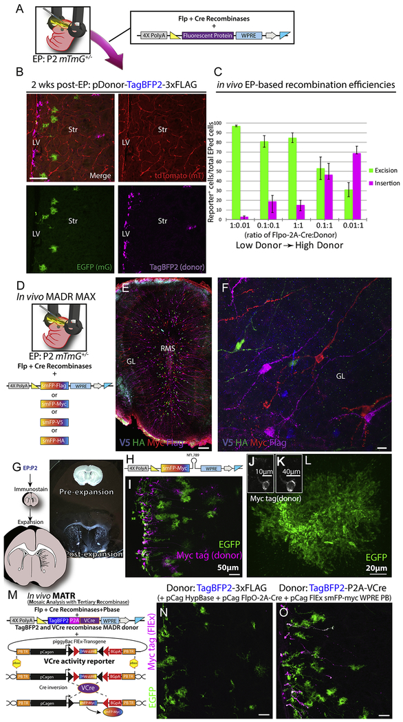
Figure 2: MADR in heterozygous mTmG allows for efficient tracing of lineages in vivo.
A) Standard postnatal EP protocol targeting the ventricular zone in P2 heterozygous Rosa26WT/mTmG pups with DNA mixture of a Flp-Cre vector and a donor plasmid
B) Postnatal EP recapitulates in vitro nucleofection experiment and yields TagBFP2+ MADR along with EGFP+ and tdTomato+ lineages at 2 weeks post-EP. Scale bar, 100μm
C) Different concentrations of recombinase and donor plasmids result in various efficiencies of both MADR and Cre-excision recombination reactions in vivo. All mixtures contained a nuclear TagBfp2 reporter plasmid. (See Supp. Fig. 2D for representative images from this quantitation). Error bars indicate standard error of the mean (SEM).
D) Schematic of plasmid delivery for combinatorial MADR MAX “brainbow” like multiplex labeling
E) Low mag image of olfactory bulb displaying multiplex smFP-based MADR MAX EPed cells and immunostaining for the smFP-linked epitope tags. Scale bar, 100μm
F) High mag image of cells from E exhibiting expression of a single smFP epitope tag per neuron. Scale bar, 10μm
G) Schematic of expansion microscopy and brightfield image example
H) MADR pDonor smFP-myc sh.Nf1 miR-E plasmid for simultaneous knockdown of Nf1 and smFP-myc labeling of transgenic cells
I) Image of EPed striatum showing two populations of reporter labeled cells—EGFP and smFP-myc (i.e. Nf1 knockdown cells).
J) Pre-expansion smFP-myc cell body
K) Post-expansion of cell in J
L) Post-expansion EGFP astrocyte displaying “super-resolution” detail.
M) Schematic of MADR pDonor-TagBFP2-P2A-VCre and FlEx VCre reporter plasmids for mosaic analysis with tertiary recombinase
N) EPed striatum with FlpO-2A-Cre, MADR pDonor-TagBFP2, HypBase and FlEx VCre reporter. Scale bar, 50μm
O) Striatum of littermate of mouse shown in N with FlpO-2A-Cre, MADR pDonor-TagBFP2–2A-VCre, HypBase and FlEx VCre reporter exhibiting VCre-dependent FlEx reporter (smFP-myc). Scale bar, 50μm

