Abstract
Major advances have occurred in recent years in our understanding of HIV-1 assembly, release and maturation, as work in this field has been propelled forwards by developments in imaging technology, structural biology, and cell and molecular biology. This increase in basic knowledge is being applied to the development of novel inhibitors designed to target various aspects of virus assembly and maturation. This Review highlights recent progress in elucidating the late stages of the HIV-1 replication cycle and the related interplay between virology, cell and molecular biology, and drug discovery.
HIV-1 is the causative agent of AIDS, one of the most devastating viral pandemics in history. Although effective treatments are available to suppress HIV-1 replication in infected patients, current drugs do not eradicate the virus, making life-long treatment necessary. Prolonged treatment is associated with drug toxicities and a risk of viral resistance. Thus, the HIV research community must continue to explore novel therapeutic strategies, including those that target steps in the viral replication cycle that are not disrupted by currently available drugs.
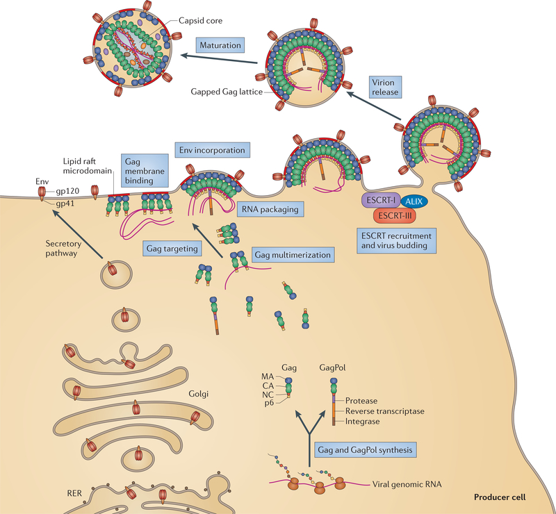
The HIV-1 replication cycle can be divided into an early and a late phase. The early phase encompasses the events that occur from virus binding to the surface of the host cell until the integration of the viral DNA into the host cell genome. These early events include virus binding to cell surface receptors; cell entry; reverse transcription of the viral RNA to DNA; uncoating of the viral capsid; nuclear import of viral DNA; and DNA integration. The late phase refers to the events that occur from gene expression to the release and maturation of new virions, and includes the transcription of viral genes; export of the viral RNAs from the nucleus to the cytoplasm; translation of viral RNAs to produce the Gag polyprotein precursor (also known as Pr55Gag), the GagPol polyprotein precursor, the viral envelope glycoproteins (Env glycoproteins), and the regulatory and accessory viral proteins; trafficking of the Gag and GagPol precursors and of the Env glycoproteins to the plasma membrane; assembly of the Gag and GagPol polyproteins at the plasma membrane; encapsidation of the viral RNA genome by the assembling Gag lattice; incorporation of the viral Env glycoproteins; budding off of the new virions from the infected cell; and particle maturation (FIG. 1).
Figure 1 |. The late stages of the HIV-1 replication cycle.
The viral envelope (Env) glycoproteins traffic via the secretory pathway, from the rough endoplasmic reticulum (RER) to the Golgi and then in vesicles until they arrive at the plasma membrane. The Gag precursor polyprotein — which contains the matrix (MA), capsid (CA), nucleocapsid (NC) and p6 domains — is synthesized in the cytosol from full-length viral RNA. The GagPol precursor polyprotein — which contains MA, CA, NC, protease, reverse transcriptase and integrase domains — is synthesized as the result of a programmed frameshifting event during the translation of Gag-encoding viral RNA. Gag recruits the viral genomic RNA, begins to multimerize and reaches the plasma membrane by a still-undefined pathway. Gag is anchored to the plasma membrane in lipid raft microdomains via insertion of its amino-terminal myristate into the lipid bilayer and by direct interactions with the phospholipid phosphatidylinositol-(4,5)-bisphosphate. The assembling particle incorporates Env and then recruits endosomal sorting complex required for transport I (ESCRT-I)via a direct association between the PTAP motif in p6 and the tumour susceptibility gene 101 (TSG101) subunit of ESCRT-I. Gag also engages in direct binding with the ESCRT-associated factor ALG2-interacting protein X(ALIX), primarily through the YPXL motif in p6. As the budding process proceeds, the ESCRT-III and vacuolar protein sorting 4 (VPS4) complexes are recruited and drive the membrane scission reaction that leads to particle release. Maturation, which leads to the formation of the conical capsid core, is triggered by proteolytic cleavage of the Gag and GagPol polyprotein precursors by the viral protease.
This Review provides an overview of what is currently known about the late stages of the HIV-1 replication cycle and how these steps are orchestrated by the Gag precursor protein, which assembles to form the virus particle at the plasma membrane. This information may, in the future, be applied to the development of novel inhibitors that target these events. Given the similarities in the replication strategies of different viruses, this information has also provided fundamental insights into the basic biology of virus replication.
Gag protein: structure and function
During the late phase of the HIV-1 replication cycle, viral genes are transcribed and viral RNAs are exported from the nucleus to the cytoplasm, where viral proteins are synthesized. Gag is produced as a 55 kDa precursor protein that forms the virus particle. At the same time, the 160 kDa GagPol polyprotein precursor — which contains the viral enzymes protease, reverse transcriptase and integrase — is also expressed at ~5% of the level of Gag, owing to a programmed ribosomal frameshifting event during Gag translation.
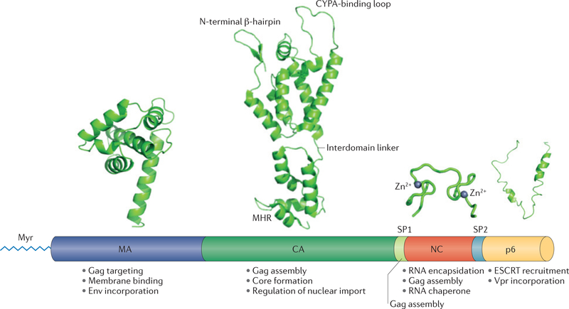
The Gag precursor contains matrix (MA), capsid (CA), nucleocapsid (NC) and p6 domains, as well as two spacer peptides, SP1 and SP2 (FIG. 2). As part of the uncleaved Gag precursor, the MA domain targets Gag to the plasma membrane and promotes incorporation of the viral Env glycoproteins into the forming virions; CA drives Gag multimerization during assembly; NC recruits the viral RNA genome into virions and facilitates the assembly process; and the p6 domain recruits the endosomal sorting complex required for transport (ESCRT) apparatus, which catalyses the membrane fission step to complete the budding process (BOX 1; FIG. 1). After virion release, the viral protease — which is encoded within the GagPol polyprotein precursor — cleaves the Gag precursor to the mature Gag proteins MA, CA, NC and p6. Cleavage of the Gag polyprotein triggers the morphological transformation in virion structure known as maturation (see below).
Figure 2 |. Gag structure and functions.
High-resolution structures of the major domains of Gag — matrix (MA), capsid (CA), nucleocapsid (NC) and p6 — are presented over a linear depiction of the Gag precursor polyprotein. The major functions of each Gag domain are indicated. MA (Protein Data Bank (PDB) accession 1HIW)37 is involved in Gag targeting and binding to the plasma membrane, and in incorporation of the envelope (Env) glycoprotein. Membrane binding requires amino-terminal myristylation (Myr). CA (PDB accession 2M8N)115 participates in Gag assembly, formation of the conical capsid core and regulation of the nuclear import of viral DNA. The CA N-terminal domain (CANTD) contains an N-terminal β-hairpin and a proline-rich loop that binds the host protein cyclophilin A (CYPA); the CA carboxy-terminal domain (CACTD) contains the major homology region (MHR). CANTD and CActd are connected by a short, flexible interdomain linker. Spacer peptide 1 (SP1) is involved in Gag assembly. NC (PDB accession 1BJ6)116 contains two zinc-finger domains; it participates in Gag assembly and RNA encapsidation, and serves as an RNA chaperone. p6 (PDB accession 2C55)8 is involved in recruitment of the endosomal sorting complex required for transport (ESCRT) components and in Vpr incorporation.
Box 1 |. Cellular factors involved in virus assembly and release
Gag partners during assembly
The steps that Gag follows to reach the plasma membrane have not been clearly defined, and likewise, the host factors that promote Gag trafficking to the site of assembly remain only partially characterized. The phospholipid phosphatidylinositol-4,5-bisphosphate (PtdIns(4,5)P2) is now well established as a host cell determinant of Gag binding to the plasma membrane (see the main text), and a number of host proteins have also been implicated in various aspects of Gag localization, membrane binding and assembly16. Further studies will be required to elucidate the specific roles of these protein factors in HIV-1 replication.
gp41 partners during envelope glycoprotein incorporation
The function of the gp41 cytoplasmic tail in envelope (Env) incorporation and virus replication is strongly cell type dependent105; it is indispensable in most T cell lines and in physiologically relevant primary T cells and macrophages, but is not required in several adherent cell lines and in the MT-4 T cell line. This cell type dependence suggests a role for host factors in Env incorporation. Several host factors have been postulated to bind matrix (MA) and/or the gp41 cytoplasmic tail and thereby promote Env incorporation44. For example, Rab11 family-interacting protein 1 isoform C (Rab11-FIP1C) was reported to promote Env trafficking to the plasma membrane and Env incorporation into virions106.
Endosomal sorting complex required for transport
Studies in yeast in the early 2000s revealed that vacuolar protein sorting 23 (Vps23), the yeast homologue of tumour susceptibility gene 101 (TSG101), is part of endosomal sorting complex required for transport I (ESCRT-I), which together with several other ESCRT complexes (ESCRT-0, ESCRT-II and ESCRT-III) and the AAA ATPase VPS4 complex, forms a highly conserved apparatus that plays a vital part in the sorting of ubiquitylated cargo proteins into vesicles. These vesicles bud into late endosomes to form multivesicular bodies (MVBs). The PTAP late domain of Gag (in which the second residue is threonine or serine) interacts with the ESCRT-I component TSG101. The YPXL late domain of Gag was found to bind ALG2-interacting protein X (ALIX), a protein that in mammals interacts directly with both ESCRT-I and ESCRT-III. The ESCRT machinery was also shown to be required for other membrane scission events in the cell, most notably the abscission step of cytokinesis107,108. Indeed, the use of primitive ESCRT-III and VPS4 homologues in cell division seems to be an ancient and primordial activity of the ESCRT machinery, as this function is conserved in the domain Archaea109. Thus, in a remarkable convergence of virology and cell biology, it became clear that retroviruses evolved to co-opt a cellular machinery that promotes membrane scission reactions topologically equivalent to virus budding (that is, those facing away from the cytoplasm) (FIG. 4). The use of the ESCRT machinery for particle release has been established not only for retroviruses but also for several other families of enveloped viruses (for example, filoviruses, paramyxoviruses and arenaviruses) and even for a virus that infects the archaeal organism Sulfolobus solfataricus 110.
Although the structure of the intact, full-length Gag precursor has been elusive owing to its large size and the presence of flexible interdomain connections, atomic-level structures are available for the individual Gag proteins (FIG. 2). MA folds into a highly globular structure comprising five α-helices, a 310 helix and a three-stranded mixed (β-sheet. A carboxy-terminal α-helix projects away from the globular core, serving to connect MA with the adjacent CA domain1. A number of basic residues cluster at the top of MA, allowing the protein to interact electrostatically with the negatively charged lipids in the inner leaflet of the plasma membrane. The amino terminus of the MA domain is co-translationally modified by the covalent attachment of a myristic acid moiety; this 14-carbon fatty acid, which can adopt a sequestered or an exposed conformation2, plays an essential part in Gag-membrane binding (see below).
The CA domain folds into two independent, largely helical domains — the N-terminal domain (CANTD) and C-terminal domain (CACTD) — connected by a flexible linker3–5. In the mature protein, the N terminus of CANTD folds back into a β-hairpin structure (FIG. 2). A proline-rich loop serves as the binding site for the host proline isomerase cyclophilin A (CYPA; also known as PPIA)4, which is incorporated into virions and may help to shield the viral capsid from antiviral innate immune defences after virus entry into the target cell. The CActd contains a dimer interface, which plays an important part in Gag multimerization5, and the major homology region (MHR), which is highly conserved among orthoretroviruses and participates in virus assembly. Nuclear magnetic resonance (NMR) data obtained with a Gag fragment containing MA and the CANTD have provided insights into how the structure of CA is affected by cleavage between MA and CA6. In this pre-cleavage structure, the N terminus of the CANTD is unfolded, suggesting that the N-terminal p-hairpin of the mature CA protein (FIG. 2) forms after MA-CA cleavage.
NC contains two zinc-finger-like domains (often referred to as zinc knuckles) that are important for genome recognition and the recruitment of viral RNA to the assembling Gag complex. In the mature protein, the zinc fingers form independently folded domains analogous to beads on a string7. The C-terminal p6 domain is largely unstructured8, consistent with its primary role as an adaptor for the host ESCRT machinery (BOX 1).
HIV-1 assembly
The path to virus assembly begins with synthesis of the Gag polyprotein in the cytosol and its translocation to the site of assembly. During its trafficking to the plasma membrane, or after its arrival there, Gag interacts with dimeric viral RNA. Virus assembly proceeds at the plasma membrane, and viral Env glycoproteins accumulate at the site. Gag then recruits the cellular ESCRT machinery, which drives the membrane scission reaction required for particle release (BOX 1; FIG. 1).
Gag trafficking to the site of assembly.
Although at times this has been the subject of controversy, it is now well established that the inner leaflet of the plasma membrane is the primary site at which HIV-1 particles assemble. However, the mechanism by which Gag is specifically targeted to the plasma membrane has been a topic of active investigation for many years. An important role for the MA domain in Gag targeting was demonstrated by the observation that mutations in MA, particularly those involving the highly basic region, caused Gag to be mistargeted to a late endosome or multivesicular body (MVB) compartment9. The phosphoinositide phosphatidylinositol-4,5-bisphosphate (PtdIns(4,5)P2), a phospholipid that is highly enriched in the inner leaflet of the plasma membrane, plays a crucial part in Gag localization10. Depletion of plasma membrane PtdIns(4,5)P2 largely recapitulates the phenotype of MA basic-domain mutants, leading to retargeting of Gag to late endosomes or MVBs. Subsequent structural studies demonstrated that MA interacts directly with the negatively charged inositol headgroup of PtdIns(4,5)P2 (REFS 11,12). Notably, NMR data revealed that binding of a soluble derivative of PtdIns(4,5)P2 to MA triggers exposure of the myristic acid moiety covalently linked to the N terminus of MA11. Myristate exposure probably facilitates its insertion into the inner leaflet of the plasma membrane, thereby stabilizing the Gag-membrane association. An additional level of regulation of Gag targeting to the plasma membrane was suggested by the observation, in liposome studies, that RNA — probably tRNA13 —inhibits Gag-membrane interactions by binding to the highly basic region of MA. However, PtdIns(4,5)P2, but not other acidic phospholipids, is able to outcompete RNA for MA binding to liposomes14,15. Collectively, these findings show that PtdIns(4,5)P2 regulates Gag targeting to the plasma membrane by mediating direct electrostatic interactions with MA, by displacing RNA bound to MA and by inducing the exposure of the N-terminal myristate, allowing it to insert into the target membrane. A number of cellular proteins have been implicated in Gag targeting to the plasma membrane and in subsequent assembly16; however, the mechanism of action of these host factors remains to be elucidated (BOX 1).
On reaching the plasma membrane, Gag induces the recruitment and coalescence of cholesterol- and sphingolipid-enriched membrane microdomains, often referred to as lipid rafts17–19 (FIG. 1). The targeting of Gag to lipid rafts in the plasma membrane and the importance of PtdIns(4,5)P2 in this plasma membrane targeting are reflected by the high levels of cholesterol, sphingolipid and PtdIns(4,5)P2 that are found on the viral membrane20,21. Lipid rafts probably serve as platforms for virus assembly, and the targeting of Gag to these microdomains may facilitate the incorporation of Env glycoproteins into virions (see below). Given that areas of cell-cell contact — referred to by virologists as ‘virological synapses’ — are also rich in lipid rafts22, the preference exhibited by Gag for lipid raft-like membrane microdomains may enhance assembly at the virological synapse, thereby promoting cell-cell viral transfer.
RNA encapsidation.
After synthesis in the nucleus, HIV-1 RNAs are transported to the cytoplasm, where they undergo two main processes: translation into viral proteins and packaging into newly assembled virus particles. A large number of viral mRNA species are synthesized, some of which are multiply spliced before nuclear export; others largely escape splicing and are exported in a partially spliced or unspliced form. Export of intron-containing viral RNAs is mediated by a highly structured cis-acting RNA element known as the Rev-responsive element (RRE), which is bound by the virus-encoded trans-acting protein Rev23. Rev, which shuttles between the nucleus and the cytoplasm, promotes nuclear export by forming a complex with the nuclear export factor CRM1 (also known as EXP1) and the GTPase RAN24.
As is the case for other retroviruses, two copies of full-length viral RNA are packaged into HIV-1 virions25. The presence of two copies of genomic RNA in each particle provides the opportunity for recombination during reverse transcription and may also allow reverse transcription to proceed if one RNA copy is damaged26,27. The bulk of recent evidence suggests that RNA dimerization occurs in the cytosol or at the plasma membrane28 and that an RNA dimer is the recognition unit for packaging into assembling virions29.
The NC domain of Gag is the primary viral determinant that drives RNA packaging. NC contains two Cys-Cys-His-Cys zinc-finger-like (or ‘zinc-knuckle’) domains (FIG. 2), which are flanked by basic residues that increase the affinity of NC for RNA. NC functions as a nucleic acid chaperone, an activity that contributes to RNA packaging, to the incorporation and placement of the tRNA primer and to reverse transcription30. Interactions between NC and RNA also help to drive Gag multimerization (see below).
Retroviral NC domains direct the packaging of viral genomic RNA by binding to a packaging signal, often referred to as the ψ-element, that is located near the 5’ end of the genomic RNA in the 5’ untranslated region (UTR). In some retroviral systems, the ψ-element is fairly well defined. By contrast, a small, well-defined ψ-element has not been identified for HIV-1. The 5’ UTR of the HIV-1 genomic RNA is a highly structured region that contains a number of folded RNA elements31, including the transactivation response element (TAR), which serves as the binding site for the viral Tat protein and the positive transcription elongation factor b (P-TEFb) complex; a polyadenylation site; the primer-binding site (PBS), which binds a molecule of tRNALys so that it is packaged into virions and can prime the initiation of reverse transcription; the dimer initiation signal (DIS), which contributes to RNA dimerization; the major splice donor (SD); and potentially other stem-loop structures. A variety of deletions and mutations in the 5’ UTR reduce packaging efficiency, and a large number of alternative structures have been proposed for this region of the viral RNA (reviewed in REF 31). Therefore, it is likely that multiple folding conformations exist for the 5’ UTR of the HIV-1 genomic RNA, and different conformations may serve distinct functions. For example, the 5’ end of the viral RNA has been proposed to undergo a conformational switch from a structure that favours translation to one that promotes packaging32. Structures are available for NC bound to a number of different segments of the 5’ UTR, although it is not clear which of these is most biologically relevant for packaging.
To follow the movement of HIV-1 RNA together with Gag in living cells, total internal reflection fluorescence (TIRF) microscopy (BOX 2) was used to visualize RNA and Gag localization near the cell surface33. In the absence of Gag, the HIV-1 RNA moved dynamically to and away from the plasma membrane. In the presence of Gag, the RNA docked stably at the plasma membrane and then became immobile as Gag co-assembled around the RNA. RNA docking in the presence of Gag occurred before Gag-RNA colocalization could be detected, suggesting that initial contacts are made between the RNA and a small (subdetectable) number of Gag molecules. These observations, which are consistent with the hypothesis that Gag and RNA first interact in the cytosol34, indicate that a few molecules of Gag recruit the viral RNA to the plasma membrane, where the RNA then nucleates particle assembly (FIG. 1). A separate study used live-cell imaging to track single molecules of HIV-1 RNA in the cytosol and observed that the overwhelming majority of RNA molecules moved by passive diffusion, whether or not Gag was also expressed35. To better understand the nature of HIV-1 Gag-RNA interactions, the technique of crosslinking immunoprecipitation (CLIP) sequencing was used (BOX 2). This analysis revealed striking changes in Gag-RNA interactions during HIV-1 assembly and maturation; Gag in the cytosol bound primarily to several distinct sites in the 5’ UTR and the RRE, whereas Gag at the plasma membrane and in the assembling virion was crosslinked to a large number of sites throughout the HIV-1 genome13. In mature virions, in which NC is thought to coat the viral genomic RNA, NC binding was observed at specific sites that again included the 5’ UTR and the RRE. These results provide a window into the dynamic nature of the interactions between HIV-1 Gag and viral RNA.
Box 2 |. Techniques to analyse virus assembly and release
Super-resolution fluorescence microscopy
The diffraction limit sets the resolution of the light microscope to approximately half the wavelength of light being measured (~250 nm for blue light). As a result, a point source emitter, such as a blue dye, is observed as being ~250 nm in diameter, preventing the resolution of objects closer together than this distance. Super-resolution techniques allow considerably higher resolutions, in the range of tens of nanometres, to be achieved. There are several types of super-resolution (or super-resolved) microscopy, such as photoactivated localization microscopy (PALM), stimulated emission depletion (STED) microscopy and stochastic optical reconstruction microscopy (STORM) (reviewed in REF 111). The use of super-resolution techniques to study HIV-1 budding and release is described in the main text.
Cryoelectron microscopy and cryoelectron tomography
Rapid freezing of the sample followed by electron microscopy analysis performed at very low temperatures enables data to be gathered on native samples in the absence of a fixative. Combined with controlled exposure to the electron beam, this minimizes damage to the sample. Analysis of a large number of identical particles allows averaging to generate a high-resolution 3D volume. A limitation of cryoelectron microscopy (cryo-EM) is that it requires the particles being analysed to be identical or nearly identical. Cryo-EM has thus been a powerful technique for the analysis of viruses with identical morphologies, but it is less useful for highly pleomorphic viruses such as HIV-1. In such cases, cryoelectron tomography (cryo-ET) is used to obtain a number of 2D electron microscopy images of an individual particle over a range of tilt angles. These 2D images are then combined to obtain a medium-resolution 3D reconstruction. Subvolume or subtomogram averaging techniques greatly increase the resolution by averaging common features present in the pleomorphic specimens. When available, atomic-resolution data from X-ray crystallography or NMR spectroscopy can be fitted into the electron microscopy density maps to produce pseudo-atomic models of large and complex structures (reviewed in REF. 112).
Total internal reflection fluorescence microscopy
Using an evanescent wave of light, total internal reflection fluorescence (TIRF) microscopy illuminates a thin section of the sample within ~100 nm of the cover slip. This method, which is often applied to single-molecule experiments, permits high-resolution information to be obtained about events occurring at the cell surface, such as virus budding (reviewed in REF. 113).
Fluorescence resonance energy transfer
In fluorescence resonance energy transfer (FRET; also called Forster resonance energy transfer), a donor fluorophore is excited by a specific wavelength of light; the donor can then transfer energy through non-radiative coupling to an acceptor fluorophore located in close proximity (in the range of 1–10 nm) to the donor, provided that the fluorescence emission spectrum of the donor overlaps with the absorption spectrum of the acceptor. Energy transfer between donor and acceptor is highly dependent on their close proximity and thus provides a sensitive measure of the distance between the two fluorophores. This method is often used to measure protein-protein and protein-nucleic acid interactions in cells or in vitro (reviewed in REF 114).
Gag assembly.
Following their arrival at the plasma membrane, the Gag protein and the full-length genomic RNA assemble into the nascent, immature virus particle together with the GagPol precursor. As mentioned above, Gag-RNA contacts help to nucleate assembly, but the main viral determinant that drives Gag multimerization is the CA domain. In the immature particle, Gag molecules are aligned and packed radially, with the MA domain bound to the inner leaflet of the viral membrane and the C terminus of Gag oriented towards the centre of the particle (FIG. 1). Recent biochemical results suggest that the MA domain is trimeric in virions36, consistent with data indicating that HIV-1 MA crystallizes as a trimer37 and forms hexamers of trimers on a model membrane monolayer38. To accommodate the curvature necessary to form a particle with a diameter of ~120 nm — the size of the HIV-1 virion — the immature Gag lattice is continuous but contains gaps that appear to be devoid of Gag protein39–41 (FIG. 1).
An extensive amount of high-resolution structural information is available for the hexameric lattice formed by CA in the mature, conical capsid (see below). However, largely because of its curved and flexible nature, the structure of the immature Gag lattice has been technically more challenging to define. Cryoelectron tomography (cryo-ET) (BOX 2) data based on subtomogram averaging provided a low-resolution view of the immature Gag lattice in virus-like particles (VLPs); these studies showed a hexameric arrangement for the immature Gag lattice, with a hole at the centre of the hexamer39,41. More recently, cryoelectron microscopy (cryo-EM) and cryo-ET techniques were combined to solve the structure of the immature Gag lattice in in vitro-assembled tubes formed by a truncated version of the Gag protein from the betaretrovirus Mason-Pfizer monkey virus (MPMV)42; the atomic-resolution HIV-1 CA crystal structures were then modelled into the electron densities of the MPMV cryo-EM reconstruction. One of the most significant features of this analysis was to reveal that the CA-CA contacts in the immature Gag lattice differ from those in the mature CA lattice. This MPMV-based model has recently been revised with the realization that, despite conservation of the tertiary structures of HIV-1 and MPMV CA proteins, the quaternary arrangements of the CA proteins from these two retroviruses differ substantially43. Even in this most recent, subnanometre-resolution structure of the immature Gag lattice, the precise structure of SP1 is not resolved43, although the observed electron densities are consistent with SP1 forming a six-helix bundle as proposed earlier41. Further studies will be required to obtain a structure of the CA-SP1 boundary region of Gag at atomic-level resolution. This is an important objective, as this region not only promotes virus assembly but is also the likely binding site for HIV-1 maturation inhibitors (see below).
Env glycoprotein incorporation.
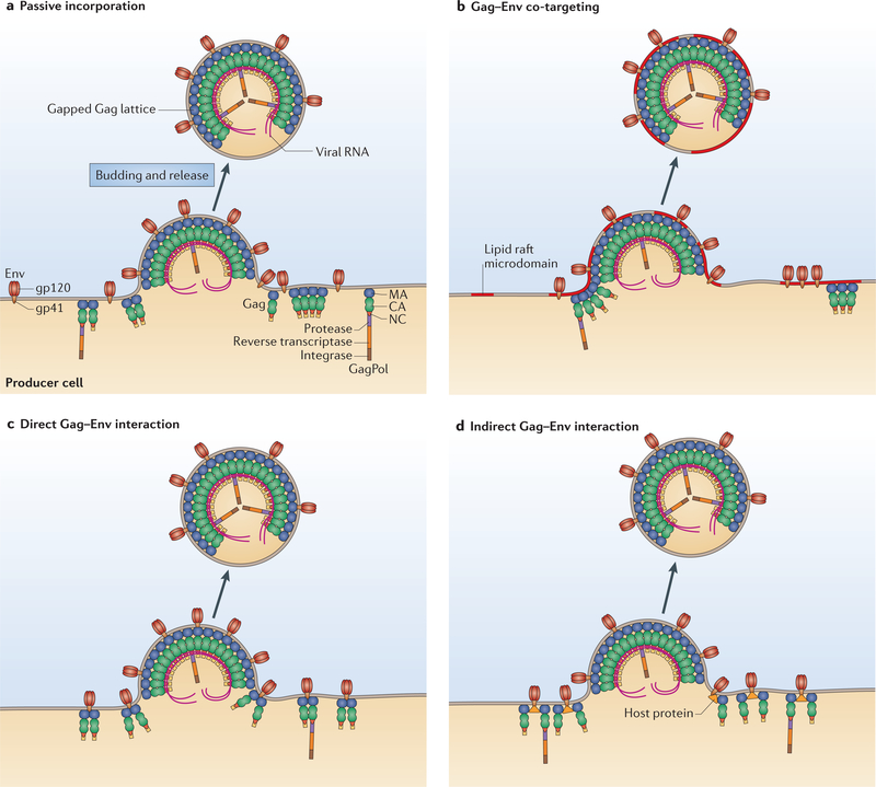
The mechanism by which the Env glycoprotein complex is incorporated into virus particles remains incompletely understood. Lentiviruses, such as HIV-1, are unique among retroviruses in bearing Env glycoproteins with very long cytoplasmic tails; these long tails must in some way be accommodated by the underlying Gag lattice during Env incorporation44,45. Env incorporation could, in theory, take place by several non-mutually exclusive mechanisms: a passive process; co-targeting of Gag and Env to a common site on the plasma membrane (for example, a lipid raft-type microdomain); direct recruitment of Env by Gag; and indirect recruitment of Env by Gag via a host cell bridging protein44 (FIG. 3).
Figure 3 |. Models of viral envelope glycoprotein incorporation.
Several non-mutually exclusive mechanisms may participate in the incorporation of the viral envelope (Env) glycoproteins into new virions. a | Env expressed on the cell surface might be incorporated passively into virions as the particles acquire host-derived membrane during assembly and budding. b | In a putative Gag-Env co-targeting mechanism, both Gag and Env are targeted to a specific site on the plasma membrane (for example, a lipid raft microdomain), allowing Env to be concentrated at sites of assembly. c | Evidence suggests that the matrix (MA) domain of Gag and the cytoplasmic tail of the gp41 subunit of Env interact directly, resulting in the retention of Env complexes at sites of budding and their recruitment into virions. d | The MA domain of Gag and the cytoplasmic tail of gp41 could interact indirectly, with a host protein bridging the two viral proteins. CA, capsid; NC, nucleocapsid. Figure adapted from REF 44, Elsevier.
Genetic data support a central role for the MA domain of Gag and the gp41 cytoplasmic tail of Env in Env glycoprotein incorporation, and suggest direct or indirect interactions between Env and MA44. Mutations in MA and the gp41 cytoplasmic tail can block Env incorporation, and an MA mutant can rescue an Env-incorporation-deficient Env mutant46. The ability of MA mutations to block Env incorporation is relieved by truncation of the gp41 cytoplasmic tail, suggesting that these MA mutants block Env incorporation by imposing a steric clash between MA and the long gp41 cytoplasmic tail. The notion that the gp41 cytoplasmic tail and Gag interact is supported by the observation that the Env complex in immature virions (in which Gag is uncleaved) is non-fusogenic; activation of fusion activity is triggered by cleavage of Gag by the viral protease47,48. Fusion activity can also be activated by truncation of the gp41 cytoplasmic tail, suggesting that an interaction between uncleaved Gag and the gp41 cytoplasmic tail locks Env in a non-fusogenic conformation. Consistent with this hypothesis, Env was able to cluster on the surface of mature but not immature virions, and this clustering was found to depend on the gp41 cytoplasmic tail49. Furthermore, Env was retained on immature particles from which the lipid envelope had been removed with detergent, and deletion of the gp41 cytoplasmic tail resulted in a loss of Env from such detergent-stripped particles, suggesting that direct or indirect contacts take place between the immature Gag lattice and the gp41 cytoplasmic tail50.
One of the major impediments to understanding HIV-1 Env incorporation is the lack of structural information about either MA in the context of an infectious virus particle, or the unusually long cytoplasmic tail of gp41. Early studies showed that HIV-1 MA crystallizes as a trimer37, a finding supported by a more recent analysis of crystals assembled on 2D membrane monolayers38. However, direct analysis of HIV-1 virions by cryo-ET has not revealed any long-range order in the MA shell43. Even less is known about the structure of the gp41 cytoplasmic tail. This region of gp41, which is approximately 150 amino acids long, could be folded in several different conformations. Peptides derived from strongly helical motifs in the cytoplasmic tail, often referred to as lentiviral lytic peptides (LLPs), bind lipid bilayers, consistent with the idea that the cytoplasmic tail interacts with the membrane. Alternatively, the gp41 cytoplasmic tail could adopt a more rod-like, helical-bundle conformation in which it projects inwards, perhaps passing through the large gap in the underlying Gag lattice38. It is plausible that the gp41 cytoplasmic tail could adopt a different conformation in immature versus mature virions and at different stages of the fusion process. Although the higher-order arrangement of MA in the context of the infected cell membrane or the virion is still unknown, as mentioned above, a recent study supports the hypothesis that MA trimers form in virions and are important for Env incorporation36. One analysis used subtomogram averaging of the putative MA lattice present in membrane-enclosed structures containing multiple CA cores, and the resultant data provided evidence for the formation of hexamers of trimers51, consistent with the arrangement of MA on PtdIns(4,5)P2-containing model membrane monolayers38.
HIV-1 release and maturation
After assembly of the immature Gag lattice at the plasma membrane, the nascent particle must undergo a membrane fission event to be released from the cell surface. This release step is mediated by the cellular ESCRT machinery (BOX 1), which is hijacked by Gag. During or shortly after budding off of the particle from the cell surface, the viral protease cleaves the Gag polyprotein precursor to trigger HIV-1 maturation.
Virion release.
The path to our current understanding of virus release began with the observation that deletion of the C-terminal p6 domain of Gag resulted in a release block and the accumulation of particles that were attached to the cell by a thin membranous stalk52. Subsequently, a detailed mutational analysis mapped the virus release function of p6 to a highly conserved PTAP motif (in which the second residue can be threonine or serine) near the N terminus of p6 (REF 53). Studies performed in other retroviral systems observed similar defects in virus release following mutation of short peptide motifs located at various positions within the Gag polyprotein. These motifs, or late domains, are composed of three distinct sequences: the above-mentioned PTAP, PPXY and YPXL54. The basis for the ability of these viral late domains to drive virus release remained a mystery until several groups independently and almost simultaneously reported55–58 that the PTAP motif of HIV-1 interacts directly with a cellular protein known as tumour susceptibility gene 101 (TSG101), which is part of ESCRT-I. The ESCRT machinery, which is made up of several multiprotein complexes and accessory factors, has a key role in a variety of membrane budding and scission processes in the cell (BOX 1).
Many retroviruses, including HIV-1, encode more than one late domain; in addition to the above-mentioned TSG101-binding PTAP motif, the p6 domain of HIV-1 Gag also bears a YPXL-type binding site for ALG2-interacting protein X (ALIX; also known as PDCD6IP) (BOX 1). Although the TSG101-binding PTAP motif plays the dominant part in HIV-1 release, the ALIX-binding site is also required for optimal virus replication59. In mammals, the ESCRT machinery is greatly expanded relative to that in other, more primitive organisms and comprises ~20 proteins54. However, HIV-1 (and, in all likelihood, other enveloped viruses) requires only a subset of these factors; most notably, ESCRT-II and several subunits of ESCRT-III seem to be dispensable for HIV-1 budding60. Ubiquitylation of cargo proteins often serves as a signal for recruitment of the ESCRT machinery, and several ESCRT components contain ubiquitin-binding domains54. Although it is well established that many retroviral Gag proteins are likewise ubiquitylated, a role for Gag ubiquitylation in ESCRT recruitment and virus release has not yet been definitively confirmed. However, evidence supports the hypothesis that ubiquitin, together with the viral late domains, can promote Gag-mediated ESCRT recruitment61,62. Recent work has demonstrated that the host protein angiomotin promotes HIV-1 budding by forming a bridge between HIV-1 Gag and the E3 ubiquitin ligase NEDD4L. Intriguingly, the virus release defect induced by angiomotin depletion seems to be imposed at an earlier stage in the budding process than that induced by disruption of the Gag-TSG101 interaction63.
For more than a decade, a wealth of data has accumulated about the structure and function of the ESCRT machinery and its role in vesicle budding, cytokinesis and virus release. Although the precise mechanism by which the ESCRT machinery drives membrane scission remains to be fully defined, it is clear that ESCRT-III plays a central part in this process. ESCRT-III has been observed to assemble into circular arrays or spirals64,65 that could help to constrict the membrane at the neck of the bud to drive scission (FIG. 4). Indeed, in an in vitro giant unilamellar system, ESCRT-III components were sufficient to drive the budding and detachment of intraluminal vesicles66. ESCRT-III also recruits the AAA ATPase vacuolar protein sorting 4 (VPS4), which may provide energy for the membrane scission event and is also required for the recycling of ESCRT components after budding has been completed.
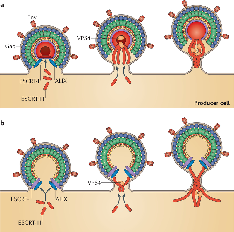
Figure 4 |. HIV-1 budding and release.
Models for membrane scission mediated by endosomal sorting complex required for transport III (ESCRT-III). In both models, the HIV-1 Gag protein interacts with ESCRT-I and ALG2-interacting protein X(ALIX), which recruit ESCRT-III. Membrane scission is driven by ESCRT-III polymerization in concert with the activity of the AAA ATPase vacuolar protein sorting 4 (VPS4). a | In the first model, ESCRT-III is localized within the budding particle70. b | In the second model, ESCRT-I and ALIX are located within the budding particle and recruit ESCRT-III and VPS4, which are mostly located outside the budding particle. Env, envelope. Part a adapted from Van Engelenburg, S. B. et al. Distribution of ESCRT machinery at HIV assembly sites reveals virus scaffolding of ESCRT subunits. Science 343,653–656 (2014). Reprinted with permission from AAAS. Part b adapted from REF 117, © Cashikar et al.
Real-time imaging has been used to address the kinetics of HIV-1 assembly and release, and the timing of ESCRT recruitment to the site of assembly. Using fluorescently tagged Gag and a combination of TIRF microscopy, fluorescence resonance energy transfer (FRET) (BOX 2) and fluorescence recovery after photobleaching (FRAP), the kinetics of assembly — that is, the time from initial Gag detection to the completion of Gag accumulation — were estimated to be in the range of 5–9 minutes, with some particles taking ~20 minutes to assemble67,68. ESCRT-III recruitment was observed late in the assembly process, consistent with its role in mediating the final stage of particle release69. To delve more deeply into the mechanism of ESCRT-mediated membrane scission, advanced microscopy techniques were applied to examine the topology of ESCRT components with respect to the budding HIV-1 particle. The application of 3D super-resolution microscopy and correlative electron microscopy led to the conclusion that the ESCRT machinery localizes within the head of the budding virion, driving membrane scission from within the interior of the bud70 (FIG. 4a). By contrast, another study using super-resolution microscopy observed the sequential accumulation of ESCRT components at the neck of the assembling virion, with the ESCRT-III component charged MVB protein 4B (CHMP4B) recruited ~10 seconds before VPS4 (REF 71) (FIG. 4b). Although these studies are all consistent with their being a central role for the assembled ESCRT-III components in driving membrane scission, details relating to the topology of the machinery and the precise role of VPS4 in the process await further enquiry.
Virus maturation.
When expressed alone, Gag is competent to drive the assembly and release of non-infectious, immature VLPs. Particle infectivity requires the proteolytic activity of the viral protease, which is expressed and brought into virions as part of the GagPol precursor. Concomitant with virus release, the viral protease cleaves a number of sites in both the Gag and GagPol polyproteins to trigger virus maturation. The HIV-1 protease is an aspartyl protease that functions as a dimer, in which the active site is located in a cleft at the dimer interface72. The efficiency with which the viral protease cleaves each of its target sites varies considerably, giving rise to a highly ordered, stepwise processing cascade73. Mutations that either increase or decrease the efficiency with which the protease cleaves a particular site or sites can be highly detrimental to maturation and particle infectivity, leading to the formation of aberrant, non-functional cores74.
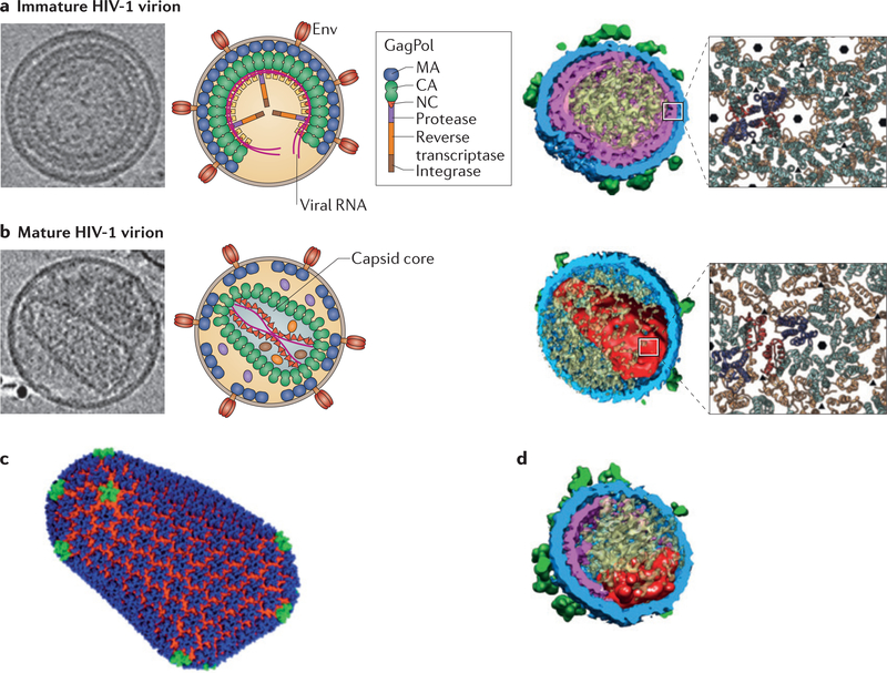
Protease-mediated Gag and GagPol processing is accompanied by a major change in virion morphology (FIG. 5a-c). In the immature particle, Gag molecules are packed in a radial manner. Following liberation of the individual Gag domains by the viral protease, the CA protein reassembles to form the conical capsid core (note that the term ‘capsid’ is used for both the CA protein or domain and the conical structure formed by the CA protein; here, I use the term ‘CA’ when referring to the protein or domain and ‘capsid’ when referring to the conical capsid core). It remains an open question whether and to what extent the immature CA lattice disassembles and reassembles to form the mature CA lattice, or whether it undergoes a transition to the mature lattice without complete disassembly51,75.
Figure 5 |. HIV-1 maturation.
During virus release, the viral protease cleaves a number of sites in both the Gag and GagPol polyproteins to trigger virus maturation, resulting in major changes in virion morphology, includin g the generation of the conical capsid core. a | A cryoelectron tomogram (left panel), an illustration (middle panel) and a cryoelectron tomography (cryo-ET) reconstruction (right panel) of the immature HIV-1 virion, together with a zoomed-in view of the immature Gag lattice (far-right inset). In the far-right panel, the capsid (CA) amino-terminal domain (CANTD) is depicted in blue and the CA carboxy-terminal domain (CActd) is in orange. b | A cryoelectron tomogram (left panel), an illustration (middle panel) and a cryo-ET reconstruction (right panel) of the mature HIV-1 virion, together with a zoomed-in view of the mature CA lattice (far-right inset; the CANTD and CACTD are coloured as in part a). c | The all-atom structure of an HIV-1 capsid core, with CA pentamers (green) in the otherwise hexameric lattice. d | A cryo-ET reconstruction of a virion produced from cells treated with the maturation inhibitor bevirimat, showing disruption of capsid formation. Env, envelope; MA, matrix; NC, nucleocapsid. Cryoelectron tomograms in part a and b reproduced from REF. 118, Elsevier. Cryo-ET reconstructions in parts a, b and d republished with permission of the American Society for Microbiology, from HIV 1 maturation inhibitor bevirimat stabilizes the immature Gag lattice. Keller, P. W. et al. 85, 4, 2015; permission conveyed through Copyright Clearance Center. Zoomed-in views in parts a and b reproduced from REF 43, Nature Publishing Group. Part c reproduced from REF 78, Nature Publishing Group.
The organization of CA in the mature HIV-1 capsid core is well defined. An early study correctly proposed that the capsid core assembles with fullerene-like geometry, such that CA forms predominantly hexameric rings to generate a lattice that is closed off at both ends by the inclusion of pentamers: seven at the wide end and five at the narrow end76 (FIG. 5c). The formation of a hexameric CA lattice with 12 pentamers is a widespread feature among retroviruses, and the final shape of the capsid core is determined by the placement of the pentamers (in this sense, the structure of the retroviral capsid core is analogous to that of a soccer ball, the outer shell of which is composed of a combination of hexamers and 12 pentamers). Many studies over the years have contributed to our understanding of the core structure77. For example, a combination of cryo-EM and molecular dynamics simulation generated an atomic model for the HIV-1 capsid core78. The inter-hexamer and intra-hexamer contacts have been defined; as mentioned above, although both immature Gag and mature CA assemble into hexameric lattices, the intermolecular interfaces in the two structures differ considerably43 (FIG. 5a,b). Notably, several studies used artificially crosslinked CA hexamers to overcome aggregation issues during the process of protein purification. However, this problem was recently solved by crystallizing unmodified CA at low protein concentrations79. The resulting structure provided a detailed view of the native CA-CA interfaces and revealed that water molecules play an important part in stabilizing inter-hexamer interactions79.
In the next round of infection, the viral capsid core provides a degree of protection for the viral RNA genome as it undergoes reverse transcription. Details of how rapidly and to what extent the capsid core disassembles — a process known as uncoating — after infection are currently being investigated80. Sensitivity to several post-entry restriction factors maps to CA, indicating that some CA protein remains associated with the reverse transcription complex after entry81. An emerging view is that at least a remnant of the hexagonal CA lattice stays intact as the reverse transcription complex traffics to the nucleus; this lattice serves as the recognition signal for host cell restriction factors and is also recognized by nuclear pore components that play an active part in the nuclear import of viral DNA. The observation that both increased and decreased core stability inhibit HIV-1 infection82 suggests that uncoating kinetics have been precisely optimized to promote infection (reviewed in REF 81).
Gag as a target for antivirals
Having recognized for many years that the Gag polyprotein precursor and the mature Gag proteins have vital roles at multiple steps in the virus replication cycle, the research community has directed considerable effort into developing inhibitors of Gag function83. These efforts have unfortunately not resulted in the highly efficacious drugs that have come from parallel efforts to inhibit the viral enzymes reverse transcriptase, protease and, most recently, integrase. However, recent progress has been noteworthy.
The capsid domain and protein as a target.
The central role of CA in several steps of the virus replication cycle makes it an attractive target for the development of anti-HIV-1 inhibitors. Disruption of CA-CA contacts by small molecules could block the assembly of immature particles or of the mature capsid core. As core stability seems to be precisely balanced, compounds that either accelerate or delay uncoating could also block infection by acting in the target cell. One could also imagine an inhibitor that works by ‘unmasking’ the core and making it more vulnerable to post-entry restriction factors that target the capsid lattice.
Although no clinically viable inhibitors targeting CA have been reported, an increasing number of compounds have been described that demonstrate antiviral activity in cell culture. Early inhibitors include the peptide CAI (for CA inhibitor) that binds the CACTD and disrupts CA dimer formation in vitro84. Although CAI is cell impermeable, circularized derivatives were developed using the technique of hydrocarbon stapling; these derivatives penetrate cells and display antiviral activity85. In silico screening identified a small-molecule inhibitor, CAP-1, that binds a pocket near the base of the CANTD by an induced-fit mechanism86,87. This pocket is also the reported binding site for several other small molecules, including BD3 and BM4 (REF. 88). Although all of these compounds dock in the same pocket in the CANTD, their binding seems to have different outcomes, either disrupting the assembly of immature VLPs or blocking core condensation. A second pocket in the CANTD serves as the binding site for several other CA-based inhibitors, the best characterized being PF74 (also known as PF-3450074)89,90. These compounds act at a post-entry, pre-integration step. Interestingly, the binding site for PF74 overlaps with that of host cell factors that regulate nuclear import (for example, nuclear pore complex protein 153 (NUP153) and cleavage and polyadenylation specificity factor 6 (CPSF6)), suggesting that compounds that occupy this pocket block infection by preventing the viral nucleoprotein complex from entering the nucleus91. PF74, NUP153 and CPSF6 make contacts with the CANTD-CACTD interface and the adjacent monomer in the CA hexamer, and thus bind with higher affinity to the intact hexamer than to monomeric CA92,93. Notably, the recently reported crystal structure of the native CA suggests that PF74 destabilizes the capsid by inducing subtle changes at inter-hexamer interfaces79. As anticipated from the many parts that CA plays during replication, compounds that bind CA can disrupt HIV-1 replication at several distinct steps, making them worthy of further development.
The nucleocapsid, matrix and p6 domains and proteins as targets.
Like CA, NC functions throughout the virus replication cycle; as a result, research aimed at blocking NC function has been underway for many years. Much of this effort has been aimed at developing compounds that eject zinc from the NC zinc fingers (see REFS 83,94). A limitation to this approach has been toxicity arising from a lack of specificity for retroviral NC versus host protein zinc-fingers. Although in theory MA and p6 could also be targeted by antivirals that disrupt their functions, progress in this area is at an early stage.
Maturation inhibitors.
The importance of highly ordered and complete Gag processing for HIV-1 maturation and infectivity indicates that Gag processing could be a target for novel antiretrovirals that block specific steps in the proteolytic cascade. Such inhibitors are fundamentally distinct from protease inhibitors, which act by targeting the protease enzyme rather than the substrate (Gag). The first-in-class ‘maturation inhibitor’ with this mode of action (that is, targeting Gag) was a dimethyl succinyl betulinic acid derivative known as PA-457 or bevirimat (FIG. 5d). This compound was shown to inhibit HIV-1 maturation by blocking the protease-mediated cleavage of the CA-SP1 processing intermediate to mature CA95,96. Selection studies demonstrated that resistance to bevirimat could be conferred by changes in the sequences surrounding the CA-SP1 cleavage site95,97, and several lines of evidence supported the hypothesis that bevirimat acts by binding the CA-SP1 region of Gag in the immature particle, thereby blocking protease-mediated cleavage at this site98,99. Advances in defining the structure of the CA-SP1 boundary region in the context of assembled immature Gag will be required for the precise determination of the bevirimat binding site, although insights into the mechanism of maturation inhibitor binding were obtained from the study of the structurally distinct compound PF-46396. Based on the study of PF-46396-resistant mutants, it seems likely that bevirimat and PF-46396 bind the same pocket in the assembled Gag complex100. Phase II clinical trials with bevirimat demonstrated the proof of concept that maturation inhibitors can significantly reduce viral loads in infected patients101; however, naturally occurring polymorphisms in SP1 compromised the activity of bevirimat102. Recent progress has been made in developing bevirimat analogues with low-nanomolar potency even against strains of HIV-1 bearing a variety of SP1 polymorphisms (Urano, E., Ablan, S. and E.O.F., unpublished observations), and it is anticipated that these potent bevirimat analogues will be tested in future clinical trials.
Outlook
Recent years have witnessed remarkable advances in our understanding of the late phase of the HIV-1 replication cycle, including virus assembly, release and maturation. However, many questions remain to be fully answered. For example, how does Gag traffic to the plasma membrane, and what is the physical and chemical nature of the sites where Gag and Env make their first encounter at the membrane? What are the mechanisms of action for the various host cell proteins that have been reported to have roles in Gag trafficking, particle assembly and Env incorporation? How does the ESCRT machinery drive membrane scission, and how does virus budding bypass the need for ESCRT-II in the budding process? What is the structure of the MA lattice in the immature and mature virion? What is the topology of the gp41 cytoplasmic tail with respect to the viral membrane, and in what manner does this tail interface with MA? What is the structure of the CA-SP1 boundary region in the assembled Gag lattice, and how do maturation inhibitors bind to this region? It is well established that the host restriction factor tetherin (also known as BST2) can block particle release from the cell surface after completion of the budding process103,104, but to what extent do other host restriction factors target virus assembly and budding? Answers to these questions will continue to fundamentally advance our understanding of the HIV-1 replication cycle and will provide information that is key to exploiting the late stages of this cycle as targets for therapeutic intervention.
Acknowledgements
The author thanks A. Ono, W.-S. Hu, S. Van Engelenburg and members of the Freed laboratory for critical review of the manuscript and helpful discussions. Work in the Freed laboratory is supported by the Intramural Research Program of the Center for Cancer Research (National Cancer Institute, US National Institutes of Health (NIH)) and by the Intramural AIDS Targeted Antiviral Program of the NIH.
Glossary
- Capsid
The electron-dense structure at the centre of the mature virus particle. The capsid is composed of an outer layer of capsid protein surrounding the viral RNA genome and the viral enzymes reverse transcriptase and integrase.
- Maturation
The morphological transition from the immature virus particle, in which uncleaved Gag proteins are aligned in a radial manner inside the viral membrane, to the mature particle, which contains a condensed, conical core.
- Envelope glycoproteins (Env glycoproteins).
The heterotrimeric complexes of surface glycoprotein (gp 1 20) and transmembrane glycoprotein (gp41) that are packaged into the viral membrane and mediate receptor-co-receptor binding and fusion in the next round of infection.
- Assembly
The process by which viral proteins and nucleic acids come together in an infected cell to produce new virus particles.
- Protease
The viral enzyme that cleaves multiple sites in Gag and GagPol during maturation.
- Reverse transcriptase
The viral enzyme that converts the single-stranded viral RNA genome to double-stranded DNA after virus entry into the cell.
- Integrase
The viral enzyme that is responsible for catalysing the insertion of the newly synthesized viral DNA into the host cell genome.
- Endosomal sorting complex required for transport (ESCRT)
A multi-complex machinery that comprises ESCRT-0, ESCRT-I, ESCRT-II and ESCRT-III, and that promotes membrane scission reactions (for example, during vesicle budding, cytokinesis and enveloped-virus budding).
- 310 helix
A structural element within a protein; it is composed of a right-handed helix with three residues per turn, in which the first and third residues hydrogen bond with each other. The 310 helix is more tightly wound than the more common α-helix.
- Cyclophilin A (CYPA)
A member of the family of peptidyl prolyl isomerases that facilitate protein folding.
- Multivesicular body (MVB)
A late endosome containing intraluminal vesicles.
- Phosphatidylinositol-4,5-bisphosphate (PtdIns(4,5)P2).
A phospholipid that plays an important part in the association of HIV-1 Gag with the inner leaflet of the plasma membrane.
- Virus-like particles (VLPs)
Non-infectious particles formed by the expression of Gag alone, or newly budded particles before maturation.
- Maturation inhibitors
Small molecules that block virus maturation by preventing a specific late step in the Gag processing cascade.
- Late domains
Small peptide motifs in retroviral Gag proteins that recruit cellular machinery (that is, endosomal sorting complexes required for transport) to the site of budding to promote virus release.
- Angiomotin
A cellular protein that is implicated in endothelial cell migration and in the formation of tight junctions.
- Fullerene-like geometry
The structural arrangement of atoms as observed in certain elemental forms of carbon; in their spherical arrangement, fullerenes (such as buckmin-sterfullerene) are composed of hexagonal rings of carbon with 1 2 pentameric rings, allowing the structure to adopt a closed conformation. An analogous arrangement of hexameric and pentameric rings is observed in the soccer ball (football, for the non-American reader) and in the retroviral core.
- Hydrocarbon stapling
A chemical method of circularizing peptides, thereby stabilizing their conformation and enhancing their cellular penetration.
Footnotes
Competing interests statement
The author declares no competing interests.
References
- 1.Massiah MA et al. Three-dimensional structure of the human immunodeficiency virus type 1 matrix protein. J. Mol. Biol 244, 198–223 (1994). [DOI] [PubMed] [Google Scholar]
- 2.Tang C et al. Entropic switch regulates myristate exposure in the HIV-1 matrix protein. Proc. Natl Acad. Sci. USA 101, 517–522 (2004). [DOI] [PMC free article] [PubMed] [Google Scholar]
- 3.Gitti RK et al. Structure of the amino-terminal core domain of the HIV-1 capsid protein. Science 273, 231–235 (1996). [DOI] [PubMed] [Google Scholar]
- 4.Gamble TR et al. Crystal structure of human cyclophilin A bound to the amino-terminal domain of HIV-1 capsid. Cell 87, 1285–1294 (1996). [DOI] [PubMed] [Google Scholar]
- 5.Gamble TR et al. Structure of the carboxyl-terminal dimerization domain of the HIV-1 capsid protein. Science 278, 849–853 (1997). [DOI] [PubMed] [Google Scholar]
- 6.Tang C, Ndassa Y & Summers MF Structure of the N-terminal 283-residue fragment of the immature HIV-1 Gag polyprotein. Nat. Struct. Biol 9, 537–543 (2002). [DOI] [PubMed] [Google Scholar]
- 7.Summers MF et al. Nucleocapsid zinc fingers detected in retroviruses: EXAFS studies of intact viruses and the solution-state structure of the nucleocapsid protein from HIV-1. Protein Sci. 1, 563–574 (1992). [DOI] [PMC free article] [PubMed] [Google Scholar]
- 8.Fossen T et al. Solution structure of the human immunodeficiency virus type 1 p6 protein. J. Biol. Chem 280, 42515–42527 (2005). [DOI] [PubMed] [Google Scholar]
- 9.Ono A & Freed EO Cell-type-dependent targeting of human immunodeficiency virus type 1 assembly to the plasma membrane and the multivesicular body. J. Virol 78, 1552–1563 (2004). [DOI] [PMC free article] [PubMed] [Google Scholar]
- 10.Ono A, Ablan SD, Lockett SJ, Nagashima K & Freed EO Phosphatidylinositol (4,5) bisphosphate regulates HIV-1 Gag targeting to the plasma membrane. Proc. Natl Acad. Sci. USA 101, 14889–14894 (2004).Demonstrates that the phospholipid PtdIns(4,5)P2 plays a central part in directing Gag to the plasma membrane.
- 11.Saad JS et al. Structural basis for targeting HIV-1 Gag proteins to the plasma membrane for virus assembly. Proc. Natl Acad. Sci. USA 103, 11364–11369 (2006).Provides structural evidence for a direct interaction between HIV-1 matrix and PtdIns(4,5)P2.
- 12.Shkriabai N et al. Interactions of HIV-1 Gag with assembly cofactors. Biochemistry 45, 4077–4083 (2006). [DOI] [PubMed] [Google Scholar]
- 13.Kutluay SB et al. Global changes in the RNA binding specificity of HIV-1 Gag regulate virion genesis. Cell 159, 1096–1109 (2014).Uses CLIP sequencing to probe interactions between Gag and RNA during assembly and maturation.
- 14.Alfadhli A, Still A & Barklis E Analysis of human immunodeficiency virus type 1 matrix binding to membranes and nucleic acids. J. Virol 83, 12196–12203 (2009). [DOI] [PMC free article] [PubMed] [Google Scholar]
- 15.Chukkapalli V, Oh SJ & Ono A Opposing mechanisms involving RNA and lipids regulate HIV-1 Gag membrane binding through the highly basic region of the matrix domain. Proc. Natl Acad. Sci. USA 107, 1600–1605 (2010). [DOI] [PMC free article] [PubMed] [Google Scholar]
- 16.Balasubramaniam M & Freed EO New insights into HIV assembly and trafficking. Physiology 26, 236–251 (2011). [DOI] [PMC free article] [PubMed] [Google Scholar]
- 17.Hogue IB, Grover JR, Soheilian F, Nagashima K & Ono A Gag induces the coalescence of clustered lipid rafts and tetraspanin-enriched microdomains at HIV-1 assembly sites on the plasma membrane. J. Virol 85, 9749–9766 (2011). [DOI] [PMC free article] [PubMed] [Google Scholar]
- 18.Nguyen DH & Hildreth JE Evidence for budding of human immunodeficiency virus type 1 selectively from glycolipid-enriched membrane lipid rafts. J. Virol 74, 3264–3272 (2000). [DOI] [PMC free article] [PubMed] [Google Scholar]
- 19.Ono A & Freed EO Plasma membrane rafts play a critical role in HIV-1 assembly and release. Proc. Natl Acad. Sci. USA 98, 13925–13930 (2001). [DOI] [PMC free article] [PubMed] [Google Scholar]
- 20.Brugger B et al. The HIV lipidome: a raft with an unusual composition. Proc. Natl Acad. Sci. USA 103, 2641–2646 (2006). [DOI] [PMC free article] [PubMed] [Google Scholar]
- 21.Chan R et al. Retroviruses human immunodeficiency virus and murine leukemia virus are enriched in phosphoinositides. J. Virol 82, 11228–11238 (2008). [DOI] [PMC free article] [PubMed] [Google Scholar]
- 22.Jolly C & Sattentau QJ Human immunodeficiency virus type 1 virological synapse formation in T cells requires lipid raft integrity. J. Virol 79, 12088–12094 (2005). [DOI] [PMC free article] [PubMed] [Google Scholar]
- 23.Zapp ML & Green MR Sequence-specific RNA binding by the HIV-1 Rev protein. Nature 342, 714–716 (1989). [DOI] [PubMed] [Google Scholar]
- 24.Cullen BR Nuclear RNA export. J. Cell Sci 116, 587–597 (2003). [DOI] [PubMed] [Google Scholar]
- 25.Chen J et al. High efficiency of HIV-1 genomic RNA packaging and heterozygote formation revealed by single virion analysis. Proc. Natl Acad. Sci. USA 106, 13535–13540 (2009). [DOI] [PMC free article] [PubMed] [Google Scholar]
- 26.Coffin JM Structure, replication, and recombination of retrovirus genomes: some unifying hypotheses. J. Gen. Virol 42, 1–26 (1979). [DOI] [PubMed] [Google Scholar]
- 27.Hu WS & Hughes SH HIV-1 reverse transcription. ColdSpringHarb. Perspect. Med 2, a006882 (2012). [DOI] [PMC free article] [PubMed] [Google Scholar]
- 28.Moore MD et al. Probing the HIV-1 genomic RNA trafficking pathway and dimerization by genetic recombination and single virion analyses. PLoS Pathog. 5, e1000627 (2009). [DOI] [PMC free article] [PubMed] [Google Scholar]
- 29.Nikolaitchik OA et al. Dimeric RNA recognition regulates HIV-1 genome packaging. PLoS Pathog. 9, e1003249 (2013).Provides compelling data supporting the hypothesis that Gag recognizes an RNA dimer.
- 30.Levin JG, Guo J, Rouzina I & Musier-Forsyth K Nucleic acid chaperone activity of HIV-1 nucleocapsid protein: critical role in reverse transcription and molecular mechanism. Prog. Nucleic Acid Res. Mol. Biol 80, 217–286 (2005). [DOI] [PubMed] [Google Scholar]
- 31.Lu K, Heng X & Summers MF Structural determinants and mechanism of HIV-1 genome packaging. J. Mol. Biol 410, 609–633 (2011). [DOI] [PMC free article] [PubMed] [Google Scholar]
- 32.Lu K et al. NMR detection of structures in the HIV-1 5’-leader RNA that regulate genome packaging. Science 334, 242–245 (2011). [DOI] [PMC free article] [PubMed] [Google Scholar]
- 33.Jouvenet N, Simon SM & Bieniasz PD Imaging the interaction of HIV-1 genomes and Gag during assembly of individual viral particles. Proc. Natl Acad. Sci. USA 106, 19114–19119 (2009). [DOI] [PMC free article] [PubMed] [Google Scholar]
- 34.Kutluay SB & Bieniasz PD Analysis of the initiating events in HIV-1 particle assembly and genome packaging. PLoS Pathog. 6, e1001200 (2010). [DOI] [PMC free article] [PubMed] [Google Scholar]
- 35.Chen J et al. Cytoplasmic HIV-1 RNA is mainly transported by diffusion in the presence or absence of Gag protein. Proc. Natl Acad. Sci. USA 111, E5205–E5213 (2014). [DOI] [PMC free article] [PubMed] [Google Scholar]
- 36.Tedbury PR, Ablan SD & Freed EO Global rescue of defects in HIV-1 envelope glycoprotein incorporation: implications for matrix structure. PLoS Pathog. 9, e1003739 (2013). [DOI] [PMC free article] [PubMed] [Google Scholar]
- 37.Hill CP, Worthylake D, Bancroft DP, Christensen AM & Sundquist WI Crystal structures of the trimeric human immunodeficiency virus type 1 matrix protein: implications for membrane association and assembly. Proc. Natl Acad. Sci. USA 93, 3099–3104 (1996).Solves the crystal structure of the HIV-1 matrix protein.
- 38.Alfadhli A, Barklis RL & Barklis E HIV-1 matrix organizes as a hexamer of trimers on membranes containing phosphatidylinositol-(4,5)-bisphosphate. Virology 387, 466–472 (2009). [DOI] [PMC free article] [PubMed] [Google Scholar]
- 39.Briggs JA et al. Structure and assembly of immature HIV. Proc. NatlAcad. Sci. USA 106, 11090–11095 (2009). [DOI] [PMC free article] [PubMed] [Google Scholar]
- 40.Fuller SD, Wilk T, Gowen BE, Krausslich HG & Vogt VM Cryo-electron microscopy reveals ordered domains in the immature HIV-1 particle. Curr. Biol 7, 729–738 (1997). [DOI] [PubMed] [Google Scholar]
- 41.Wright ER et al. Electron cryotomography of immature HIV-1 virions reveals the structure of the CA and SP1 Gag shells. EMBO J. 26, 2218–2226 (2007).Provides early insights into the structure of the immature Gag lattice.
- 42.Bharat TA et al. Structure of the immature retroviral capsid at 8 A resolution by cryo-electron microscopy. Nature 487, 385–389 (2012). [DOI] [PubMed] [Google Scholar]
- 43.Schur FK et al. Structure of the immature HIV-1 capsid in intact virus particles at 8.8 A resolution. Nature 517, 505–508 (2015).Presents what is currently the most refined model for the structure of the Gag lattice in the immature HIV-1 virion.
- 44.Checkley MA, Luttge BG & Freed EO HIV-1 envelope glycoprotein biosynthesis, trafficking, and incorporation. J. Mol. Biol 410, 582–608 (2011). [DOI] [PMC free article] [PubMed] [Google Scholar]
- 45.Tedbury PR & Freed EO The role of matrix in HIV-1 envelope glycoprotein incorporation. Trends Microbiol. 22, 372–378 (2014). [DOI] [PMC free article] [PubMed] [Google Scholar]
- 46.Murakami T & Freed EO Genetic evidence for an interaction between human immunodeficiency virus type 1 matrix and a-helix 2 of the gp41 cytoplasmic tail. J. Virol 74, 3548–3554 (2000). [DOI] [PMC free article] [PubMed] [Google Scholar]
- 47.Murakami T, Ablan S, Freed EO & Tanaka Y Regulation of human immunodeficiency virus type 1 Env-mediated membrane fusion by viral protease activity. J. Virol 78, 1026–1031 (2004). [DOI] [PMC free article] [PubMed] [Google Scholar]
- 48.Wyma DJ et al. Coupling of human immunodeficiency virus type 1 fusion to virion maturation: a novel role of the gp41 cytoplasmic tail. J. Virol 78, 3429–3435 (2004). [DOI] [PMC free article] [PubMed] [Google Scholar]
- 49.Chojnacki J et al. Maturation-dependent HIV-1 surface protein redistribution revealed by fluorescence nanoscopy. Science 338, 524–528 (2012). [DOI] [PubMed] [Google Scholar]
- 50.Wyma DJ, Kotov A & Aiken C Evidence for a stable interaction of gp41 with Pr55Gag in immature human immunodeficiency virus type 1 particles. J. Virol 74, 9381–9387 (2000). [DOI] [PMC free article] [PubMed] [Google Scholar]
- 51.Frank GA et al. Maturation of the HIV-1 core by a non-diffusional phase transition. Nat. Commun 6, 5854 (2015). [DOI] [PMC free article] [PubMed] [Google Scholar]
- 52.Gottlinger HG, Dorfman T, Sodroski JG & Haseltine WA Effect of mutations affecting the p6 gag protein on human immunodeficiency virus particle release. Proc. Natl Acad. Sci. USA 88, 3195–3199 (1991).Shows for the first time that the p6 domain of HIV-1 Gag has a central role in virus release.
- 53.Huang M, Orenstein JM, Martin MA & Freed EO p6Gag is required for particle production from full-length human immunodeficiency virus type 1 molecular clones expressing protease. J. Virol 69, 6810–6818 (1995). [DOI] [PMC free article] [PubMed] [Google Scholar]
- 54.Votteler J & Sundquist WI Virus budding and the ESCRT pathway. Cell Host Microbe 14, 232–241 (2013) [DOI] [PMC free article] [PubMed] [Google Scholar]
- 55.Demirov DG, Ono A, Orenstein JM & Freed EO Overexpression of the N-terminal domain of TSG101 inhibits HIV-1 budding by blocking late domain function. Proc. Natl Acad. Sci. USA 99, 955–960 (2002). [DOI] [PMC free article] [PubMed] [Google Scholar]
- 56.Garrus JE et al. Tsg101 and the vacuolar protein sorting pathway are essential for HIV-1 budding. Cell 107, 55–65 (2001).Uses RNA-mediated interference to demonstrate that TSG101 plays an important part in HIV-1 budding.
- 57.Martin-Serrano J, Zang T & Bieniasz PD HIV-1 and Ebola virus encode small peptide motifs that recruit Tsg101 to sites of particle assembly to facilitate egress. Nat. Med 7, 1313–1319 (2001). [DOI] [PubMed] [Google Scholar]
- 58.VerPlank L et al. Tsg101, a homologue of ubiquitin-conjugating (E2) enzymes, binds the L domain in HIV type 1 Pr55Gag. Proc. Natl Acad. Sci. USA 98, 7724–7729 (2001).Together with references 55–57, establishes the role for the ESCRT machinery in virus budding.
- 59.Fujii K et al. Functional role of Alix in HIV-1 replication. Virology 391, 284–292 (2009). [DOI] [PMC free article] [PubMed] [Google Scholar]
- 60.Morita E et al. ESCRT-III protein requirements for HIV-1 budding. CellHost Microbe 9, 235–242 (2011). [DOI] [PMC free article] [PubMed] [Google Scholar]
- 61.Joshi A, Munshi U, Ablan SD, Nagashima K & Freed EO Functional replacement of a retroviral late domain by ubiquitin fusion. Traffic 9, 1972–1983 (2008). [DOI] [PMC free article] [PubMed] [Google Scholar]
- 62.Sette P, Nagashima K, Piper RC & Bouamr F Ubiquitin conjugation to Gag is essential for ESCRT-mediated HIV-1 budding. Retrovirology 10, 79 (2013). [DOI] [PMC free article] [PubMed] [Google Scholar]
- 63.Mercenne G, Alam SL, Arii J, Lalonde MS & Sundquist WI Angiomotin functions in HIV-1 assembly and budding. eLife 4, e03778 (2015). [DOI] [PMC free article] [PubMed] [Google Scholar]
- 64.Hanson PI, Roth R, Lin Y & Heuser JE Plasma membrane deformation by circular arrays of ESCRT-III protein filaments. J. CellBiol 180, 389–402 (2008). [DOI] [PMC free article] [PubMed] [Google Scholar]
- 65.Shen QT et al. Structural analysis and modeling reveals new mechanisms governing ESCRT-III spiral filament assembly. J. Cell Biol 206, 763–777 (2014). [DOI] [PMC free article] [PubMed] [Google Scholar]
- 66.Wollert T, Wunder C, Lippincott-Schwartz J & Hurley JH Membrane scission by the ESCRT-III complex. Nature 458, 172–177 (2009). [DOI] [PMC free article] [PubMed] [Google Scholar]
- 67.Ivanchenko S et al. Dynamics of HIV-1 assembly and release. PLoS Pathog. 5, e1000652 (2009). [DOI] [PMC free article] [PubMed] [Google Scholar]
- 68.Jouvenet N, Bieniasz PD & Simon SM Imaging the biogenesis of individual HIV-1 virions in live cells. Nature 454, 236–240 (2008).Applies advanced microscopy techniques to visualize the kinetics of individual HIV-1 particle assembly in real time.
- 69.Jouvenet N, Zhadina M, Bieniasz PD & Simon SM Dynamics of ESCRT protein recruitment during retroviral assembly. Nat. Cell Biol 13, 394–401 (2011). [DOI] [PMC free article] [PubMed] [Google Scholar]
- 70.Van Engelenburg SB et al. Distribution of ESCRT machinery at HIV assembly sites reveals virus scaffolding of ESCRT subunits. Science 343, 653–656 (2014). [DOI] [PMC free article] [PubMed] [Google Scholar]
- 71.Bleck M et al. Temporal and spatial organization of ESCRT protein recruitment during HIV-1 budding. Proc. Natl Acad. Sci. USA 111, 12211–12216 (2014). [DOI] [PMC free article] [PubMed] [Google Scholar]
- 72.Wlodawer A & Erickson JW Structure-based inhibitors of HIV-1 protease. Annu. Rev Biochem 62, 543–585 (1993). [DOI] [PubMed] [Google Scholar]
- 73.Pettit SC et al. The p2 domain of human immunodeficiency virus type 1 Gag regulates sequential proteolytic processing and is required to produce fully infectious virions. J. Virol 68, 8017–8027 (1994). [DOI] [PMC free article] [PubMed] [Google Scholar]
- 74.Kaplan AH et al. Partial inhibition of the human immunodeficiency virus type 1 protease results in aberrant virus assembly and the formation of noninfectious particles. J. Virol 67, 4050–4055 (1993). [DOI] [PMC free article] [PubMed] [Google Scholar]
- 75.Keller PW et al. A two-pronged structural analysis of retroviral maturation indicates that core formation proceeds by a disassembly-reassembly pathway rather than a displacive transition. J. Virol 87, 13655–13664 (2013). [DOI] [PMC free article] [PubMed] [Google Scholar]
- 76.Li S, Hill CP, Sundquist WI & Finch JT Image reconstructions of helical assemblies of the HIV-1 CA protein. Nature 407, 409–413 (2000).Proposes that the HIV-1 core is arranged on the principles of fullerene geometry, with a hexameric lattice closed off at both ends by a total of 12 pentameric ‘defects.
- 77.Sundquist WI & Krausslich HG HIV-1 assembly, budding, and maturation. Cold Spring Harb. Perspect. Med 2, a006924 (2012). [DOI] [PMC free article] [PubMed] [Google Scholar]
- 78.Zhao G et al. Mature HIV-1 capsid structure by cryoelectron microscopy and all-atom molecular dynamics. Nature 497, 643–646 (2013).Provides an all-atom model for the HIV-1 capsid core.
- 79.Gres AT et al. X-ray crystal structures of native HIV-1 capsid protein reveal conformational variability. Science 10.1126/science.aaa5936 (2015). [DOI] [PMC free article] [PubMed] [Google Scholar]
- 80.Hulme AE, Perez O & Hope TJ Complementary assays reveal a relationship between HIV-1 uncoating and reverse transcription. Proc. Natl Acad. Sci. USA 108, 9975–9980 (2011). [DOI] [PMC free article] [PubMed] [Google Scholar]
- 81.Campbell EM & Hope TJ HIV-1 capsid: the multifaceted key player in HIV-1 infection. Nat. Rev. Microbiol 13,471–483 (2015). [DOI] [PMC free article] [PubMed] [Google Scholar]
- 82.Forshey BM, von Schwedler U, Sundquist WI & Aiken C Formation of a human immunodeficiency virus type 1 core of optimal stability is crucial for viral replication. J. Virol 76, 5667–5677 (2002). [DOI] [PMC free article] [PubMed] [Google Scholar]
- 83.Tedbury PR & Freed EO HIV-1 Gag: an emerging target for antiretroviral therapy. Curr. Top. Microbiol. Immunol 389,171–201 (2015). [DOI] [PMC free article] [PubMed] [Google Scholar]
- 84.Ternois F, Sticht J, Duquerroy S, Krausslich HG & Rey FA The HIV-1 capsid protein C-terminal domain in complex with a virus assembly inhibitor. Nat. Struct. Mol. Biol 12, 678–682 (2005). [DOI] [PubMed] [Google Scholar]
- 85.Zhang H et al. A cell-penetrating helical peptide as a potential HIV-1 inhibitor. J. Mol. Biol 378, 565–580 (2008). [DOI] [PMC free article] [PubMed] [Google Scholar]
- 86.Kelly BN et al. Structure of the antiviral assembly inhibitor CAP-1 complex with the HIV-1 CA protein. J. Mol. Biol 373, 355–366 (2007). [DOI] [PMC free article] [PubMed] [Google Scholar]
- 87.Tang C et al. Antiviral inhibition of the HIV-1 capsid protein. J. Mol. Biol 327, 1013–1020 (2003). [DOI] [PubMed] [Google Scholar]
- 88.Lemke CT et al. Distinct effects of two HIV-1 capsid assembly inhibitor families that bind the same site within the N-terminal domain of the viral CA protein. J. Virol 86, 6643–6655 (2012). [DOI] [PMC free article] [PubMed] [Google Scholar]
- 89.Blair WS et al. HIV capsid is a tractable target for small molecule therapeutic intervention. PLoS Pathog. 6, e1001220 (2010). [DOI] [PMC free article] [PubMed] [Google Scholar]
- 90.Shi J, Zhou J, Shah VB, Aiken C & Whitby K Small-molecule inhibition of human immunodeficiency virus type 1 infection by virus capsid destabilization. J. Virol 85, 542–549 (2011). [DOI] [PMC free article] [PubMed] [Google Scholar]
- 91.Matreyek KA & Engelman A Viral and cellular requirements for the nuclear entry of retroviral preintegration nucleoprotein complexes. Viruses 5, 2483–2511 (2013). [DOI] [PMC free article] [PubMed] [Google Scholar]
- 92.Price AJ et al. Host cofactors and pharmacologic ligands share an essential interface in HIV-1 capsid that is lost upon disassembly. PLoS Pathog. 10, e1004459 (2014). [DOI] [PMC free article] [PubMed] [Google Scholar]
- 93.Bhattacharya A et al. Structural basis of HIV-1 capsid recognition by PF74 and CPSF6. Proc. Natl Acad. Sci. USA 111, 18625–18630 (2014). [DOI] [PMC free article] [PubMed] [Google Scholar]
- 94.Garg D & Torbett BE Advances in targeting nucleocapsid-nucleic acid interactions in HIV-1 therapy. Virus Res. 193, 135–43 (2014). [DOI] [PMC free article] [PubMed] [Google Scholar]
- 95.Li F et al. PA-457: a potent HIV inhibitor that disrupts core condensation by targeting a late step in Gag processing. Proc. Natl Acad. Sci. USA 100, 13555–13560 (2003).Provides the first description of the mechanism of action of an HIV-1 maturation inhibitor.
- 96.Zhou J et al. Small-molecule inhibition of human immunodeficiency virus type 1 replication by specific targeting of the final step of virion maturation. J. Virol 78, 922–929 (2004). [DOI] [PMC free article] [PubMed] [Google Scholar]
- 97.Adamson CS et al. In vitro resistance to the human immunodeficiency virus type 1 maturation inhibitor PA-457 (Bevirimat). J. Virol 80, 10957–10971 (2006). [DOI] [PMC free article] [PubMed] [Google Scholar]
- 98.Nguyen AT et al. The prototype HIV-1 maturation inhibitor, bevirimat, binds to the CA-SP1 cleavage site in immature Gag particles. Retrovirology 8, 101 (2011). [DOI] [PMC free article] [PubMed] [Google Scholar]
- 99.Zhou J, Huang L, Hachey DL, Chen CH & Aiken C Inhibition of HIV-1 maturation via drug association with the viral Gag protein in immature HIV-1 particles. J. Biol. Chem 280, 42149–42155 (2005). [DOI] [PubMed] [Google Scholar]
- 100.Waki K et al. Structural and functional insights into the HIV-1 maturation inhibitor binding pocket. PLoS Pathog. 8, e1002997 (2012). [DOI] [PMC free article] [PubMed] [Google Scholar]
- 101.Salzwedel K, Martin DE & Sakalian M Maturation inhibitors: a new therapeutic class targets the virus structure. AIDS Rev. 9, 162–172 (2007). [PubMed] [Google Scholar]
- 102.Adamson CS, Sakalian M, Salzwedel K & Freed EO Polymorphisms in Gag spacer peptide 1 confer varying levels of resistance to the HIV-1 maturation inhibitor bevirimat. Retrovirology 7, 36 (2010). [DOI] [PMC free article] [PubMed] [Google Scholar]
- 103.Neil SJ, Zang T & Bieniasz PD Tetherin inhibits retrovirus release and is antagonized by HIV-1 Vpu. Nature 451, 425–430 (2008). [DOI] [PubMed] [Google Scholar]
- 104.Van Damme N et al. The interferon-induced protein BST-2 restricts HIV-1 release and is downregulated from the cell surface by the viral Vpu protein. Cell Host Microbe 3, 245–252 (2008).Together with reference 103, shows that tetherin blocks the release of mature particles from the plasma membrane and is counteracted by Vpu.
- 105.Murakami T & Freed EO The long cytoplasmic tail of gp41 is required in a cell type-dependent manner for HIV-1 envelope glycoprotein incorporation into virions. Proc. Natl Acad. Sci. USA 97, 343–348 (2000).Demonstrates that the cytoplasmic tail of HIV-1 gp41 is required for Env incorporation in physiologically relevant cell types.
- 106.Qi M et al. Rabl 1-FIP1C and Rab14 direct plasma membrane sorting and particle incorporation of the HIV-1 envelope glycoprotein complex. PLoS Pathog. 9, e1003278 (2013).Determines that the host cell factor Rab11-FIP1C plays an important part in HIV-1 Env trafficking and incorporation into virions.
- 107.Carlton JG & Martin-Serrano J Parallels between cytokinesis and retroviral budding: a role for the ESCRT machinery. Science 316, 1908–1912 (2007). [DOI] [PubMed] [Google Scholar]
- 108.Morita E et al. Human ESCRT and ALIX proteins interact with proteins of the midbody and function in cytokinesis. EMBO J. 26, 4215–4227 (2007). [DOI] [PMC free article] [PubMed] [Google Scholar]
- 109.Samson RY, Obita T, Freund SM, Williams RL & Bell SD A role for the ESCRT system in cell division in archaea. Science 322, 1710–1713 (2008). [DOI] [PMC free article] [PubMed] [Google Scholar]
- 110.Snyder JC, Samson RY, Brumfield SK, Bell SD & Young MJ Functional interplay between a virus and the ESCRT machinery in archaea. Proc. Natl Acad. Sci. USA 110, 10783–10787 (2013). [DOI] [PMC free article] [PubMed] [Google Scholar]
- 111.Sengupta P, Van Engelenburg S & Lippincott-Schwartz J Visualizing cell structure and function with point-localization superresolution imaging. Dev. Cell 23, 1092–1102 (2012). [DOI] [PMC free article] [PubMed] [Google Scholar]
- 112.Milne JL et al. Cryo-electron microscopy—a primer for the non-microscopist. FEBS J. 280, 28–45 (2012). [DOI] [PMC free article] [PubMed] [Google Scholar]
- 113.Martin-Fernandez ML, Tynan CJ & Webb SEA ‘pocket guide’ to total internal reflection fluorescence. J. Microsc 252, 16–22 (2013). [DOI] [PMC free article] [PubMed] [Google Scholar]
- 114.Sekar RB & Periasamy A Fluorescence resonance energy transfer (FRET) microscopy imaging of live cell protein localizations. J. Cell Biol 160, 629–633 (2003). [DOI] [PMC free article] [PubMed] [Google Scholar]
- 115.Deshmukh L et al. Structure and dynamics of full-length HIV-1 capsid protein in solution. J. Am. Chem. Soc 135, 16133–16147 (2013). [DOI] [PMC free article] [PubMed] [Google Scholar]
- 116.Morellet N et al. Structure of the complex between the HIV-1 nucleocapsid protein NCp7 and the single-stranded pentanucleotide d(ACGCC). J. Mol. Biol 283, 419–434 (1998). [DOI] [PubMed] [Google Scholar]
- 117.Cashikar AG et al. Structure of cellular ESCRT-III spirals and their relationship to HIV budding. eLife 3, e02184 (2014). [DOI] [PMC free article] [PubMed] [Google Scholar]
- 118.Ganser-Pornillos BK, Yeager M & Sundquist WI The structural biology of HIV assembly. Curr. Opin. Struct. Biol 18, 203–217 (2008). [DOI] [PMC free article] [PubMed] [Google Scholar]


