Abstract
Each cell comprising an intact, healthy, confluent epithelial layer ordinarily remains sedentary, firmly adherent to and caged by its neighbors, and thus defines an elemental constituent of a solid-like cellular collective [1,2]. After malignant transformation, however, the cellular collective can become fluid-like and migratory, as evidenced by collective motions that arise in characteristic swirls, strands, ducts, sheets, or clusters [3,4]. To transition from a solid-like to a fluid-like phase and thereafter to migrate collectively, it has been recently argued that cells comprising the disordered but confluent epithelial collective can undergo changes of cell shape so as to overcome geometric constraints attributable to the newly discovered phenomenon of cell jamming and the associated unjamming transition (UJT) [1,2,5–9]. Relevance of the jamming concept to carcinoma cells lines of graded degrees of invasive potential has never been investigated, however. Using classical in vitro cultures of six breast cancer model systems, here we investigate structural and dynamical signatures of cell jamming, and the relationship between them [1,2,10,11]. In order of roughly increasing invasive potential as previously reported, model systems examined included MCF10A, MCF10A.Vector; MCF10A.14–3-3ζ; MCF10.ErbB2, MCF10AT; and MCF10CA1a [12–15]. Migratory speed depended on the particular cell line. Unsurprisingly, for example, the MCF10CA1a cell line exhibited much faster migratory speed relative to the others. But unexpectedly, across different cell lines higher speeds were associated with enhanced size of cooperative cell packs in a manner reminiscent of a peloton [9]. Nevertheless, within each of the cell lines evaluated, cell shape and shape variability from cell-to-cell conformed with predicted structural signatures of cell layer unjamming [1]. Moreover, both structure and migratory dynamics were compatible with previous theoretical descriptions of the cell jamming mechanism [2,10,11,16,17]. As such, these findings demonstrate the richness of the cell jamming mechanism, which is now seen to apply across these cancer cell lines but remains poorly understood.
Keywords: unjamming, collective migration, cell shape, breast carcinoma, cooperativity
Introduction
Recent evidence suggests that the mature, uninjured, non-malignant, confluent epithelial layer approaches a disordered collective phase that is quiescent, non-migratory, and jammed [3,18–24]. This jammed phase is characterized by cells that swap places with immediately neighboring cells only rarely and become virtually locked in place as if the cellular collective as a whole were frozen and solid-like. By contrast, the epithelial layer that is maturing, healing, or remodeling exhibits a collective phase that is dynamic, migratory, and unjammed. This unjammed phase is characterized by cellular rearrangements in which cells swap places with immediate neighbors frequently and cooperatively, and often migrate in striking multicellular packs and swirls reminiscent of fluid flow, as if the cellular collective as a whole were melted [1,2,20,23]. Other factors impacting the transition between a solid-like jammed phase and a fluid-like unjammed phase are thought to be cellular crowding, cell-cell adhesion, cortical tension, cellular propulsion, and the angular persistence of that propulsion [6,10,11,21,25–27].
Across a wide range of systems, both in vivo and in vitro, it has been shown, further, that as the epithelial layer becomes progressively more unjammed cell shapes become progressively more elongated and more variable [1,2,28]. In order for the cellular collective to flow as a fluid, constituent cells undergo changes of cell shape so as to overcome geometrical constraints imposed by interactions with their neighbors [1,2,29]. Together, collective migratory dynamics and structural changes comprise the hallmarks of an unjamming transition (UJT) [2,21]. Cell unjamming is increasingly implicated in biological processes as diverse as wound healing [20,22,23], embryonic development [1,30–32], tissue remodeling [2,27,33,34], internal tumor dynamics [3,6,28,35], and cancer invasion [3,7,36,37]. In those connections, both the newly discovered UJT and the better known epithelial-to-mesenchymal transition (EMT) endow epithelial cells with plasticity and migratory capacity, and in the cellular collective which is crowded, solid-like, and truly epithelial in character, the UJT can occur in the absence of EMT [9]. During EMT or partial EMT, for example, E-cadherin expression is reduced, cell-cell junctions are disrupted, and barrier function is compromised. During UJT, by contrast, E-cadherin expression is sustained, cell-cell junctions remain intact, and barrier function remains uncompromised [9].
In breast cancer, prostate cancer, and lung cancer, cellular invasion and metastasis are dominated not by dispersal of individual cells but rather by collective migration of cellular cohorts, including multicellular sheets, packs, clusters, or strands [38,39]. In such cohorts, carcinoma cells often remain mutually cohesive, they coordinate their multicellular movements, and they often continue to express key epithelial markers such as E-cadherin [40–43]. In the acquisition of such collective migratory capacity, UJT has been recently proposed as being at play [3,6,7,35–37]. But whether the jamming/unjamming framework applies across diverse carcinoma cell lines, and the extent to which this conceptual framework brings any novel perspectives, have yet to be assessed. To fill that gap, here we use classical in vitro breast cancer model systems with graded degrees of invasive potential, and in each model we measured structural and dynamical features that are the hallmarks of cellular jamming. First, we used control epithelial MCF10A and its derivatives, MCF10AT and MCF10CA1a cell lines, which were derived by forced expression of mutated H-Ras followed by repeated selection from xenograft tumors; these cell lines exhibit increasingly transformed phenotypes and therefore comprise a well-characterized model of breast carcinoma progression [13–15]. In addition, we used MCF10A.Vector, MCF10A.ErbB2, and MCF10A.14–3-3ζ cell lines, which were generated by stable transfection of control vector, ErbB2, and 14–3-3ζ; overexpression of ErbB2 or 14–3-3ζ has been associated with metastatic recurrence in breast cancer patients [12]. Together, these breast carcinoma cell lines provide reliable in vitro models with diverse levels of invasiveness [12–15,44–46]. In order of roughly increasing invasiveness as previously reported, the cell lines examined included control epithelial MCF10A (referred to as 10A in the following text) and MCF10A.Vector (referred to as 10A.Vector), non-invasive MCF10A.14–3-3ζ which lacks E-cadherin expression (referred to as 10A.14–3-3ζ), tumorigenic but less invasive MCF10A.ErbB2 (referred to as 10A.ErbB2) and MCF10AT (referred to as 10AT), and highly invasive MCF10CA1a (referred to as 10CA1a) [12–15].
These model systems clearly lack a host of factors that pertain in vivo, most notably immune cells, cancer stem cells, connective tissue, vascularity, and three-dimensionality. Despite these inherent limitations, these reduced cancer model systems have been widely used in a variety of contexts but have remained poorly understood [12,20,45–51]. It is interesting, therefore, to determine whether these reduced two-dimensional cell layers retain the cell jamming mechanism that has been established both theoretically [2,11,25] and experimentally in non-cancerous epithelial layers [1,2] as well as in three dimensional cancer organoids [28,35,52]. As shown below, the jamming mechanism helps to explain differences in collective migratory behaviors across these cell lines and place them into a generic framework that spans system dimensionality. As such, they point to jamming-related migratory mechanisms that have yet to be examined in more complex systems.
Materials and Methods
Cell culture and monolayer preparation
Each human breast carcinoma cell line was cultured using protocols as previously reported [12,15,53] and incubated at 37° and 5% CO2. For monolayer preparation, each cell line was seeded on a flat polyacrylamide gel (Young’s modulus of 1.2 kPa, thickness of 100 μm) [2,18]. A polydimethyl siloxane (PDMS) membrane with a rectangular opening (8 × 8 mm) was deposited on the gel. After coating the gel with type 1 collagen (BD Biosciences), cells were seeded within the rectangular opening. The PDMS membrane was then removed and cells were allowed to grow to confluence for four days. 10A cell line was received from the Physical Sciences-Oncology Network (PS-ON) Bioresource Core Facility (PBCF) at ATCC. 10AT and 10CA1a cell lines were kind gifts from the lab of Dr. Jeffrey A. Nickerson in the University of Massachusetts Medical School. 10A.Vector, 10A.14–3-3ζ and 10A.ErbB2 cell lines were kind gifts from the lab of Dr. Dihua Yu in MD Anderson Cancer Center.
Immunofluorescence staining
On specific days of cell culture as described in Figure 1, the cells were fixed with 3% paraformaldehyde (Sigma-Aldrich) in phosphate buffered saline (PBS), permeabilized with 1% Triton X-100 (Sigma-Aldrich) in PBS, and blocked with 10% bovine serum albumin (BSA; Sigma-Aldrich) in PBS. Cells were then labeled for beta-catenin [18,20,54] (Supplementary Figure S1). Primary antibody against beta-catenin (AHO0462, Thermofisher) was diluted at 1:100 in 10% BSA in PBS. For nuclei visualization, cells were counterstained with DAPI (62248, Thermofisher) at 1:5,000 in PBS.
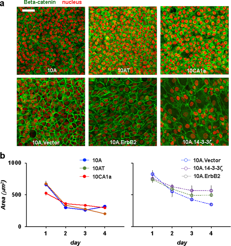
Figure 1. Confluent layers of six breast carcinoma cell lines with graded degree of invasive potential.
a. MCF10A cell lines at day 3 stained with DAPI (red) and beta-catenin (green). All cell layers became fully confluent by day 3 of cell culture. Scale bar = 50μm. b. Left panel: time dependence of cell area from control epithelial 10A cells (filled blue), tumorigenic but less invasive 10AT cells (mocha) and invasive 10CA1a cells (red) (Each datum pools all cells from 6~7 fields of view for a given day of cell culture). Right panel: time dependence of cell area from control epithelial 10A.vector cells (open blue), non-invasive 10A.14–3-3ζ cells (purple) and tumorigenic but less invasive 10A.ErbB2 cells (clover green). Cell area decreases with increasing days of cell culture but plateaued at day 3 for most of cell lines. Error bars represent the standard error of the mean.
Cell shapes
To measure individual cell shapes, nuclei and beta-catenin were imaged with a Leica DMI8 confocal fluorescence microscope using either a 40x or 63x oil objective (Leica). Cell-cell boundaries labelled by beta-catenin were traced using the semi-automatic, watershed-based SeedWater Segmenter (SWS), as previously described [55]. Briefly, SWS takes an edge-labeled image of a confluent cellular tissue and performs a watershed segmentation based on user-given seeds. Here as input seeds we use the nuclei markers from the DAPI channel. To characterize cell shape and shape variation from cell-to-cell we used the mean of aspect ratio, AR, which was obtained from the moment of inertia tensor, and the standard deviation of the aspect ratio SD(AR), as described previously [1,2]. For example, the more elongated cell shape profile, the higher its AR. In a previous study, we had also used as an index of shape the metric q, which is the cell perimeter divided by the square root of area [2]. Although AR and q were roughly correlated in the data set described here, the observed range of AR spanned roughly four-fold whereas that of q spanned less than two-fold (Supplementary Figure S2). To better resolve small changes in cell shape, here we used AR.
Cell shape distributions
We fitted cell aspect ratios measured across all individual cells to the k-gamma distribution using maximum likelihood estimation following the method previously described [1]. Briefly, the aspect ratios were shifted and rescaled by defining x = (AR −1) / (< AR > −1) (Figure 3) where <…> represents taking the mean. With this parametrization, the likelihood function is the product , where ρ(x k,) is the k-Gamma density function kk xk−1e−kx / Γ(k). The value of k that maximizes L(k) is the maximum likelihood estimator of k.
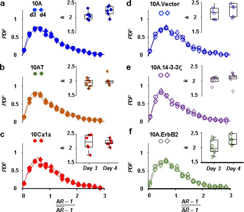
Figure 3. Across different carcinoma model lines, shape variability collapses to a family of PDFs that is common to all.
a-f. PDFs of the rescaled parameter x = (AR − 1) / (< AR > −1) of breast carcinoma model cell lines at days 3 and 4, where < ⋯ > denotes the mean, followed a k-gamma distribution (average k = 2.13 ± 0.26, p = 0.371 for difference in k between cell lines). Insets: k estimated for each cell line and culture day (Each datum represents a fitted k value determined by maximum likelihood estimation, for each individual field of view).
Migratory dynamics
Time-lapse movies were captured in an environment control chamber (37°C, 5% CO2) installed on an inverted optical microscope (Leica, DMI 6000B). Phase-contrast images were acquired at 3 min intervals for 25 hrs. To minimize the effect of cell proliferation, we tracked cell migratory dynamics starting on day 3 post-seeding, when all of cell lines reached confluence and projected area per cell had attained a plateau value for most of the cell lines (Figure 1). Six movies were analyzed for each cell type. For analysis, each movie was divided into 5 of 300min time-windows. Time-lapse movies of phase contrast images were registered to sub-pixel resolution using a discrete Fourier transform method [56]. Flow fields were calculated from the registered movies using Matlab’s Optical Flow Farneback function. The average speed was calculated by averaging the optical flow output over the whole frame and the entire time window.
Cooperative packs were determined from optical flow fields, using a community-finding algorithm as described previously [9]. In brief, velocities determined using optical flow were locally averaged over space (10×10 μm) and time (15 min) to assign an average displacement vector to each position. This allowed the determination of an orientation with respect to the laboratory coordinate system. We applied a uniform speed threshold equal to the mean speed on each image. To assign neighbors for the i-th cell, all immediate neighbors with an orientation within δθ = 20° were grouped. This orientation grouping was performed recursively, allowing the number of neighbors mi to grow and the orientation to shift gradually across a group. We continued to look for neighbors for all the new members of the set until we were unable to find a neighbor with similar orientation for any new member. We determined the mean pack-size per cell by counting, for each cell, the number of cells in its pack, and averaging. The absolute pack size depended on thresholds chosen for both δθ and speed, but using different thresholds did not change overall rank ordering within and across cell types over time (Supplementary Figure S3). To determine the persistence length, trajectories were seeded from the movie’s first frame using a square grid with spacing comparable to the cell size and obtained from forwards-integration of the flow fields; for our field of view (1020 μm x 1020 μm) there were about 10000 trajectories. Persistence length was determined by fitting where s is the distance along the path between two points, R is the displacement between the same two points, <…> represents an average and P is the persistence length.
Western blot analysis
On day 3 after seeding on a polyacrylamide gel, we prepared cell lysates by scraping the gel surface and detected the level of proteins by western blot analysis. To determine the degree of the EMT, we compared the level of epithelial and mesenchymal marker proteins, including E-cadherin, vimentin, and N-cadherin. GAPDH was used as a loading control. Following primary and secondary antibodies were purchased from Cell Signaling Technology: E- cadherin (3195s), Vimentin (5741s), N-Cadherin (13116s), GAPDH (5174s) and HRP-conjugated anti-rabbit IgG (7074s). We used Image J to quantify the level of target proteins and to compare their levels between cell lines.
Statistical methods
All of the data was analyzed in Matlab. To determine statistical significance, we ran a t-test for each data set and considered significant when p < 0.05. Average difference (a.d.) in variable y between cell type i and j was determined by computing where < yi > represents the average of variable y for cell type i.
Results
By day 3 after seeding, the layer of each cell line had become fully confluent and cell sizes had stabilized (Figure 1). Therefore, we focused on days 3 and 4 of culture.
Cell Shape and Shape Variation
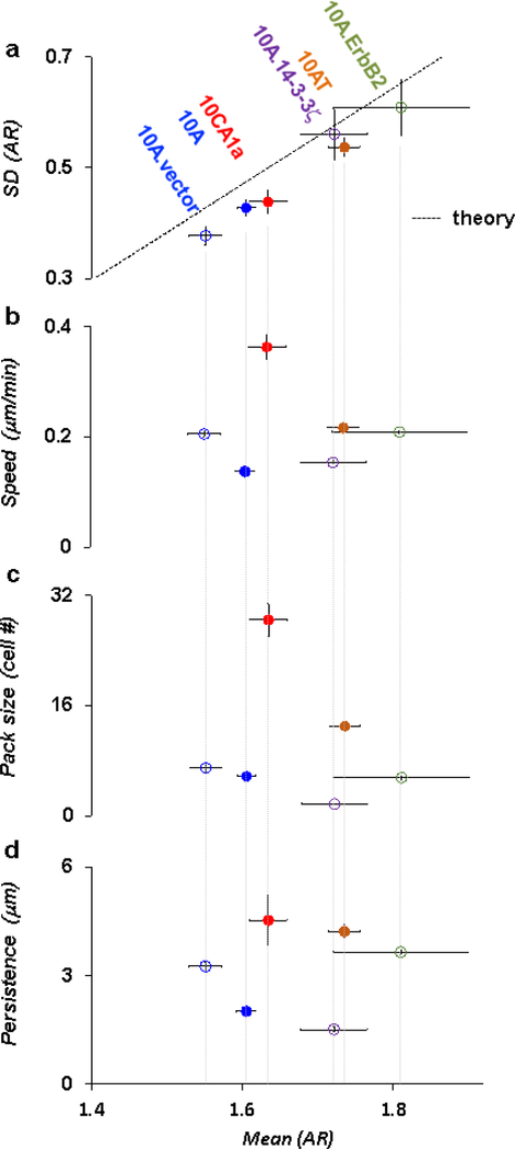
Data from all cell lines, taken together, defined a positive relationship between the mean of AR and the standard deviation (SD) of AR (Figure 2a; Supplementary Figure S4). This relationship mirrors that reported by Atia et al. for a variety of collective epithelial systems, wherein cell shapes became progressively more elongated and more variable as the collective system became progressively more unjammed [1]. Along this relationship the cell lines studied here ordered roughly with transforming potential, the notable exception being 10CA1a. Compared to control epithelial 10A.vector cells, 10A.ErbB2 and 10A.14–3-3ζ cells tended to be more elongated (p = 0.015 and a.d. = 16% for AR between 10A.vector vs. 10A.ErbB2, p = 0.005 and a.d. = 11% between 10A.vector vs. 10A.14–3-3ζ). Compared to control epithelial 10A cells, 10AT cells tended to be elongated (p < 0.0005 and a.d. = 8% for AR, 10A vs. 10AT), but 10CA1a cells were not significantly elongated (p = 0.32 and a.d. = 2% for AR,10A vs. 10CA1a). Based on structural signatures as reflected in these ARs, the most jammed system was 10A.vector, the most unjammed was 10A.ErbB2, while others were intermediate.
Figure 2. Structural signatures of cell layer unjamming and migratory dynamics.
a. AR and SD (AR) of breast carcinoma model cell lines at culture day 3 (colored). Each datum pools all cells from 6~7 field of views for a given day of cell culture. Solid line depicts the theoretical prediction of cell unjamming [1]. Carcinoma cell lines aligned roughly with transforming potential along the unjamming line, the notable exception being 10CA1a. Compared to control 10A.vector cells (open blue), 10A.ErbB2 (clover green) and 10A.14–3-3ζ (purple) cells tended to be more elongated (p = 0.015 and a.d. = 16% for AR between 10A.vector vs. 10A.ErbB2, p = 0.005 and a.d. = 11% between 10A.vector vs. 10A.14–3-3ζ). Compared to control 10A cells (filled blue), 10AT cells (mocha) tended to be elongated (p < 0.0005 and a.d. = 8% for AR, 10A vs. 10AT), but 10CA1a cells (red) were not significantly elongated (p = 0.32 and a.d. = 2% for AR,10A vs. 10CA1a). Each datum represents 800~1500 cells measured in 6 fields of view for each cell type. b. For most of cell lines except for 10AT, migratory dynamics did not conform with expectations based on structural signatures. For tumorigenic but less invasive 10AT cells, migratory dynamics changed in concert with expectations based on structural signatures; as AR and SD (AR) progressively increased in tandem, cellular speeds progressively increased as well (p < 0.0005 and a.d. = 45% for speeds between 10A and 10AT). For tumorigenic but less invasive 10A.ErbB2 cells, however, structures at day 3 signified a more unjammed state than the control vector whereas migratory dynamics did not change significantly (p = 0.28 and a.d. = 2% for speeds between 10A.vector and 10A.ErbB2). For 10A.14–3-3ζ cells, which tend to be non-invasive and lack E-cadherin [12], structures signified a more unjammed state than the control vector whereas cellular speeds were lower (p < 0.0005 and a.d. = 29% for speed between 10A.vector and 10A.14–3-3ζ). For the highly invasive 10CA1a cells, structures signified a moderately jammed state whereas cellular speeds were excessively high and therefore discordant again with structures (p < 0.0005 and a.d. = 90% for speed between 10A and 10CA1a). c. Compared to control epithelial cells, 10AT cells that exhibited faster migratory dynamics showed larger cooperative pack sizes (p < 0.0005 and a.d. = 77% for pack sizes between 10A.vector and 10AT). 10A.14–3-3ζ cells that exhibited slower migratory dynamics also showed smaller cooperative pack sizes (p < 0.0005 and a.d. =120% for pack sizes between 10A.vector and 10A.14–3-3ζ). 10CA1a cells, which exhibited inordinately fast migratory dynamics, showed anomalously large pack sizes (p < 0.0005 and a.d. = 132% between10CA1a and 10A). Each datum represents averages of 6 movies over 300min time-window for each cell type. d. Persistence in migratory dynamics exhibited poor correspondence with structures. Error bars in a,b,c,d represent the standard error of the mean.
Cell shapes in all cases collapsed to a universal distribution function given by a k-gamma distribution [1,29], where k did not depend on cell type (average k = 2.13 ± 0.26, p = 0.371 for difference in k between cell lines; Figure 3). These k values of breast carcinoma models conformed well to values previously reported for other epithelial tissues [1]. Moreover, the cell aspect ratio did not co-vary with projected cell area (Supplementary Figure S5).
Taken together, these results demonstrate that in all carcinoma model cell lines tested, cell shape and shape variability conformed well to predicted structural signatures that have been linked to cell layer unjamming [1]; as cell aspect ratio increased shape variations increased in concert, and did so in close coordination with theoretical expectations of cellular unjamming (dotted line in Figure 2a).
Migratory Dynamics
Across all cell lines at day 3 we assessed the relationship between structural signatures of cellular unjamming and cell migratory dynamics [7,37]. When comparing 10AT cells to their control 10A cells, migratory dynamics changed roughly in concert with expectations based on structural signatures in the sense that as AR and SD (AR) progressively increased in tandem, cellular speeds progressively increased as well (p < 0.0005 and a.d. = 45% for speeds between 10A and 10AT; Figure 2b). For tumorigenic but less invasive 10A.ErbB2 cells, however, structures at day 3 signified a more elongated state than the control 10A.vector whereas migratory dynamics did not change significantly (p = 0.28 and a.d. = 2% for speeds between 10A.vector and 10A.ErbB2). For 10A.14–3-3ζ cells, which tend to be non-invasive and lack E-cadherin [12], structures signified a more elongated state than the control 10A.vector whereas cellular speeds were lower (p < 0.0005 and a.d. = 29% for speed between 10A.vector and 10A.14–3-3ζ; Figure 2b). For the highly invasive 10CA1a cells, structures signified only an intermediate degree of elongation compared with the other cell lines whereas cellular speeds were the highest of all cell lines tested and therefore discordant again with a simple prediction of cellular speed based on structure (p < 0.0005 and a.d. = 90% for speed between 10A and 10CA1a). Overall, these data indicated discordance between elongated structures and enhanced migratory dynamics (by linear regression on all cell lines, Speed = −0.24AR + 0.63, R2 = 0.09, p = 0.62).
Cooperativity and Persistence
To examine more closely collective migratory dynamics of these carcinoma cell lines at day 3, we quantified the size of cooperative packs and persistence of single-cell trajectories. Compared to control epithelial 10A cells, 10AT cells, which were more elongated, showed larger cooperative pack sizes (p < 0.0005 and a.d. = 77% for pack sizes between 10A and 10AT; Figure 2c). Compared to control epithelial 10A.vector cells, 10A.14–3-3ζ cells, which were more elongated but exhibited slower migratory dynamics, showed smaller cooperative pack sizes (p < 0.0005 and a.d. =120% for pack sizes between 10A.vector and 10A.14–3-3ζ). 10CA1a cells, which were not elongated but exhibited inordinately fast migratory dynamics, also showed anomalously large pack sizes (p < 0.0005 and a.d. = 132% between10CA1a and 10A). These data indicated discordance between elongated structures and enhanced pack sizes (by linear regression on all cell lines, Pack size = −19.33AR + 42.64, R2 = 0.04, p = 0.71). Similarly, structures and persistence in migratory dynamics also exhibited poor correspondence (Figure 2d; by linear regression, Persistence = 1.41AR +82, R2 = 0.01, p = 0.83).
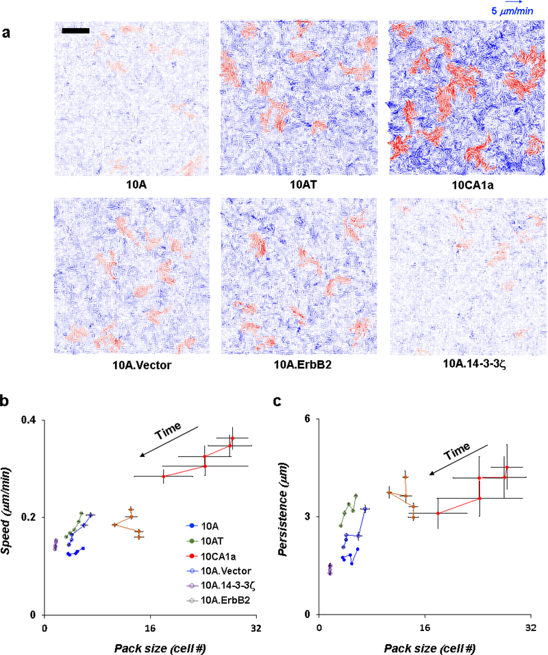
As such, across cell lines we found no clear relationship between migratory speed and AR, between pack size and AR, or between persistence and AR. However, when we compared migratory speed and persistence directly to pack size several systematic trends emerged (Figure 4b,c). Within each cell type, as the layer matured with time the migration speeds systematically slowed, pack sizes either stabilized or decreased and the persistence decreased, presumably because the layer in culture settled slowly but progressively into lower and lower energy states, and thus progressed toward a jammed phase. In glassy systems such time-dependent behavior is traditionally referred to as aging [27,57–60]. Interestingly, when comparing across different cell types, however, migration speeds increased systematically with pack size (by linear regression on data from all cell lines and different time points, Speed = 0.006Pack size + 0.13, R2 = 0.7, p < 0.005); faster migratory dynamics were associated with enhanced cooperativity and larger pack size. Similarly, migratory persistence also roughly increased with pack size (by linear regression on data from all cell lines and time points, Persistence = 0.084Pack size + 2.12, R2 = 0.33, p < 0.005).
Figure 4. Faster migratory dynamics are accompanied by larger cooperative packs.
a. Speed maps of carcinoma model cell lines (at day 3). Cellular motions were seen to organize into oriented migratory packs. Velocity vectors (blue) are shown for 15min of period, with members of the 10 largest cooperative packs highlighted in red for each movie. Scale bar is 200μm. b. Speed and cooperative pack sizes in migratory dynamics measured for 25hrs at day 3 of cell culture. Six movies were analyzed for each cell type. For analysis, each 25hrs movie was divided into five 300min time-windows. (Each datum depicts averages of 6 movies over 300min time-windows centered at 150min, 450min, 750min, 1050min and 1350min.) In each case, as time increased, cells slowed down and the pack size decreased or stabilized. Across all cases, cells tended to migrate faster as they exhibited larger packs (by linear regression on data from all cell lines and time points, Speed = 0.006Pack size + 0.13, R2 = 0.7, p < 0.005). c. Persistence and cooperative pack sizes in migratory dynamics. In each case, as time increased the persistence decreased. Across all cases, migratory persistent and pack sizes roughly exhibited a positive relationship (by linear regression on data from all cell lines and time points, Persistence = 0.084Pack size + 2.12, R2 = 0.33, p < 0.005). Error bars represent the standard error of the mean.
Expression of EMT Markers
To test whether the results of migratory dynamics might be attributable in part to EMT or pEMT, we measured expression of EMT marker proteins including vimentin and N-cadherin [61]. It is worth noting that the expression profiles of EMT marker proteins in the 10A panel–that is 10A, 10AT, and CA1a–are inconsistent in the literature, perhaps reflecting the heterogeneous nature of the cell lines [49,62–64]. Western blotting analysis confirmed that with the exception of the 10A.14–3-3ζ cell line, as expected, E-cadherin was detectable in all cell lines (Supplementary Figure S6a). Vimentin was detected highest in 10A.14–3-3ζ cells, which migrated the slowest, and undetectable in control epithelial 10A and 10A.Vector cells (Supplementary Figure S6b). N-cadherin was again detected highest in 10A.14–3-3ζ cells, and was weakly detected in other carcinoma cells (Supplementary Figure S6c). Moreover, the level of detectable N-cadherin protein in 10CA1a cells, which migrated the fastest, was not different from that in control 10A cells (p = 0.37 and a.d. = 20%). Taken together, the level of EMT marker proteins did not explain migratory dynamics.
Discussion
Our central finding is that each of the breast carcinoma cell lines tested demonstrated geometric and dynamic signatures conforming with the cell jamming/unjamming framework [1,2]. Much as described in other epithelial systems in proximity to a jamming transition [1], cell shape variation, as expressed by SD(AR), increased in proportion with mean cell shape (AR), and underlying distributions of AR were well-described by the k-gamma distribution (Figures 2 and 3). Within each cell type, as the cell layer matured migratory speeds progressively slowed, pack sizes decreased or stabilized, persistence decreased, and AR progressively decreased (Figure 4 and Supplementary Figure S4). But across cell types, paradoxically, we found no systematic relationship between migratory speeds and cell shapes, and no relationship between pack sizes and cell shapes. As described below, this paradox is reconciled by strong systematic positive relationships between migratory speed, persistence, and pack size (Figure 4). These structural and dynamical observations support the hypothesis of a unifying role of cell unjamming in these systems.
Expectations based on jamming
The transition between jammed solid-like versus unjammed fluid-like phases defines a phase diagram that is thought to be controlled by three factors: cellular propulsion, directional persistence of that propulsion, and a shape index reflecting the competition between cell-cell adhesion and cortical tension. [2,10,11,16] Regardless of which factor or combination of factors drives a transition between the solid-like jammed phase and the fluid-like unjammed phase, theory holds that the transition is invariably marked by the same characteristic changes of cell shape and shape variation [11]. To explain these behaviors, theories of cell jamming as developed thus far, paint a physical picture in which rearrangements among neighboring cells are seen to be impeded by local energy barriers [2,10,11,16,17,25]. These energy barriers, in turn, are defined by a combination of cell-cell adhesion and cortical tension but can be overcome through the agency of cellular propulsion. As the directional persistence of that propulsion increases, the ability to overcome energy barriers is further enhanced as the cells form collective packs, allowing for unjamming to occur at even lower levels of cellular propulsion [11].
When propulsive forces are negligible, it follows from theory described above that increases in cell-cell adhesion or decreases in cortical tension can cause energy barriers to diminish or even vanish altogether [2,10,11,16,17,25]. When that occurs, the jammed cell layer will necessarily unjam. But because propulsive forces remain negligible, resulting migratory speeds may remain vanishingly small. This case corresponds to unjamming wherein energy barriers do not impede migration but migration remains small nevertheless. But when cell-cell adhesion remains small, or cortical tension remains large, or both, energy barriers to rearrangement become appreciable and thereby tend to impede cellular rearrangements. Nevertheless, when propulsive forces become sufficiently large these forces can then overcome those energy barriers, fluidize the cellular collective, and therefore drive collective cellular migration; this form of fluidization is thought to be accompanied by changing geometric signatures of the cells[11]. Alternatively, highly directional persistence in propulsive forces can also promote cellular unjamming through the formation of cooperative packs; this form of fluidization does not necessarily change geometric signatures of unjamming. Through either increasing propulsive forces, increased directional persistence, or both, it is thus possible for unjamming to occur wherein finite energy barriers persist but are overcome. Because unjamming can occur through changes in energy barriers, propulsive drive, or cooperativity, it is not surprising that migration speed and AR need not be directly related when comparing across cell types. Instead, it is necessary to compare both geometric and dynamic signatures of jamming to assess the state of the cell layer.
In both inert granular systems and living confluent epithelia, jamming of the collective has been linked both empirically and theoretically to the manner in which constituent elements pack space [1,29,65–70]. In particular, as a collective system approaches the jammed phase, all possible micro-canonical configurations become equally likely, distributional entropy becomes maximized, and k-gamma distributions arise [1,29,66]. In that connection, the thermodynamic temperature is defined as 1/(∂E/∂S), where E is thermal energy and S is entropy. The jammed systems in question here are not thermodynamic, however, and instead are athermal. In such cases it has been found to be useful to define an ‘effective temperature’ in which energy is replaced by regional volumes in the case of granular matter, or cell shapes (elongations) in the case of homogeneous epithelial layers. In that sense it would be reasonable to say that more jammed systems are effectively ‘cooler’ and more unjammed systems are effectively ‘hotter’.
Discordance between cell shapes and speeds
With maturation of each of the cell lines examined, we identified structural and dynamic signatures consonant with a progressive approach to the jammed phase. In inert glassy systems similar changes with time are often observed and are referred to as aging [57–60]. Specifically, with the passage of time we observed in each cell type that migratory speeds tended to slow, cellular aspect ratios tended to diminish, and migratory persistence tended to decrease (Figure 4 and Supplementary Figure S4). Together, these changes are indicative of approaching to a jamming transition [2,20,23].-Across different cell types, however, striking dynamic differences became evident and, compared with structural changes, seemed paradoxical.
Literature on migration speeds across the MCF10A panel is somewhat fragmented and inconsistent, with rank ordering of migratory speed across the panel varying with the particular assays used and the degree of layer confluence [49,50,71]. Nevertheless, the consensus view is that 10CA1a comprises the most invasive phenotype [14,15]. Indeed, in our hands 10CA1a exhibited by far the highest migratory speeds (Figure 2b). Accordingly, we had expected 10CA1a to be the most unjammed and, therefore, to express the highest ARs, but this proved not to be the case. Instead we found that although 10CA1a was by far the fastest among all cell types tested, its structure (i.e., AR) suggested only an intermediate degree of unjamming compared with other cell lines of the panel (Figure 2a).
Shape, speed, and cooperativity
While structure of the 10CA1a cells indicate only a modestly jammed phase, they formed the largest packs, thus signifying across the panel the greatest degree of cooperativity (Figure 2c). Similarly, the relatively non-invasive 10A.14–3-3ζ cells are surprisingly slow given their AR but form smaller packs that, in theory, are less capable of overcoming energy barriers to rearrangements. Across cell types overall, migratory speed, persistence, and pack size were strongly related (Figure 4) much as reported elsewhere [9]. Importantly, these changes were not accounted for by EMT. We found no systematic variation of cell behavior with EMT character (Supplementary Figure S6). Contrary to long held assumptions, E-cadherin is now known to play a key role in metastasis [43], but we found no relationship between E-cadherin expression and pack size.
Acquisition of collective migratory capacity is crucial in metastatic dispersal of carcinoma cells away from the primary tumor mass [4]. The unjamming transition from a jammed solid-like non-migratory to an unjammed fluid-like migratory phenotype has been recently proposed as being at play in cancer progression [3,6,7,26,36,37] but across carcinoma cell lines of graded degrees of invasive potential the relevance of the cell jamming/unjamming framework had remained untested. Within each of six MCF10A carcinoma model systems spanning diverse levels of invasive potential, here we show that cell shape and shape variability in each case conform with expectations based upon the cell unjamming mechanism [1]. Across these model systems, moreover, higher speeds were associated with enhanced size of cooperative cell packs in a manner reminiscent of a peloton. As such, the jamming mechanism points to a key role for cooperative migratory character.
Supplementary Material
Highlights:
Within each of 6 different breast carcinoma models, cell shape and shape variability conformed with expectations based upon the cell unjamming mechanism.
Across these different cell lines, cell shape information alone was not sufficient to predict migratory dynamics.
Across cell lines, higher speeds were associated with enhanced size of cooperative cell packs in a manner reminiscent of a peloton.
Acknowledgements
The authors thank Jeffrey A. Nickerson and Dihua Yu for providing breast carcinoma cell lines. The authors thank Peter Friedl for helpful discussions. The authors acknowledge Youngjoo Lee and Maxim Desmond for helping with sample preparation and imaging.
Funding: This work was supported by the National Institutes of Health grants U01 CA244086, P01HL120839, and T32 HL 007118.
Footnotes
The authors declare no conflict of interest.
Publisher's Disclaimer: This is a PDF file of an unedited manuscript that has been accepted for publication. As a service to our customers we are providing this early version of the manuscript. The manuscript will undergo copyediting, typesetting, and review of the resulting proof before it is published in its final form. Please note that during the production process errors may be discovered which could affect the content, and all legal disclaimers that apply to the journal pertain.
References
- [1].Atia L, Bi D, Sharma A, Mitchel JA, Gweon B, Koehler SA, DeCamp SJ, Lan B, Kim JH, Hirsch R, Pegoraro AF, Lee KH, Starr JR, Weitz DA, Martin AC, Park JA, Butler JP, Fredberg JJ, Geometric constraints during epithelial jamming, Nature Physics (2018). [DOI] [PMC free article] [PubMed] [Google Scholar]
- [2].Park JA, Kim JH, Bi D, Mitchel JA, Qazvini NT, Tantisira K, Park CY, McGill M, Kim SH, Gweon B, Notbohm J, Steward R Jr., Burger S, Randell SH, Kho AT, Tambe DT, Hardin C, Shore SA, Israel E, Weitz DA, Tschumperlin DJ, Henske EP, Weiss ST, Manning ML, Butler JP, Drazen JM, Fredberg JJ, Unjamming and cell shape in the asthmatic airway epithelium, Nat Mater 14 (2015) 1040–1048. 10.1038/nmat4357. [DOI] [PMC free article] [PubMed] [Google Scholar]
- [3].Palamidessi A, Malinverno C, Frittoli E, Corallino S, Barbieri E, Sigismund S, Beznoussenko GV, Martini E, Garre M, Ferrara I, Tripodo C, Ascione F, Cavalcanti-Adam EA, Li Q, Di Fiore PP, Parazzoli D, Giavazzi F, Cerbino R, Scita G, Unjamming overcomes kinetic and proliferation arrest in terminally differentiated cells and promotes collective motility of carcinoma, Nature Materials (2019). 10.1038/s41563-019-0425-1. [DOI] [PubMed] [Google Scholar]
- [4].Friedl P, Gilmour D, Collective cell migration in morphogenesis, regeneration and cancer, Nat Rev Mol Cell Biol 10 (2009) 445–457. 10.1038/nrm2720. [DOI] [PubMed] [Google Scholar]
- [5].Rodriguez-Franco P, Brugues A, Marin-Llaurado A, Conte V, Solanas G, Batlle E, Fredberg JJ, Roca-Cusachs P, Sunyer R, Trepat X, Long-lived force patterns and deformation waves at repulsive epithelial boundaries, Nature Materials 16 (2017) 1029–1037. 10.1038/nmat497210.1038/nmat4972http://www.nature.com/nmat/journal/vaop/ncurrent/abs/nmat4972.html#supplementary-informationhttp://www.nature.com/nmat/journal/vaop/ncurrent/abs/nmat4972.html#supplementary-information . [DOI] [PMC free article] [PubMed] [Google Scholar]
- [6].Pawlizak S, Fritsch A, Grosser A, Ahrens D, Thalheim T, Riedel S, Kießling T, Oswald L, Zink M, Manning ML, Käs J, Testing the differential adhesion hypothesis across the epithelial−mesenchymal transition, New Journal of Physics 17 (2015) 083049. [Google Scholar]
- [7].Haeger A, Krause M, Wolf K, Friedl P, Cell jamming: collective invasion of mesenchymal tumor cells imposed by tissue confinement, Biochim Biophys Acta 1840 (2014) 2386–2395. 10.1016/j.bbagen.2014.03.020. [DOI] [PubMed] [Google Scholar]
- [8].Shellard A, Mayor R, Supracellular migration – beyond collective cell migration, Journal of Cell Science 132 (2019) jcs226142 10.1242/jcs.226142. [DOI] [PubMed] [Google Scholar]
- [9].Mitchel JA, Das A, O’Sullivan MJ, Stancil IT, DeCamp SJ, Koehler S, Butler JP, Fredberg JJ, Nieto MA, Bi D, Park J-A, The unjamming transition is distinct from the epithelial-tomesenchymal transition, bioRxiv (2019) 665018 10.1101/665018. [DOI] [PMC free article] [PubMed] [Google Scholar]
- [10].Bi D, Lopez JH, Schwarz JM, Manning ML, A density-independent rigidity transition in biological tissues, Nat Phys 11 (2015) 1074–1079. 10.1038/nphys3471. [DOI] [Google Scholar]
- [11].Bi D, Yang X, Marchetti MC, Manning ML, Motility-Driven Glass and Jamming Transitions in Biological Tissues, Physical Review X 6 (2016) 021011. [DOI] [PMC free article] [PubMed] [Google Scholar]
- [12].Lu J, Guo H, Treekitkarnmongkol W, Li P, Zhang J, Shi B, Ling C, Zhou X, Chen T, Chiao PJ, Feng X, Seewaldt VL, Muller WJ, Sahin A, Hung MC, Yu D, 14–3-3zeta Cooperates with ErbB2 to promote ductal carcinoma in situ progression to invasive breast cancer by inducing epithelial-mesenchymal transition, Cancer Cell 16 (2009) 195–207. S1535–6108(09)00256–6 [pii] 10.1016/j.ccr.2009.08.010. [DOI] [PMC free article] [PubMed] [Google Scholar]
- [13].Dawson PJ, Wolman SR, Tait L, Heppner GH, Miller FR, MCF10AT: a model for the evolution of cancer from proliferative breast disease, The American journal of pathology 148 (1996) 313–319. [PMC free article] [PubMed] [Google Scholar]
- [14].Santner SJ, Dawson PJ, Tait L, Soule HD, Eliason J, Mohamed AN, Wolman SR, Heppner GH, Miller FR, Malignant MCF10CA1 cell lines derived from premalignant human breast epithelial MCF10AT cells, Breast cancer research and treatment 65 (2001) 101–110. [DOI] [PubMed] [Google Scholar]
- [15].Imbalzano KM, Tatarkova I, Imbalzano AN, Nickerson JA, Increasingly transformed MCF-10A cells have a progressively tumor-like phenotype in three-dimensional basement membrane culture, Cancer cell international 9 (2009) 7 10.1186/1475-2867-9-7. [DOI] [PMC free article] [PubMed] [Google Scholar]
- [16].Merkel M, Manning M, A geometrically controlled rigidity transition in a model for confluent 3D tissues., arXiv (2017). [Google Scholar]
- [17].Giavazzi F, Paoluzzi M, Macchi M, Bi D, Scita G, Manning ML, Cerbino R, Marchetti MC, Flocking transitions in confluent tissues, Soft Matter 14 (2018) 3471–3477. 10.1039/c8sm00126j. [DOI] [PMC free article] [PubMed] [Google Scholar]
- [18].Kim JH, Serra-Picamal X, Tambe DT, Zhou EH, Park CY, Sadati M, Park JA, Krishnan R, Gweon B, Millet E, Butler JP, Trepat X, Fredberg JJ, Propulsion and navigation within the advancing monolayer sheet, Nature Materials (2013). 10.1038/nmat3689. [DOI] [PMC free article] [PubMed] [Google Scholar]
- [19].Serra-Picamal X, Conte V, Vincent R, Anon E, Tambe D, Bazellieres E, Butler J, Fredberg J, Trepat X, Mechanical waves during tissue expansion, Nature Physics 8 (2012) 628–634. [Google Scholar]
- [20].Tambe DT, Corey Hardin C, Angelini TE, Rajendran K, Park CY, Serra-Picamal X, Zhou EH, Zaman MH, Butler JP, Weitz DA, Fredberg JJ, Trepat X, Collective cell guidance by cooperative intercellular forces, Nature Materials 10 (2011) 469–475. nmat3025 [pii] 10.1038/nmat3025. [DOI] [PMC free article] [PubMed] [Google Scholar]
- [21].Sadati M, Taheri Qazvini N, Krishnan R, Park CY, Fredberg JJ, Collective migration and cell jamming, Differentiation (2013). 10.1016/j.diff.2013.02.005. [DOI] [PMC free article] [PubMed] [Google Scholar]
- [22].Trepat X, Fredberg JJ, Plithotaxis and emergent dynamics in collective cellular migration, Trends Cell Biol 21 (2011) 638–646. S0962–8924(11)00127–9 [pii], 10.1016/j.tcb.2011.06.006. [DOI] [PMC free article] [PubMed] [Google Scholar]
- [23].Angelini TE, Hannezo E, Trepat X, Marquez M, Fredberg JJ, Weitz DA, Glass-like dynamics of collective cell migration, Proc Natl Acad Sci U S A 108 (2011) 4714–4719. 10.1073/pnas.1010059108. [DOI] [PMC free article] [PubMed] [Google Scholar]
- [24].Trepat X, Wasserman M, Angelini T, Millet E, Weitz D, Butler J, Fredberg J, Physical forces during collective cell migration., Nature Physics 5 (2009) 426–430. [Google Scholar]
- [25].Bi D, Lopez J, Schwarz J, Manning M, Energy barriers and cell migration in densely packed tissues†, Soft Matter (2014). [DOI] [PubMed] [Google Scholar]
- [26].Nnetu KD, Knorr M, Pawlizak S, Fuhs T, Käs JA, Slow and anomalous dynamics of an MCF-10A epithelial cell monolayer, Soft Matter 9 (2013). 10.1039/c3sm50806d. [DOI] [Google Scholar]
- [27].Garcia S, Hannezo E, Elgeti J, Joanny JF, Silberzan P, Gov NS, Physics of active jamming during collective cellular motion in a monolayer, Proc Natl Acad Sci U S A 112 (2015) 15314–15319. 10.1073/pnas.1510973112. [DOI] [PMC free article] [PubMed] [Google Scholar]
- [28].Grosser S, Lippoldt J, Oswald L, Merkel M, Sussman DM, Renner F, Morawetz E, Pawlizak S, Fritsch A, Horn L, Aktas B, Manning M, Kaes J, Elongated Cells Fluidize Malignant Tissues, Physical Review X In press (2019). [Google Scholar]
- [29].Aste T, Di Matteo T, Emergence of Gamma distributions in granular materials and packing models, Physical Review E 77 (2008) 021309. [DOI] [PubMed] [Google Scholar]
- [30].Mongera A, Rowghanian P, Gustafson HJ, Shelton E, Kealhofer DA, Carn EK, Serwane F, Lucio AA, Giammona J, Campas O, A fluid-to-solid jamming transition underlies vertebrate body axis elongation, Nature 561 (2018) 401–405. 10.1038/s41586-018-0479-2. [DOI] [PMC free article] [PubMed] [Google Scholar]
- [31].Spurlin JW, Siedlik MJ, Nerger BA, Pang MF, Jayaraman S, Zhang R, Nelson CM, Mesenchymal proteases and tissue fluidity remodel the extracellular matrix during airway epithelial branching in the embryonic avian lung, Development 146 (2019). 10.1242/dev.175257. [DOI] [PMC free article] [PubMed] [Google Scholar]
- [32].Tetley RJ, Staddon MF, Heller D, Hoppe A, Banerjee S, Mao Y, Tissue fluidity promotes epithelial wound healing, Nature Physics (2019). 10.1038/s41567-019-0618-1. [DOI] [PMC free article] [PubMed] [Google Scholar]
- [33].Krishnan R, Canovic EP, Iordan AL, Rajendran K, Manomohan G, Pirentis AP, Smith ML, Butler JP, Fredberg JJ, Stamenovic D, Fluidization, resolidification, and reorientation of the endothelial cell in response to slow tidal stretches, Am J Physiol Cell Physiol 303 (2012) C368–375. 10.1152/ajpcell.00074.2012. [DOI] [PMC free article] [PubMed] [Google Scholar]
- [34].Leggett SE, Neronha ZJ, Bhaskar D, Sim JY, Perdikari TM, Wong IY, Motility-limited aggregation of mammary epithelial cells into fractal-like clusters, Proc Natl Acad Sci U S A (2019). 10.1073/pnas.1905958116. [DOI] [PMC free article] [PubMed] [Google Scholar]
- [35].Staneva R, El Marjou F, Barbazan J, Krndija D, Richon S, Clark AG, Vignjevic DM, Cancer cells in the tumor core exhibit spatially coordinated migration patterns, Journal of Cell Science 132 (2019) jcs220277. 10.1242/jcs.220277. [DOI] [PubMed] [Google Scholar]
- [36].Oswald L, Grosser S, Smith DM, Kas JA, Jamming transitions in cancer, Journal of physics D: Applied physics 50 (2017) 483001 10.1088/1361-6463/aa8e83. [DOI] [PMC free article] [PubMed] [Google Scholar]
- [37].van Helvert S, Storm C, Friedl P, Mechanoreciprocity in cell migration, Nature Cell Biology 20 (2018) 8–20. 10.1038/s41556-017-0012-0. [DOI] [PMC free article] [PubMed] [Google Scholar]
- [38].Friedl P, Locker J, Sahai E, Segall JE, Classifying collective cancer cell invasion, Nat Cell Biol 14 (2012) 777–783. 10.1038/ncb2548. [DOI] [PubMed] [Google Scholar]
- [39].Christiansen JJ, Rajasekaran AK, Reassessing epithelial to mesenchymal transition as a prerequisite for carcinoma invasion and metastasis, Cancer Res 66 (2006) 8319–8326. 10.1158/000-85472.CAN-06-0410. [DOI] [PubMed] [Google Scholar]
- [40].Cheung KJ, Gabrielson E, Werb Z, Ewald AJ, Collective invasion in breast cancer requires a conserved basal epithelial program, Cell 155 (2013) 1639–1651. 10.1016/j.cell.2013.11.029. [DOI] [PMC free article] [PubMed] [Google Scholar]
- [41].Fischer KR, Durrans A, Lee S, Sheng J, Li F, Wong ST, Choi H, El Rayes T, Ryu S, Troeger J, Schwabe RF, Vahdat LT, Altorki NK, Mittal V, Gao D, Epithelial-to-mesenchymal transition is not required for lung metastasis but contributes to chemoresistance, Nature 527 (2015) 472–476. 10.1038/nature15748. [DOI] [PMC free article] [PubMed] [Google Scholar]
- [42].Zheng XF, Carstens JL, Kim J, Scheible M, Kaye J, Sugimoto H, Wu CC, LeBleu VS, Kalluri R, Epithelial-to-mesenchymal transition is dispensable for metastasis but induces chemoresistance in pancreatic cancer, Nature 527 (2015) 525–+. 10.1038/nature16064. [DOI] [PMC free article] [PubMed] [Google Scholar]
- [43].Padmanaban V, Krol I, Suhail Y, Szczerba BM, Aceto N, Bader JS, Ewald AJ, E-cadherin is required for metastasis in multiple models of breast cancer, Nature (2019). 10.1038/s41586-019-1526-3. [DOI] [PMC free article] [PubMed] [Google Scholar]
- [44].So JY, Lee HJ, Kramata P, Minden A, Suh N, Differential Expression of Key Signaling Proteins in MCF10 Cell Lines, a Human Breast Cancer Progression Model, Mol Cell Pharmacol 4 (2012) 31–40. [PMC free article] [PubMed] [Google Scholar]
- [45].Pathak A, Kumar S, Transforming potential and matrix stiffness co-regulate confinement sensitivity of tumor cell migration, Integr Biol (Camb) 5 (2013) 1067–1075. 10.1039/c3ib40017d. [DOI] [PMC free article] [PubMed] [Google Scholar]
- [46].Baker EL, Srivastava J, Yu D, Bonnecaze RT, Zaman MH, Cancer cell migration: integrated roles of matrix mechanics and transforming potential, PLoS One 6 (2011) e20355 10.1371/journal.pone.0020355. [DOI] [PMC free article] [PubMed] [Google Scholar]
- [47].Nieman MT, Prudoff RS, Johnson KR, Wheelock MJ, N-cadherin promotes motility in human breast cancer cells regardless of their E-cadherin expression, J Cell Biol 147 (1999) 631–643. Doi 10.1083/Jcb.147.3.631. [DOI] [PMC free article] [PubMed] [Google Scholar]
- [48].West AV, Wullkopf L, Christensen A, Leijnse N, Tarp JM, Mathiesen J, Erler JT, Oddershede LB, Dynamics of cancerous tissue correlates with invasiveness, Scientific reports 7 (2017) 43800 10.1038/srep43800. [DOI] [PMC free article] [PubMed] [Google Scholar]
- [49].Chaudhury A, Cheema S, Fachini JM, Kongchan N, Lu G, Simon LM, Wang T, Mao S, Rosen DG, Ittmann MM, Hilsenbeck SG, Shaw CA, Neilson JR, CELF1 is a central node in post-transcriptional regulatory programmes underlying EMT, Nat Commun 7 (2016) 13362 10.1038/ncomms13362. [DOI] [PMC free article] [PubMed] [Google Scholar]
- [50].Weiger MC, Vedham V, Stuelten CH, Shou K, Herrera M, Sato M, Losert W, Parent CA, Real-time motion analysis reveals cell directionality as an indicator of breast cancer progression, PLoS One 8 (2013) e58859 10.1371/journal.pone.0058859. [DOI] [PMC free article] [PubMed] [Google Scholar]
- [51].Choong LY, Lim S, Chong PK, Wong CY, Shah N, Lim YP, Proteome-wide profiling of the MCF10AT breast cancer progression model, PLoS ONE 5 (2010) e11030 10.1371/journal.pone.0011030. [DOI] [PMC free article] [PubMed] [Google Scholar]
- [52].Han Y, Pegoraro A, Li H, Li K, Yuan Y, Xu G, Gu Z, Sun J, Hao Y, Gupta S, Li Y, Tang Y, Tang X, Teng L, Fredberg J, Guo M, Cell swelling, softening and invasion in a 3D breast cancer model, Nature Physics In press (2019). [DOI] [PMC free article] [PubMed] [Google Scholar]
- [53].Debnath J, Muthuswamy SK, Brugge JS, Morphogenesis and oncogenesis of MCF-10A mammary epithelial acini grown in three-dimensional basement membrane cultures, Methods 30 (2003) 256–268. [DOI] [PubMed] [Google Scholar]
- [54].He S, Carman CV, Lee JH, Lan B, Koehler S, Atia L, Park CY, Kim JH, Mitchel JA, Park JA, Butler JP, Lu Q, Fredberg JJ, The tumor suppressor p53 can promote collective cellular migration, PLoS One 14 (2019) e0202065 10.1371/journal.pone.0202065. [DOI] [PMC free article] [PubMed] [Google Scholar]
- [55].Mashburn DN, Lynch HE, Ma X, Hutson MS, Enabling user-guided segmentation and tracking of surface-labeled cells in time-lapse image sets of living tissues, Cytometry A 81 (2012) 409–418. 10.1002/cyto.a.22034. [DOI] [PMC free article] [PubMed] [Google Scholar]
- [56].Guizar-Sicairos M, Thurman ST, Fienup JR, Efficient subpixel image registration algorithms, Opt Lett 33 (2008) 156–158. Doi 10.1364/Ol.33.000156. [DOI] [PubMed] [Google Scholar]
- [57].Jabbari-Farouji S, Mizuno D, Atakhorrami M, MacKintosh FC, Schmidt CF, Eiser E, Wegdam GH, Bonn D, Fluctuation-dissipation theorem in an aging colloidal glass, Phys Rev Lett 98 (2007) 108302. [DOI] [PubMed] [Google Scholar]
- [58].Viasnoff V, Lequeux F, Rejuvenation and overaging in a colloidal glass under shear, Phys Rev Lett 89 (2002) 065701. [DOI] [PubMed] [Google Scholar]
- [59].Ramos L, Cipelletti L, Ultraslow dynamics and stress relaxation in the aging of a soft glassy system, Phys. Rev. Lett 87 (2001) 245503. [DOI] [PubMed] [Google Scholar]
- [60].Abou B, Bonn D, Meunier J, Aging dynamics in a colloidal glass, Phys Rev E 64 (2001) 021510. [DOI] [PubMed] [Google Scholar]
- [61].Zeisberg M, Neilson EG, Biomarkers for epithelial-mesenchymal transitions, J Clin Invest 119 (2009) 1429–1437. 10.1172/JCI36183. [DOI] [PMC free article] [PubMed] [Google Scholar]
- [62].Shinde A, Libring S, Alpsoy A, Abdullah A, Schaber JA, Solorio L, Wendt MK, Autocrine Fibronectin Inhibits Breast Cancer Metastasis, Mol Cancer Res 16 (2018) 1579–1589. 10.1158/1541-7786.MCR-18-0151. [DOI] [PMC free article] [PubMed] [Google Scholar]
- [63].Fritz AJ, Ghule PN, Boyd JR, Tye CE, Page NA, Hong D, Shirley DJ, Weinheimer AS, Barutcu AR, Gerrard DL, Frietze S, van Wijnen AJ, Zaidi SK, Imbalzano AN, Lian JB, Stein JL, Stein GS, Intranuclear and higher-order chromatin organization of the major histone gene cluster in breast cancer, J Cell Physiol 233 (2018) 1278–1290. 10.1002/jcp.25996. [DOI] [PMC free article] [PubMed] [Google Scholar]
- [64].Meyer MJ, Fleming JM, Ali MA, Pesesky MW, Ginsburg E, Vonderhaar BK, Dynamic regulation of CD24 and the invasive, CD44posCD24neg phenotype in breast cancer cell lines, Breast Cancer Res 11 (2009) R82 10.1186/bcr2449. [DOI] [PMC free article] [PubMed] [Google Scholar]
- [65].Aste T, Matteo TD, Saadatfar M, Senden TJ, Matthias S, Harry LS, An invariant distribution in static granular media, EPL (Europhysics Letters) 79 (2007) 24003. [Google Scholar]
- [66].Martiniani S, Schrenk KJ, Ramola K, Chakraborty B, Frenkel D, Numerical test of the Edwards conjecture shows that all packings are equally probable at jamming, Nat Phys 13 (2017) 848–851. 10.1038/nphys416810.1038/nphys4168http://www.nature.com/nphys/journal/vaop/ncurrent/abs/nphys4168.html#supplementary-informationhttp://www.nature.com/nphys/journal/vaop/ncurrent/abs/nphys4168.html#supplementary-information . [Google Scholar]
- [67].Edwards SF, Oakeshott RBS, Theory of powders, Physica A: Statistical Mechanics and its Applications 157 (1989) 1080–1090. 10.1016/0378-4371(89)90034-4. [DOI] [Google Scholar]
- [68].Atkinson S, Stillinger FH, Torquato S, Existence of isostatic, maximally random jammed monodisperse hard-disk packings, Proc Natl Acad Sci U S A 111 (2014) 18436–18441. 10.1073/pnas.1408371112. [DOI] [PMC free article] [PubMed] [Google Scholar]
- [69].Chen D, Aw WY, Devenport D, Torquato S, Structural Characterization and Statistical-Mechanical Model of Epidermal Patterns, Biophys J 111 (2016) 2534–2545. 10.1016/j.bpj.2016.10.036. [DOI] [PMC free article] [PubMed] [Google Scholar]
- [70].Donev A, Connelly R, Stillinger FH, Torquato S, Underconstrained jammed packings of nonspherical hard particles: Ellipses and ellipsoids, Physical Review E 75 (2007) 051304. [DOI] [PubMed] [Google Scholar]
- [71].Lin HH, Qraitem M, Lian Y, Taylor SR, Farkas ME, Analyses of BMAL1 and PER2 Oscillations in a Model of Breast Cancer Progression Reveal Changes With Malignancy, Integr Cancer Ther 18 (2019) 1534735419836494 10.1177/1534735419836494. [DOI] [PMC free article] [PubMed] [Google Scholar]
Associated Data
This section collects any data citations, data availability statements, or supplementary materials included in this article.


