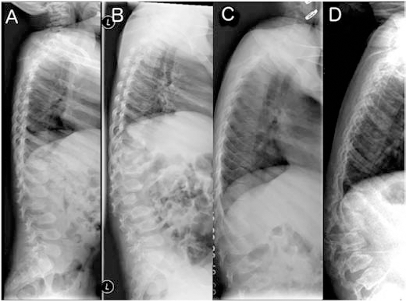
Figure 2:

Lumbar spine radiographs in late infantile patients. A, B. Patient GSL0013 at 4 years and 9 years. The vertebral bodies have a pear-shaped ovoid appearance. Ossification of the anterior segments of the vertebral bodies is defective in the thoraco-lumbar junction leading to their hook-shape or an aspect of dorsal displacement. Note progression in 5 years consistent with clinical disease course C, D. Patient GSL007 at 6 and 10 years. Similar changes and progression are seen over time.
