Abstract
Leiomyoma is a benign tumor of smooth muscle origin. Primary leiomyoma of the adrenal gland is very rare. Adrenal tumors are often diagnosed during the ultrasound or computerized tomography (CT) study as incidentaloma. According to the literature review, up to 2018, the largest size of adrenal leiomyoma which had ever been reported was 12 × 10 × 8 cm in dimension (Maher et al.). Herein, we report the largest adrenal leiomyoma with the tumor mass of 600 g (14,1x11,4x10,1 cm) from a female patient admitted to our hospital.
Keywords: Adrenal leiomyoma, Adrenal tumor, Leiomyoma, Incidentaloma
Introduction
Leiomyoma is a benign tumor of smooth muscle origin. Adrenal tumors are often diagnosed during the ultrasound or computerized tomography (CT) study as incidentaloma. Leiomyoma is often difficult to distinguish from malignant tumor either through clinical examination or imaging studies due to the absence of any significant symptoms.1 Primary leiomyoma of the adrenal is very rare. Several case reports have stated that there were only 20 patients described in English literature with the majority of these exceptional cases found incidentally.2 We present a case of the largest adrenal leiomyoma of the right adrenal which has ever been reported.
Case presentation
A 34-year-old female presented with persistent pain on the right of the abdomen since 5 months ago. The pain had worsened and radiated to her back in the past month and reduced during the rest period. The patient complained of weight loss and fatigue in the last 2–3 months. She had a history of hepatitis B infection. From the physical examinations and urological examinations were normal.
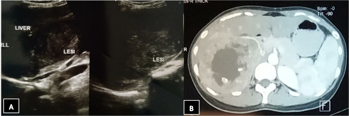
The abdominal ultrasonography (USG) had shown heterogeneous density due to suspected necrotic component with clear limits in the right hepatic lobe, pancreatic head, and right kidney upper pole. Conclusion of USG examination was suspected mass or abscess in the upper right kidney with mild splenomegaly.
An abdominal CT scan with contrast revealed that there was right adrenal mass with hypervascularization and central necrosis which extending through inferior vena cava (IVC). Pheochromocytoma with the differential diagnosis of adrenal cortical carcinoma or adrenal metastasis was suspected (Fig. 1).
Fig. 1.
A. USG results of the right and left kidney, B. Computed tomography (CT) scan reveal mass arising from the right kidney with the diameter of 14 cm.
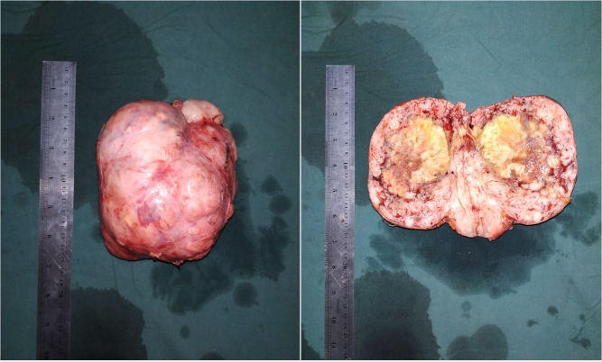
The patient was performed adrenalectomy. From intraoperative finding, right adrenal tissue was removed which weighed 600 g and tumor compressed IVC. Based on the gross examination, the adrenal mass was whitish, lamellar, lobulated with partial necrosis and blood clot (Fig. 2). Normal adrenal parenchyma was not found.
Fig. 2.
Macroscopic appearance of the tumor revealed whitish, lamellar, lobulated mass with partial necrosis and blood clot.
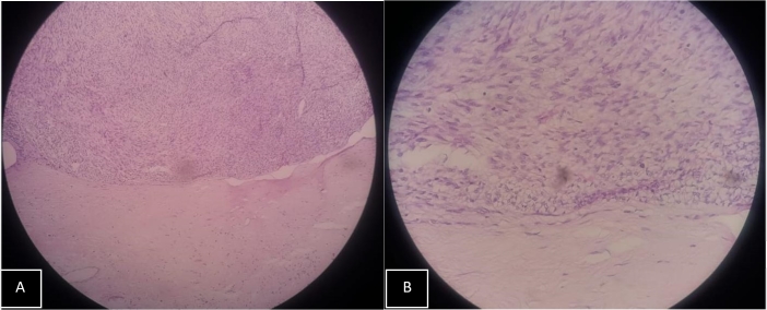
Histopathological examination using light microscope with 100x and 400x magnification with hematoxylin staining was performed. The tumor had a sharply defined border; and a dense proliferation of spindle cells with oval, fusiform, and normochromic nuclei was found. The mass was diagnosed as leiomyoma of the right adrenal (Fig. 3).
Fig. 3.
A. Microscopic appearance of the tumor with 100x magnification; B. Microscopic appearance of the tumor with 400x magnification.
Discussion
Leiomyoma is a benign tumor with smooth muscle differentiation. Previous reports had indicated that the leiomyoma adrenal have wide range of age, with median age was 38 years, and female showed higher predilection (66,6%).1 The increasing trend of adrenal masses increased because the accuracy of imaging studies, such as USG, CT scans, and MRI.3 In those individuals, the only visible signs and symptoms were flank pain, with or without radiation of pain; palpable flank mass; and hematuria. Adrenal tumors may cause increased secretion of adrenal hormones, thus causing endocrinological symptoms (associated with normetanephrine secretion). Non-specific symptoms may be present including pain, fatigue, and/or weight loss.2 Our case was concurrent with the presence of signs and symptoms of previous cases in terms of severity. The persistent abdominal pain shown in the patient might denote its relative severity. No urine-related complaints. A non-specific sign found as weight loss in the last 2–3 months.
In particular, diagnostic efforts should be aimed at assessing the effects of hormone production and differentiating between the benign and malignant lesions. Imaging findings of leiomyoma as a benign tumor are: well-defined margin and no signs of invasion into the surrounding parenchyma.4 Non-contrast CT scan may reveal hyperdense mass compared to the healthy parenchyma. In CT scan with contrast, leiomyoma may show a lower enhancement compared to the surrounding parenchyma with a relatively homogeneous enhancement. In large tumors, heterogeneous enhancement may be found. One of the most notable drawback in using imaging studies is its inability to differentiate from functioning or nonfunctioning masses. Cases of nonfunctioning adrenal mass may be differentiated by lack of symptoms pertaining to endocrinological abnormalities.5 The patient did not complain any significant symptoms other than the pain on the abdomen. No other signs and symptoms of excessive adrenal hormone secretion were found.
A case report by Meher; presented with cachectic symptoms, abdominal swelling, and dragging sensation for 6 months. CT scans had indicated a malignant of the adrenals with dimensions were 12.2 × 10.3 × 8.0 cm and weighing 91 g. The histopathological report revealed well-circumscribed and encapsulated benign spindle cells arranged in fascicles and whorls with nonfunctioning mass.2 Both dimensions and weight of our case were larger than previous reports; similar signs and symptoms, CT scan, and histopathological findings were reported. Comparing to the aforementioned case reported, the adrenal leiomyoma dimension of 14,1x11,4x10,1 cm and mass of 600 g, we may conclude that our case is the largest adrenal leiomyoma reported so far in the present. There are no previous case reports in Indonesia which related to this case.
Indications for surgery of adrenal mass as follows: symptoms of biochemically functioning tumors and/or masses with >6 cm in diameter. Further follow-up after the management may be required in masses less than 4 cm and/or nonfunctioning. The consensus regarding the frequency and duration of the follow-up, however, does not exist yet. Repeat imaging is recommended at 6 and 12 months after initial identification of the mass.5 The right adrenalectomy was performed successfully despite the extremely large adrenal mass. The only indication for surgery in this patient was based on the CT scan findings that reported the large size of adrenal mass. Furthermore, the size of the mass presented an additional need for prompt biopsy in order to assess the malignancy of the lesion.
Conclusion
Adrenal leiomyomas are extremely rare cases of benign adrenals tumor with only approximately 20 cases were reported in medical literature. The patient had been admitted to the Urology Department with minimal symptoms despite the massive adrenal mass found during the supporting examinations for the case. Despite the size of the mass, the surgery was uneventful. The lack of biochemical activity from the tumor may contribute to the favorable prognosis of this case despite the extreme size of the adrenal mass.
References
- 1.Corti MCL, Véliz L, Campitelli A. Adrenal leiomyoma: a rare tumor presented as an incidentaloma in a patient with AIDS. Mathews J HIV AIDS. 2016;1(2):6. [Google Scholar]
- 2.Meher D., Dutta D., Giri R., Kar M. 2015. Adrenal Leiomyoma Mimicking Adrenal Malignancy: Diagnostic Challenges and Review of Literature. [Google Scholar]
- 3.Alteer M., Ascott-Evans B., Conradie M. Leiomyoma: a rare cause of adrenal incidentaloma. J Endocrinol Metab Diabetes S Afr. 2013;18(1):71–74. [Google Scholar]
- 4.Kerkhofs T.M., Roumen R.M., Demeyere T.B., van der Linden A.N., Haak H.R. Adrenal tumors with unexpected outcome: a review of the literature. Internet J Endocrinol. 2015;2015:710514. doi: 10.1155/2015/710514. [DOI] [PMC free article] [PubMed] [Google Scholar]
- 5.Bhat H.S., Tiyadath B.N. Management of adrenal masses. Indian J Surg Oncol. 2017;8(1):67–73. doi: 10.1007/s13193-016-0597-y. [DOI] [PMC free article] [PubMed] [Google Scholar]


