Leukemia stem cells (LSC) are responsible for the occurrence, therapeutic resistance, and relapse of leukemia. LSC are typically found in a dormant state and thus are less likely to respond to standard chemotherapeutic agents, which preferentially eradicate actively cycling cells and can have significant toxicity to normal hematopoietic cells.1–3 Identifying agents that target both LSC and leukemia cells is thus an important step in the development of novel therapies for leukemia. Despite the uncertainty over the phenotype, biological properties, and hierarchical organization of B-cell acute lymphoblastic leukemia (B-ALL) LSC,4–7 CD34 and CD19 together are robust markers for B-ALL LSC.5,8 Notably, these LSC have been shown to occur more frequently in high-risk B-ALL than in other hematopoietic malignancies,3,9 making it more difficult to target them. An in vitro study indicated that Tenovin-6 induces apoptosis in B-ALL cells and eliminates CD133+ B-ALL LSC.10 LSC of patient-derived ALL are sensitive to TNF-related apoptosis inducing ligand (TRAIL),11 and the sensitivity of B-ALL LSC to recombinant human CD19L-sTRAIL is greater.12 Nevertheless, attempts at targeting B-ALL LSC or precursor cells have been limited compared to efforts combating acute myeloid leukemia stem cells. Recent immunotherapies have shown promise in long-term remissions for B-ALL, but have potential side-effects. Chimeric antigen receptor T-cell (CAR-T) therapy, for example, targets antigens on the surface of B cells, and attacks not only leukemic B cells, but also normal B cells, consequently preventing them from making the antibodies needed to protect against infection.13 Targeting both LSC and leukemia cells for B-ALL while also sparing normal hematopoietic stem cells and their progeny cells is therefore critical. Yet it remains difficult, primarily due to the relative lack of safe agents capable of precisely targeting both the leukemia cells and their LSC in human B-ALL.
Bafilomycin A1, a natural macrolide antibiotic from Streptomyces griseus, selectively inhibits the vacuolar H+-ATPase and prevents acidification of organelles that contain this enzyme. The disruption of vesicular acidification by bafilomycin A1 has been shown to keep autophagic vesicles from maturing by inhibiting the fusion between autolysosomes and lysosomes, ultimately disrupting functional autophagy. Although bafilomycin A1 at doses of 0.1-1 mM is widely used as an in vitro autophagy inhibitor,14 its unfavorable toxicity profile has limited its use as a clinical intervention in vivo. Our previous study, however, indicated that bafilomycin A1 at low doses targets patient-derived B-ALL cells and extends the lifespan of NOD-SCID mice engrafted with primary BALL cells.15 We thus hypothesized that LSC of B-ALL patients may also be susceptible to low-dose bafilomycin A1.
To test this hypothesis, we first briefly examined the effect of bafilomycin A1 on CD34+ bone marrow cells from B-ALL patients (Online Supplementary Figure S1). The cells were sorted from primary samples of B-ALL patients and a control group of healthy volunteer donors, because B-ALL LSC in high-risk ALL with chromosomal translocations t(9;22) and t(4;11) are present in primitive lymphoid-restricted CD34+ but CD19− cells.4 The CD34+ cell population from B-ALL patients exhibited an obvious sensitivity to 1 nM bafilomycin A1 in vitro, while normal primitive bone marrow cells from the control group were unaffected (Online Supplementary Figure S1). To examine the difference in self-renewal potency or proliferation between CD34+ and CD34− cell populations from the B-ALL patients, we performed a colony-forming unit assay, and the result showed that CD34+CD19+ B-ALL cells have a stronger self-renewal capacity or proliferation than CD34−CD19+ B-ALL cells (Online Supplementary Figure S2). IgM is a marker for B-cell terminal differentiation. Detection of its expression revealed that the CD34+CD19+ B-ALL cells virtually did not express IgM, in contrast, CD34−CD19+ B-ALL cells significantly expressed IgM (Online Supplementary Figure S3A). In addition, after 3 days of culturing the cells with a modified recipe (Online Supplementary Table S2), CD34+CD19+ B-ALL cells were differentiated into CD34− cells (Online Supplementary Figure S3B). Examination of the culture condition for the B-ALL LSC with Giemsa staining and flow cytometry showed no significant difference in the morphology and viability of CD34+ CD19+ B-ALL cells between day 0 and day 3 of cell culturing (Online Supplementary Figure S4A,B), suggesting that our method is valid for culturing B-ALL LSC. Collectively, these data support the concept that CD34+CD19+ cells are in an earlier stage than CD34− cells in the B-ALL hierarchy.
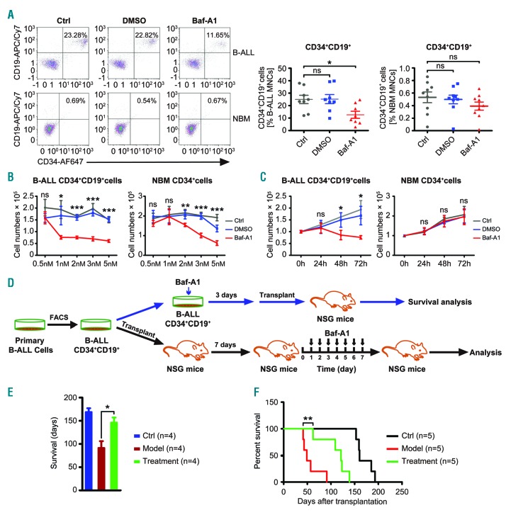
Patients’ samples used in this study shared the same expression pattern in double-positive CD34CD19, which are reliable B-ALL LSC markers (Online Supplementary Table S1).5,8 Upon in vitro treatment with 1 nM bafilomycin A1, the percentage of the CD34+CD19+ population derived from patients (n=8) was significantly reduced, while normal B-cell stem and progenitor cells (Figure 1A) were unaffected. Indeed, 1 nM bafilomycin A1 was sufficient to induce clear cytotoxicity after 48 h or 72 h of treatment in B-ALL LSC from patients, but not in normal hematopoietic stem cells isolated from healthy donors (Figure 1B, C).
Figure 1.
Low-dose bafilomycin A1 extends the lifespan of humanized leukemia mice engrafted with CD34+CD19+cells derived from patients with B-cell acute lymphoblastic leukemia. (A) Flow cytometric analysis of the frequency of CD34+CD19+ cells in primary mononuclear cells from eight patients (ALL#1-8) with B-cell acute lymphoblastic leukemia (B-ALL) and normal bone marrow (NBM) mononuclear cells from nine healthy donors after 1 nm bafilomycin A1 treatment. (B) Reduction of primary B-ALL CD34+CD19+ cells by bafilomycin A1 is dose-dependent. Left, ALL patients (#9-14), n=6; right, normal hematopoietic cells from healthy donors, n=6. (C) Reduction of primary B-ALL CD34+CD19+ cells is time-dependent. Left, 24 h and 48 h n=3, ALL#12,15,16; 72 h n=6, ALL#9-14; right, 24 h and 48 h n=3, 72 h n=6, NBM CD34+ cells from healthy donors. (D) Schematic experimental design for NSG mice engrafted with B-ALL CD34+CD19+ samples after bafilomycin A1 treatment either ex vivo (n=4, ALL#14,17-19) or in vivo (n=5, ALL#20-24). (E) Ex vivo treatment with 1 nm bafilomycin A1 prolonged the lifespan of the NSG mice engrafted with B-ALL CD34CD19 cells (n=4, ALL#14,17-19). (F) Survival curve reflecting time to lethal leukemia burden in NSG mice injected with 1-5x10⁶ B-ALL CD34+CD19+ cells, and 7 days later, treated with vehicle or bafilomycin A1 (0.1 mg/kg) (n=5/group, ALL#20-24). Ctrl: control; DMSO: dimethylsulfoxide; Baf-A1: bafilomycin A1.
To determine whether bafilomycin A1 treatment in vitro weakens the capacity of primary LSC in B-ALL patients to initiate disease in mice, primary CD34+CD19+ cells were treated for 72 h with bafilomycin A1 at 1 nM, and then transplanted into humanized immunodeficient NSG mice (NOD-SCID IL-2Rγ−/−). The animals were then monitored until death from overt leukemia (Figure 1D). In vitro bafilomycin A1 treatment of the LSC before transplantation significantly reduced the mice’s leukemic burden (Figure 1E).
To determine the effect of bafilomycin A1 on B-ALL patient-derived LSC in vivo, we tested the compound’s ability to suppress the primary LSC in the humanized mouse model. We transplanted primary CD34+CD19+ B-ALL cells from B-ALL patients into NSG mice. The LSC were allowed to complete homing and proliferate for 7 days, followed by 7 successive days of injection with 0.1 mg/kg bafilomycin A1. The animals were then monitored until their death from palpable leukemia (Figure 1D). Bafilomycin A1 significantly extended the survival of the treated mice, delaying the onset of disease compared to that in control mice (Figure 1F).
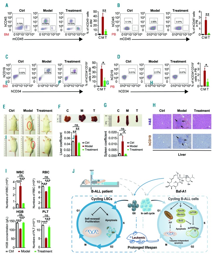
To examine whether in vivo treatment with bafilomycin A1 at low doses improved the pathology of the humanized leukemia model, we analyzed blood cells from three groups of mice (control mice, disease model and bafilomycin A1-treated disease model) using human CD45, CD34 and CD19 antibodies alone or in combination. Flow cytometric results showed that bafilomycin A1 significantly reduced the number of both human leukemia cells and B-ALL CD34+CD19+ cells in the bone marrow and peripheral blood (Figure 2A-D). The mice in the B-ALL model group showed signs of mental dysfunction, hind limb paralysis, and back curvature, whereas the mice in the bafilomycin A1 treatment group did not have spinal curvature (Figure 2E). Additionally, mice from the disease model group had serious hepatosplenomegaly compared to mice in the control group, while both the size and weight of livers and spleens in the bafilomycin A1-treated group were significantly normalized (Figure 2F, G). Livers from the disease model group were also significantly infiltrated. In contrast, considerably fewer ALL cells infiltrated the livers of bafilomycin A1-treated mice (Figure 2H). These data suggest that bafilomycin A1 dramatically inhibited B-ALL engraftment by diminishing the patient-derived CD34+CD19+ LSC in vivo. Peripheral blood counts showed that in the disease model group, the number of white blood cells increased, while in the bafilomycin A1-treated group they were significantly normalized (Figure 2I). In the B-ALL model group, the red blood cell count was significantly reduced, together with hemoglobin level and particularly the platelet count, whereas these blood parameters in the bafilomycin A1-treated group were significantly normalized (Figure 2I). These data suggest that bafilomycin A1 was well-tolerated, and that the treatment reduced the leukemia burden without causing significant anemia or thrombocytopenia.
Figure 2.
Low-dose bafilomycin A1 improves the pathology of mice engrafted with primary human B-acute lymphoblastic leukemia CD34+CD19+ cells. (A, B) Representative FACS scatter plots showing the proportions (%) of human CD45 cells in the bone marrow (BM) or peripheral blood (PB) from NSG xenografts following in vivo treatment with vehicle or 0.1 mg/kg bafilomycin A1 (n=5/group, ALL#21-23,25-26). (C, D) Flow cytometric analysis of B-cell acute lymphoblastic leukemia (B-ALL) CD34+CD19+ cells in the BM or PB from NSG mice treated with vehicle or 0.1 mg/kg bafilomycin A1 (n=5/group). (E) In vivo bafilomycin A1 (0.1 mg/kg) treatment after transplantation significantly reduced leukemic burden (n=5/group). (F) Upper panel, photographs of livers recovered from NSG mice engrafted with B-ALL CD34+CD19+ cells after the indicated days of treatment with bafilomycin A1 (0.1 mg/kg) or vehicle. Lower panel, the liver coefficient (the ratio of the weight of liver to the total body weight). (G) Upper panel, photographs of spleens recovered from NSG mice engrafted with B-ALL CD34+CD19+ cells after the indicated days of treatment with bafilomycin A1 or vehicle. Lower panel, the spleen coefficient (the ratio of the weight of spleen to the total body weight). (H) Upper panel, hematoxylin and eosin–stained sections from livers of NSG mice engrafted with B-ALL CD34+CD19+ cells after treatment with vehicle or bafilomycin A1 (0.1 mg/kg). Leukemic infiltration is indicated by arrows. Lower panel, immunohistochemistry of livers stained for cells expressing human CD19; leukemic infiltration is indicated by arrows. (I) Analysis at days 40-90 for the indicated PB cells, as measured by complete blood count. (J) A proposed working model of bafilomycin A1 targeting both B-ALL cells and their leukemia stem cells. Ctrl and C: control; M: model; T: treatment; Baf-A1: bafilomycin A1; WBC, white blood cells; RBC, red blood cells; HGB, hemoglobin; PLT, platelets.
To evaluate the safety of bafilomycin A1 in vivo, the compound or vehicle was administered daily to NSG mice by intraperitoneal injection for 7 days at a dose of 0.1 mg/kg. The organ coefficients of treated mice were not significantly different (Online Supplementary Figure S5A). Flow cytometric analysis indicated that bafilomycin A1 at this low dose had no negative impact on hematopoietic stem and progenitor cells, or on total hematopoietic cells in NSG mice (Online Supplementary Figure S5B, C). In addition, peripheral blood counts were unchanged (Online Supplementary Figure S5D). Bafilomycin A1 at a dose of 0.1 mg/kg did not cause toxicity in the mice, as shown by unchanged liver size, the levels of serum aspartate amino-transferase and alanine aminotransferase, which represent normal liver function, and urea and creatinine, which represent normal kidney function (Online Supplementary Figure S5E, F). In addition, hematoxylin and eosin staining showed no liver injuries in NSG mice after treatment (Online Supplementary Figure 5G). Thus, while bafilomycin A1 is potent, it is a safe compound.
To understand how bafilomycin A1 at a dose of 1 nM diminishes B-ALL LSC, we analyzed the cell cycle of the CD34+CD19+ cells by Hoechst 33342 and Ki-67 staining and found a significant decrease in G0 phase (Online Supplementary Figure S6A). Further analysis with Hoechst and Pyronin Y staining also showed that bafilomycin A1 induced quiescent B-ALL CD34+CD19+ cells to the cell cycle, sparing normal hematopoietic stem cells (Online Supplementary Figure S6B). Next, we used the intracellular fluorescent label carboxyfluorescein diacetate succin-imidyl ester (CFSE) to track proliferating primary LSC in patients’ samples. The results showed that treatment with bafilomycin A1 for 72 h effectively inhibited cell division of B-ALL CD34+CD19+ cells only, while sparing normal hematopoietic stem cells (Online Supplementary Figure S7A). We also used a CCK-8 assay to measure the LSC proliferation at multiple time points. At 24 h of treatment, bafilomycin A1 did not alter the proliferation of B-ALL LSC compared with the control group (Online Supplementary Figure S7B). Presumably, the B-ALL LSC primarily remained in the quiescent state until 24 h of bafilomycin A1 treatment, and quiescent LSC are not sensitive to bafilomycin A1. However, the proliferation of B-ALL LSC was selectively inhibited, as shown by the data recorded at 48, 72, and 96 h of bafilomycin A1 treatment, while the normal hematopoietic stem cells were spared (Online Supplementary Figure S7B). This is largely because at and after 24 h of bafilomycin A1 treatment, the quiescent LSC were significantly induced to the cell cycle from G0 phase (Online Supplementary Figure S6). This possibly made it easier for the compound to target the cycling B-ALL LSC. Indeed, annexin V-FITC/propidi-um iodide assay showed that bafilomycin A1 caused apoptotic death in the primary B-ALL LSC, but did not induce apoptosis in normal bone marrow CD34+ cells (Online Supplementary Figure S8). As a result, a colony-forming unit assay showed impaired self-renewal capacity in bafilomycin A1-treated human B-ALL CD34+CD19+ cells, but not in normal bone marrow CD34+ cells (Online Supplementary Figure S9). Finally, flow cytometric analysis of IgM-stained B-ALL LSC in the presence of bafilomycin A1 showed that the reduction of LSC number was not caused by the induction of terminal differentiation, since bafilomycin A1 only induced marginal cell differentiation of human B-ALL CD34+CD19+ cells (Online Supplementary Figure S10). We therefore propose that bafilomycin A1 drives quiescent B-ALL LSC to the cell cycle and subsequently inhibits and kills the LSC by induction of apop-tosis. A cartoon representing the putative mechanism underlying the action of bafilomycin A1 on B-ALL LSC is shown in the left panel of Figure 2J. The right panel of Figure 2J illustrates the mechanism by which bafilomycin A1 targets B-ALL cells, previously reported by our group.15
In summary, in contrast to previous studies on recombinant proteins targeting primary B-ALL LSC, we show that bafilomycin A1, a natural compound, at low doses attenuated CD34+CD19+ LSC of B-ALL patients. The reduced leukemogenesis in the humanized mouse model is caused primarily by induction of quiescent LSC to the cell cycle, leading to apoptotic cell death and inhibition of proliferation of the LSC upon treatment with bafilomycin A1. Therefore, our data suggest that bafilomycin A1 not only preferentially targets the LSC derived from B-ALL patients, but is also well-tolerated by normal primitive hematopoietic cells. The capacity to target both leukemia cells and LSC makes bafilomycin A1 a potentially very promising candidate for drug development for B-ALL therapy.
Acknowledgments
the authors thank the patients, healthy blood donors and clinical teams who were involved in the study. Primary leukemia samples used in this study were provided by the First Affiliated Hospital of Soochow University.
Footnotes
Funding; this work was supported by the National Natural Science Foundation of China with grants N. 81570126, N. 91649113, and N. 31771640 (to JW), N. 81730003 (to DW), N. 81800152 (to NY), by Jiangsu Province Natural Science Foundation grant BK20160330 (to NY), Suzhou Municipality Science and Technology grant SYS201703 (to NY), by the Astronaut Center of China N. ACCKJZYX-14-128, and the Priority Academic Program Development of Jiangsu Higher Education Institutions.
Information on authorship, contributions, and financial & other disclosures was provided by the authors and is available with the online version of this article at www.haematologica.org.
References
- 1.Lapidot T, Sirard C, Vormoor J, et al. A cell initiating human acute myeloid leukaemia after transplantation into SCID mice. Nature. 1994;367(6464):645–648. [DOI] [PubMed] [Google Scholar]
- 2.Ebinger S, Özdemir EZ, Ziegenhain C, et al. Characterization of rare, dormant, and therapy-resistant cells in acute lymphoblastic leukemia. Cancer Cell. 2016;30(6):849–862. [DOI] [PMC free article] [PubMed] [Google Scholar]
- 3.Rehe K, Wilson K, Bomken S, et al. Acute B lymphoblastic leukaemia propagating cells are present at high frequency in diverse lymphoblast populations. EMBO Mol Med. 2013;5(1):38–51. [DOI] [PMC free article] [PubMed] [Google Scholar]
- 4.Hotfilder M, Rottgers S, Rosemann A, et al. Leukemic stem cells in childhood high-risk ALL/t(9;22) and t(4;11) are present in primitive lymphoidrestricted CD34+CD19-cells. Cancer Res. 2005;65(4):1442–1449. [DOI] [PubMed] [Google Scholar]
- 5.le Viseur C, Hotfilder M, Bomken S, et al. In childhood acute lymphoblastic leukemia, blasts at different stages of immunophenotypic maturation have stem cell properties. Cancer Cell. 2008;14(1):47–58. [DOI] [PMC free article] [PubMed] [Google Scholar]
- 6.Aoki Y, Watanabe T, Saito Y, et al. Identification of CD34+ and CD34- leukemia-initiating cells in MLL-rearranged human acute lymphoblastic leukemia. Blood. 2015;125(6):967–980. [DOI] [PMC free article] [PubMed] [Google Scholar]
- 7.Lang F, Wojcik B, Bothur S, et al. Plastic CD34 and CD38 expression in adult B–cell precursor acute lymphoblastic leukemia explains ambiguity of leukemia-initiating stem cell populations. Leukemia. 2017;31(3):731–734. [DOI] [PMC free article] [PubMed] [Google Scholar]
- 8.Kong Y, Yoshida S, Saito Y, et al. CD34+CD38+CD19+ as well as CD34+CD38-CD19+ cells are leukemia-initiating cells with self-renewal capacity in human B-precursor ALL. Leukemia. 2008;22(6): 1207–1213. [DOI] [PubMed] [Google Scholar]
- 9.Morisot S, Wayne AS, Bohana-Kashtan O, et al. High frequencies of leukemia stem cells in poor-outcome childhood precursor-B acute lymphoblastic leukemias. Leukemia. 2010;24(11):1859–1866. [DOI] [PMC free article] [PubMed] [Google Scholar]
- 10.Jin Y, Cao Q, Chen C, et al. Tenovin-6-mediated inhibition of SIRT1/2 induces apoptosis in acute lymphoblastic leukemia (ALL) cells and eliminates ALL stem/progenitor cells. BMC Cancer. 2015;15:226. [DOI] [PMC free article] [PubMed] [Google Scholar]
- 11.Alves CC, Terziyska N, Grunert M, et al. Leukemia-initiating cells of patient-derived acute lymphoblastic leukemia xenografts are sensitive toward TRAIL. Blood. 2012;119(18):4224–4228. [DOI] [PubMed] [Google Scholar]
- 12.Uckun FM, Myers DE, Qazi S, et al. Recombinant human CD19L-sTRAIL effectively targets B cell precursor acute lymphoblastic leukemia. J Clin Invest 2015;125(3):1006–1018. [DOI] [PMC free article] [PubMed] [Google Scholar]
- 13.Hay KA, Hanafi LA, Li D, Riddell SR, Maloney DG, Turtle CJ, et al. Kinetics and biomarkers of severe cytokine release syndrome after CD19 chimeric antigen receptor-modified T-cell therapy. Blood. 2017;130(21):2295–2306. [DOI] [PMC free article] [PubMed] [Google Scholar]
- 14.Klionsky DJ, Elazar Z, Seglen PO, et al. Does bafilomycin A1 block the fusion of autophagosomes with lysosomes¿ Autophagy. 2008:4(7):849–850. [DOI] [PubMed] [Google Scholar]
- 15.Yuan N, Song L, Zhang S, et al. Bafilomycin A1 targets both autophagy and apoptosis pathways in pediatric B-cell acute lymphoblastic leukemia. Haematologica. 2015;100(3):345–356. [DOI] [PMC free article] [PubMed] [Google Scholar]


