Abstract
Background
The calcium/calcineurin signaling pathway is mediated by the transcription factors NFAT (nuclear factor of activated T cells) in mammals and Crz1 (calcineurin-responsive zinc finger 1) in yeasts and other lower eukaryotes. A previous microarray analysis identified a putative Crz1-binding motif in promoters of its target genes in Candida albicans, but it has not been experimentally demonstrated.
Methods
An inactivation mutant for CaCRZ1 was generated through CRISPR/Cas9 approach. Transcript profiling was carried out by RNA sequencing of the wild type and the inactivation mutant for CaCRZ1 in response to 0.2 M CaCl2. Gene promoters were scanned by the online MEME (Multiple Em for Motif Elicitation) software. Gel electrophoretic mobility shift assay (EMSA) and chromatin immunoprecipitation (ChIP) analysis were used for in vitro and in vivo CaCrz1-binding experiments, respectively.
Results
RNA sequencing reveals that expression of 219 genes is positively, and expression of 59 genes is negatively, controlled by CaCrz1 in response to calcium stress. These genes function in metabolism, cell cycling, protein fate, cellular transport, signal transduction, transcription, and cell wall biogenesis. Forty of these positively regulated 219 genes have previously been identified by DNA microarray analysis. Promoter analysis of these common 40 genes reveals a consensus motif [5′-GGAGGC(G/A)C(T/A)G-3′], which is different from the putative CaCrz1-binding motif [5′-G(C/T)GGT-3′] identified in the previous study, but similar to Saccharomyces cerevisiae ScCrz1-binding motif [5′-GNGGC(G/T)CA-3′]. EMSA and ChIP assays indicate that CaCrz1 binds in vitro and in vivo to both motifs in the promoter of its target gene CaUTR2. Promoter mutagenesis demonstrates that these two CaCrz1-binding motifs play additive roles in the regulation of CaUTR2 expression. In addition, the CaCRZ1 gene is positively regulated by CaCrz1. CaCrz1 can bind in vitro and in vivo to its own promoter, suggesting an autoregulatory mechanism for CaCRZ1 expression.
Conclusions
CaCrz1 differentially binds to promoters of its target genes to regulate their expression in response to calcium stress. CaCrz1 also regulates its own expression through the 5′-TGAGGGACTG-3′ site in its promoter.
Video abstract
Keywords: Candida albicans, Crz1, Calcium signaling, Transcription profiling, RNA sequencing, Promoter
Plain English summary
Calcium ions regulate many cellular processes in both prokaryotes and eukaryotes, from bacteria to humans. Regulation of intracellular calcium homeostasis is highly conserved in eukaryotic cells. Gene expression in response to calcium stress is controlled by the calcium/calcineurin signalling through the transcription factors NFAT (the nuclear factor of activated T cells) in mammals and Crz1 (calcineurin-responsive zinc finger 1) in yeasts and other lower eukaryotes. Extracellular calcium stress causes an increase in cytosolic calcium, which leads to the binding of calcium ions to calmodulin that triggers activation of the protein phosphatase, calcineurin. Activated calcineurin dephosphorylates Crz1 in the cytosol, which leads to nuclear localization of Crz1 and its binding to promoters of its target genes to regulate their expression. Candida albicans is one of the most important human yeast pathogens. A previous microarray analysis identified a putative CaCrz1-binding motif in promoters of its target genes in C. albicans, but it has not been experimentally demonstrated. Using a new technology, RNA sequencing, we have identified 219 genes that are positively, and 59 genes that are negatively, controlled by CaCrz1 in response to calcium stress in this study. We have also revealed and demonstrated experimentally a novel consensus CaCrz1-binding motif [5′-GGAGGC(G/A)C(T/A)G-3′] in promoters of its target genes. In addition, we have discovered that CaCrz1 can bind to its own promoter, suggesting an autoregulatory mechanism for CaCRZ1 expression. These findings would contribute to our further understanding of molecular mechanisms regulating calcium homeostasis.
Backgound
Calcium ions regulate many cellular processes in both prokaryotes and eukaryotes, from bacteria to humans [1–5]. Intracellular calcium homeostasis is maintained by calcium transporters and sequestrators in the plasma and organelle membranes in eukaryotes. Regulation of calcium homeostasis is highly conserved in eukaryotic cells. Gene expression in response to calcium stress is controlled by the calcium/calcineurin signalling through the transcription factor Crz1 in fungi or the nuclear factor of activated T cells (NFAT) in mammals [6, 7]. In Saccharomyces cerevisiae, an increase in cytosolic calcium triggers the calmodulin/Ca2+ binding and activation of the protein phosphatase, calcineurin. Activated calcineurin dephosphorylates ScCrz1 in the cytosol, which leads to nuclear localization of ScCrz1 and its binding to promoters of its target genes, including the vacuolar calcium pump gene ScPMC1, the ER/Golgi calcium pump gene ScPMR1 and the ScRCH1 gene encoding the negative regulator of calcium uptake in the plasma membrane [8–10]. A genome-scale genetic screen has revealed additional genes that are involved in the regulation of calcium homeostasis in budding yeast [11].
Candida albicans remains as one of leading human fungal pathogens in immunocompromised patients [12–14]. Functional counterparts of calcium homeostasis and calcium/calcineurin signaling components have been characterized in C. albicans [15–21]. The calcium/calcineurin signaling functions in ion homeostasis, cell wall biogenesis, morphogenesis and drug resistance in C. albicans [22–24]. C. albicans cells lacking calcineurin show significantly reduced virulence in a murine model of systemic infection and fail to survive in the presence of membrane stress [25–27]. However, C. albicans cells lacking CaCRZ1, the major target of calcineurin, are partially virulent in the CAF4–2 strain background and even not virulent in the BWP17 background in the mouse model of systemic infection [28, 29]. Therefore, other targets are responsible for the calcineurin-mediated virulence in C. albicans. We have recently screened the GRACE (gene replacement and conditional expression) library of 2358 conditional mutants and identified a total of 21 genes whose conditional repression leads to the sensitivity of C. albicans cells to high levels of extracellular calcium [30–32]. In addition to 3 reported genes, CRZ1, MIT1 and RCH1 [16, 20, 28, 33], the rest newly-identified 18 calcium tolerance-related genes are involved in tricarboxylic acid cycle, cell wall integrity pathway, cytokinesis, pH homeostasis, magnesium transport, and DNA damage response.
Microarray analysis indicates that calcium-induced upregulation of 60 genes with a putative CaCrz1-binding motif [5′-G(C/T)GGT-3′] is dependent on both calcineurin and CaCrz1 in C. albicans [28]. Both microarray and RNA sequencing are used to measure genome-wide transcriptomic changes in different organisms, and they complement to each other in transcriptome profiling [34–36]. However, RNA sequencing approach is much more sensitive than the microarray, with the dynamic range of the former reaching at least 8000-fold in comparison to the latter only at around 60-fold in expression levels of genes detected [37].Therefore, we have examined the regulatory function of CaCrz1 in gene expression with the RNA sequencing technology in this study. We show that expression of 219 genes is positively controlled, and expression of 59 genes is negatively controlled, by CaCrz1 in the SN148 background in response to calcium stress. Furthermore, we have revealed an additional CaCrz1-binding motif in promoters of its target genes and demonstrated that CaCrz1 binds to both motifs in the promoter of its target gene CaUTR2.
Methods
Strains and media
C. albicans strains and plasmids used in this study were described in Table 1. Primers used in this study were listed in Additional file 1: Table S1. Strains were grown and maintained at 30 °C in YPD medium or SD medium (0.67% yeast nitrogen base without amino acids, 2% glucose, and auxotrophic amino acids as needed). Chemicals were obtained from Sigma (USA) and Sangon Biotech (Shanghai, China).
Table 1.
Strains and plasmids used in this study
| Name | Genotype or Description | Source |
|---|---|---|
| Strain | ||
| SN148 | Mata/α arg4/arg4 leu2/leu2 his1/his1 ura3::imm434/ura3::imm434 | [38] |
| HHCA184 | SN148 crz1/crz1 ENO1/eno1:: natMX4 | This study |
| HHCA185 | SN148 crz1/crz1 ENO1/eno1:: natMX4 | This study |
| HHCA187 | SN148 crz1/crz1 ENO1/eno1:: natMX4 | This study |
| HHCA1 | SN148 RPS1/rps1::CIp10 | This study |
| HHCA2 | HHCA184 RPS1/rps1::CIp10 | This study |
| HHCA3 | HHCA184 RPS1/rps1::CIp10-CaCRZ1 | This study |
| Plasmid | ||
| pV1093 | AmpR NatR | [39] |
| pV1093-sgCRZ1 | AmpR NatR | This study |
| CIp10 | AmpR URA | [38] |
| CIp10-sgCRZ1 | AmpR URA | This study |
Construction of CRISPR mutant for CaCRZ1
C. albicans strain SN148 was used as the parent strain to construct the CRISPR inactivation mutant for CaCRZ1 through the CRISPR [Clustered Regularly Interspaced Short Palindromic Repeat)/Cas9] approach (Additional file 1: Figure S1). We designed SgRNA primers CRZ1-sgF and CRZ1-sgR near the start codon of CaCRZ1 using the online software Benchling (https://benchling.com/academic) as well as the repair DNA primers CRZ1-RFand CRZ1-RR containing 40-bp homologous regions flanking the SgRNA sequence (Additional file 1 : Figure. S1). Primers CRZ1-sgF and CRZ1-sgR were annealed, cut with BsmBI and cloned into the BsmBI site of pV1093 (Additional file 1: Figure S1A-S1B), which generated the recombinant plasmid pV1093-SgRNA. SgRNA sequence in pV1093-SgRNA was confirmed by DNA sequencing. Primers CRZ1-RF and CRZ1-RR were annealed for PCR amplification of the repair DNA fragment of about 100 bp. The repair DNA and the recombinant plasmid pV1093-SgRNA linearized by both SacI and KpnI were used together to transform cells of C. albicans strain SN148 (Additional file 1: Figure S1C). Potential correct CRISPR mutants for CaCRZ1 were detected with diagnostical PstI-digestion of 1-kb PCR products, containing the SgRNA region, amplified with primers CRZ1-CF and CRZ1-CR from genomic DNA samples of transformants (Additional file 1: Figure S1D-S1E). Mutated sites in CaCRZ1 alleles in those potential correct CRISPR mutants were further confirmed by DNA sequencing.
DNA manipulation
To clone the full-length gene CaCRZ1 into the integration vector CIp10 [40], a DNA fragment containing the 758-bp promoter, the 2196-bp open reading frame (ORF) and the 336-bp terminator region of CaCRZ1 was amplified with primers CRZ1-clonF and CRZ1-clonR, and cloned between KpnI and XhoI sites in the CIp10, which yielded CIp10-CaCRZ1. To do complementation experiment, the wild type and the crz1/crz1 mutant strains were integrated with the StuI-linearized plasmids CIp10 or CIp10-CaCRZ1, respectively, as described [41].
To express the His6-tagged CaCrz1 expression plasmid in bacterial cells, we first optimized the codon usage by mutating all five CTG codons in CaCRZ1 to TCT codon (L22S), AGC codon (L24S), TCC codons (L601S, L649S and L686S) (Additional file 1: Fig. S2). The codon-optimized open reading frame (ORF) of CaCRZ1 was artificially synthesized and cloned into the vector pET28a(+), which yielded pET28a(+)-CRZ1 that expressing the codon-optimized and N-terminally Hisx6 tagged full-length CaCrz1 (His6-CaCrz1) protein. The pET28a(+)-CRZ1 was introduced and expressed in BL21(DE3) bacterial cells as described [42–44].
To construct a lacZ reporter plasmid, the bacterial lacZ gene was first amplified with a pair of primers lacZ_ORF_F(XhoI) and lacZ_ORF_R(KpnI) from the plasmid pGP8 [15, 28], and cloned into the KpnI and XhoI sites of CIp10 to yield CIp10-lacZ. The terminator of CaACT1 was amplified from the SN148 genomic DNA with two primers ACT1_T_F(KpnI) and ACT1_T_R(KpnI), and cloned into the KpnI site of CIp10-lacZ to yield CIp10-lacZ-TACT1. The CaUTR2 promoter was amplified from the SN148 genomic DNA with a pair of primers UTR2_P_F(XhoI) and UTR2_P_R(XhoI) and cloned into the XhoI site of CIp10-lacZ-TACT1 to yield CIp10-UTR2-lacZ.
To mutate the putative CaCrz1-binding motif identified in our study, the underlined sequence in the 5′-TCT(− 343) CAACGCCTCA(− 333)AAA-3′ region of CaUTR2 promoter was mutated to be 5′-TCT(− 343)TCTAGA(− 333)AAA-3′ (we designated this mutation as UTR2(HΔ)), which contains a XbaI site. This was accomplished by a fusion PCR strategy. We first amplified the upstream (A) and downstream (B) fragments of the CaUTR2 promoter with two pairs of primers UTR2_exF/ UTR2_(HΔ)_R and UTR2_inR/ UTR2_(HΔ)_F, respectively. These two fragments (A and B) were then fused by PCR with the two primers UTR2_P_F(XhoI) and UTR2_P_R(XhoI), and cloned into the XhoI site of CIp10-lacZ-TACT1 to yield CIp10-UTR2(HΔ)-lacZ. Similarly, to mutate the putative CaCrz1-binding motif identified in the previous study [28], the underlined sequence in the (5′-TTGT(− 377)GGGCTT(− 371)TGA-3′ region of CaUTR2 promoter was mutated to be (5′-TTGT(− 377)TCTAGAT(− 371)TGA-3′ (we designated this mutation as UTR2(MΔ)), which contains a XbaI site. The upstream (C) and downstream (D) fragments of the CaUTR2 promoter were first PCR amplified with two pairs of primers UTR2_exF/ UTR2_(MΔ)_R and UTR2_inR/UTR2_(MΔ) _F, respectively. These two fragments (C and D) were then fused by PCR with the two primers UTR2_P_F(XhoI) and UTR2_P_R(XhoI), and cloned into the XhoI site of CIp10-lacZ-TACT1 to yield CIp10-UTR2(MΔ)-lacZ. To create the CIp10-UTR2(HMΔ)-lacZ with mutations for both UTR2(HΔ) and UTR2(MΔ) in the CaUTR2 promoter, the two DNA fragments (A and D) were fused by PCR with primers UTR2_P_F(XhoI)/ UTR2_P_R(XhoI), and cloned into the XhoI site of CIp10-lacZ-TACT1. Inserts in all recombinant plasmids were confirmed by DNA sequencing.
RNA sequencing and data analysis
To identify genes regulated by CaCrz1, the wild type SN148 and its isogenic CRISPR mutant for CaCRZ1 were grown to log-phase at 30 °C before they were treated with 0.2 M CaCl2 for 2 h. Total RNA samples were extracted Qiagen RNeasy minikit protocol, and RNA integrity was evaluated using an Agilent 2100 Bioanalyzer (Agilent Technologies, USA) as described [45]. RNA-seq libraries were constructed using Illumina’s miSEQ RNA Sample Preparation Kit (Illumina Inc., USA). RNA sequencing, data analysis and sequence assembly were performed by the Quebec Genome Innovation Center at McGill University (Montreal, Canada) [31, 38]. Preparation of the paired-end libraries and sequencing were performed following standard Illumina methods and protocols. The mRNA-seq library was sequenced using an Illumina miSEQ sequencing platform. Clean reads from RNA-Seq data were assembled into full-length transcriptome with the reference genome (http://www.candidagenome.org/). Functional categories of genes were carried out by the Munich Information Center for Protein Sequences (MIPS) analysis.
Galactosidase activity assay
To measure the UTR2 promoter-driven β-galactosidase activity in the wild type and the crz1/crz1 mutant, we integrated the StuI-linearized plasmids containing the lacZ reporters for CaUTR2 promoter into the RPS1 locus of these strains as described [16, 28]. The β-galactosidase activity was determined using the substrate ONPG as described [46, 47]. Data are mean ± SD from six independent experiments. Significant differences were analysed by GraphPad Prism version 4.00. P values of < 0.05 were considered to be significant.
Results
Construction of the CRISPR mutant for CaCRZ1
To further study the regulatory functions of CaCrz1 in gene expression, we constructed three independent CRISPR mutants for CaCRZ1 in the SN148 genetic background (Additional file 1: Figure S1A-S1E). These mutants were sensitive to 0.4 M CaCl2, and their calcium sensitivity was suppressed by the specific inhibitor of calcineurin, cyclosporine A. In addition, they were sensitive to 0.05% SDS, but not to antifungal drugs including clotrimazole, ketoconazole, fluconazole and terbinafine (Additional file 1: Figure S1F). These results agree with previous reports [21, 28, 29]. We chose one of these CRISPR mutants (HHCA184) for our RNA sequencing, and its calcium-sensitive phenotype could be partially reversed by the introduction of the CaCRZ1 gene back to its genome (Fig. 1). To examine if the two mutated CaCRZ1 alleles in the CRISPR mutant (HHCA184) were still able to express the CaCrz1 proteins in C. albicans cells, we chromosomally integrated the HA tag at the C-terminus of CaCrz1 in both the mutant and the wild type strain SN148. Through western blot analysis, we failed to detect the expression of CaCrz1-HA in the mutant, although we detected two forms of CaCrz1-HA proteins in the wild type, which might correspond to the phosphorylated form and dephosphorylated form of CaCrz1 (Fig. 2). Taken together, our data demonstrate that we have successfully constructed the CRISPR mutant for CaCRZ1.
Fig. 1.
Phenotypes of CRISPR mutant for CaCRZ1. Cells of the wild-type SN148, the CRISPR mutant and the complemented strain were grown at 30 °C in liquid YPD overnight, serially diluted by 10 times and spotted on YPD plates with or without supplemented reagents as indicated, respectively. Plates were incubated for 2–5 days at 30 °C. CsA, cyclosporine A
Fig. 2.
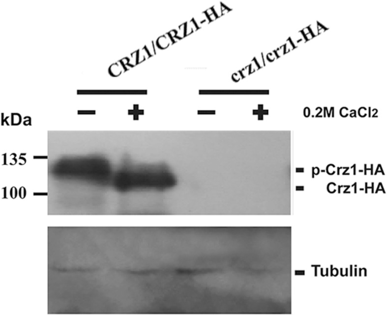
Expression of the C-terminally HA-tagged CaCrz1 protein in C. albicans cells. The wild type strain SN148 (CRZ1/CRZ1) and the CRISPR mutant for CaCRZ1 (crz1/crz1) carrying their chromosomally C-terminally HA-tagged wild-type and mutated CaCRZ1 alleles, respectively, were grown to log-phase in YPD medium at 30 °C before their cells were collected for total protein extraction. Expression of CaCrz1-HA proteins was detected by Western blot analysis with anti-HA monoclonal antibody. Expression of tubulin was detected using anti-tubulin antibody, which served as an internal expression control
Transcriptomic profiling of cells lacking CaCRZ1
Next, we carried out transcript profiling for the wild type and the crz1/crz1 mutant, growing in log phase in YPD medium at 30 °C in the absence or presence of 0.2 M CaCl2. Transcripts for two alleles of 6211 genes at various expression levels were detected in these two strains (SuppInfo 1; GEO Accession number: GSE123122). As compared to the wild type cells without 0.2 M CaCl2 treatment, there are 828 genes upregulated in the wild type cells with 0.2 M CaCl2 treatment, among which 219 genes are positively regulated, and 59 genes are negatively regulated, by CaCrz1 (SuppInfo 2; SuppInfo 3). These genes positively regulated by CaCrz1 play roles in metabolism (13), cellular transport (23), transcription (7), signal transduction (3), protein fate (17), cell rescue (9), cell cycle (6), cell fate/development/cell type differentiation (14) and cell wall biogenesis (34), with almost half of them (93) being of unknown functions (Table 2). In contrast, these genes negatively regulated by CaCrz1 function in metabolism [20], cellular transport [5], transcription [11] and cell wall biogenesis [3], with one third of them [20] being of unknown functions (Table 3). The CaCRZ1 gene itself is positively regulated by CaCrz1, which is identified in both the previous microarray study and our current study (Table 2).
Table 2.
Functional category of 219 genes positively regulated by CaCrz1 in response to 0.2 M CaCl2
| Systemic name | Standard name | Systemic name | Standard name | Systemic name | Standard name | Systemic name | Standard name | Systemic name | Standard name |
|---|---|---|---|---|---|---|---|---|---|
| Metabolism (13) | |||||||||
| C1_04010C | C2_03640W | UGA11 | C5_00220W | ROT2 | C1_14060W | C1_11240C | CHO1 | ||
| C2_01630W | C2_09150W | MIT1 | CR_00620C | ARG1 | C1_08330C | ADH2 | C7_02500C | DPP3 | |
| C3_05810C | SKN1 | C1_01620C | |||||||
| Cellular transport (23) | |||||||||
| CR_03450W | HXT5 | C5_04440C | SFC1 | C3_03060W | C3_05270C | HGT5 | C7_02910W | ENA21 | |
| C1_09220W | C2_09770C | INP51 | C1_04630C | C7_00100W | FRP2 | ||||
| CR_05310W | C4_03110W | C3_07230W | C1_01100W | CCH1 | C1_09400C | FTH1 | |||
| C2_03800C | CR_09170C | SSU1 | C2_06470W | RTA2 | C2_07730W | YVC1 | C1_06610C | HAK1 | |

|
CR_07100W | FLC2 | CR_09680C | RTA4 | |||||
| Transcription (7) | |||||||||
| CR_03890W | WOR3 | C7_00970C | YOX1 | C3_05780C | CRZ1 | C1_05340C | ZCF2 | C7_04230W | NRG1 |
| CR_02300C | C4_04210C | SOH1 | |||||||
| Signal transduction (3) | |||||||||
| C5_02290W | PDE1 | C4_06480C | CEK1 | C7_00360W | DFI1 | ||||
| Protein fate (folding, modification, destination) (17) | |||||||||
| C1_13220C | AKR1 | C5_01440C | C2_00930C | VPS24 | C5_01210W | VPS1 | C4_03890W | PTP2 | |
| CR_00290W | C5_05060C | C4_04660C | C4_05810W | ||||||
| C6_03500C | SAP4 | C2_01670C | STT3 | C4_00070C | C7_03250C | PDI1 | CR_00260W | KIN2 | |
| C1_08170C | BUL1 | C2_08790W | JEM1 | ||||||
| Cell rescue (9) | |||||||||
| CR_06040W | CR_01730W | IFU5 | C2_02060C | FMO1 | C2_00680C | SOD5 | C1_02700C | ||
| C2_09220W | DDR48 | C3_00480C | DOT5 | C2_05660W | PNG2 | CR_05390W | PST3 | ||
| Cell Cycle (6) | |||||||||
| C1_09870W | HCM1 | C6_03260W | C1_05170C | CUE5 | C1_08570C | PCL2 | C3_03850C | SOL1 | |
| C5_01680C | CCN1 | ||||||||
| Cell fate/development/cell type differentiation (14) | |||||||||
| C4_03510C | HWP2 | C7_00120W | C1_00850W | IHD2 | C2_03040W | PLC2 | C3_05190C | MCA1 | |
| C2_07930C | VRP1 | C1_07770W | FGR6–3 | C4_00600C | MUC1 | C2_05260W | BUD14 | ||
| C6_00940C | C2_00080C | FAV3 | C4_01010C | DAG7 | C1_01440C | POX18 | |||
| Cell wall biosynthesis (34) | |||||||||
| C5_02630C | MNN1 | CR_00740C | BMT3 | C3_01730C | UTR2 | CR_04440C | RBR1 | C3_02140C | |
| C4_06540W | MNN4 | C2_01560W | BMT5 | CR_10480W | PGA1 | C5_02460C | ECM331 | ||
| C1_04900W | MNN15 | C3_03450C | BMT7 | C1_09080C | PGA6 | CR_03790C | KRE1 | C4_02720C | |
| C2_01300C | MNN24 | CR_00180C | CHT1 | CR_08510W | PGA13 | C6_01690W | ACF2 | ||
| C2_03690C | MNN42 | C2_02010C | CHT4 | CR_02280W | PGA23 | C5_04110W | SCW11 | C2_05040C | |
| C4_06990W | MNN46 | CR_09020C | CHS2 | CR_04900C | PGA39 | C1_00220W | PHR2 | C4_05100C | MYO5 |
| C3_01830C | MNT2 | C4_02900C | CRH11 | C2_00100C | PGA52 | C1_04000C | KTR4 | ||
| Unknown (93) | |||||||||
| CR_00380W | C3_07360W | DLD2 | CR_10570C | YHB4 | C3_04100W | C4_03590C | OSH3 | ||
| C4_00410W | C3_07470W | C1_11970C | C2_09050C | C5_04330W | |||||
| C2_08620W | C4_06470W | C1_12060C | C2_10150W | C5_04470C | |||||
| C1_03870C | C5_03970W | C1_13240W | C2_10160W | C5_04480C | |||||
| CR_07160C | C2_08960C | C1_13590W | C2_10720C | C5_04540C | |||||
| C1_08610C | C1_00760W | C1_13810W | C3_02710W | C6_01250W | |||||
| C3_01550C | TOS1 | C1_01510W | C2_00110W | C3_04190W | C6_02210W | ||||
| C5_04960W | C1_02370C | C2_00130W | C3_06670C | C6_04420W | |||||
| C7_01700W | C1_04440W | C2_00750W | C3_06680C | C7_00310C | |||||
| C1_03150C | C1_04470C | C2_00920W | C4_04190C | C7_00350C | |||||
| C1_09800C | TVP18 | C1_05450W | C2_00940W | C4_04200C | C7_01390W | ||||
| C1_05920W | C2_02220C | C4_05000W | C7_02370W | ||||||
| CR_00420W | C1_07990C | C2_02900W | C4_05250W | C7_03310W | |||||
| C1_08830C | C2_02910W | C4_05800C | CR_01020C | ||||||
| CR_08470W | C1_10060C | C2_03570C | C4_07260W | CR_02880W | |||||
| C4_00860C | C1_10580C | C2_04750W | C5_00410W | CR_03780C | |||||
| C3_02570W | C1_11260C | C2_05120C | C5_03430W | CR_06550C | |||||
| C4_03870C | C1_11270W | C2_06630C | C5_04030W | CR_08990C | |||||
| CR_05460W | C4_00980C | MRV1 | C2_08910C | ||||||
Table 3.
Functional category of 59 genes negatively regulated by CaCrz1 in response to 0.2 M CaCl2
| Systemic name | Standard name | Systemic name | Standard name | Systemic name | Standard name | Systemic name | Standard name | Systemic name | Standard name |
|---|---|---|---|---|---|---|---|---|---|
| Metabolism (20) | |||||||||
| C6_00760W | C1_13870W | MET3 | C4_00490W | C1_03820W | PDR16 | CR_05340C | IFE2 | ||
| C6_00620W | FCA1 | C7_00490C | C5_05150C | C4_06950W | |||||
| C7_01600W | C1_04880C | MRPL37 | C7_00950W | YML6 | C3_02030W | C4_04820C | |||
| C7_01440W | CR_01390W | MGE1 | C3_05440C | C7_02120C | C7_01020C | ||||
| CR_10120C | |||||||||
| Cellular transport (5) | |||||||||
| CR_02920C | AQY1 | C6_03790C | HGT10 | C2_01020W | HGT6 | C6_04610C | NAG3 | C6_03390W | |
| Transcription (11) | |||||||||
| C4_05880W | GAT1 | C2_00280C | CR_10690W | POP3 | CR_02030C | CR_01710W | |||
| C2_09460C | C2_05230C | RPF2 | C5_01480W | FYV5 | C6_02910W | POP4 | C6_01040C | ||
| C5_00980W | TRY3 | ||||||||
| Cell wall biogenesis (3) | |||||||||
| CR_04420C | RBR2 | CR_01930C | BIO2 | C4_00720W | CSP2 | ||||
| Unknown (20) | |||||||||
| CR_09350C | C2_06440C | C6_00720C | COX15 | C5_01785W | CR_06330C | ||||
| C1_11320C | C3_00120W | CR_06920W | C4_03300C | C1_00970W | |||||
| C3_03490W | RSN1 | C3_04510W | C1_04600C | C1_14480W | C3_00410C | ||||
| C4_06960W | C1_10500W | C1_09820C | C7_03210W | C1_10250C | |||||
Among the 219 genes positively regulated by CaCrz1, a total of 40 genes have also been identified by DNA microarray analysis in the previous study (Table 2; 28). Through the online MEME (Multiple Em for Motif Elicitation) software Suite 5.0.2 (http://meme-suite.org/), we scanned promoters of these shared 40 genes and identified a consensus sequence [5′-GGAGGC(G/A)C(T/A)G-3′], which is different from the putative CaCrz1-binding consensus sequence [5′-G(C/T)GGT-3′] previously identified through DNA microarray [28], but similar to S. cerevisiae ScCrz1-binding motif [5′-GNGGC(G/T)CA-3′] [48]. Therefore, CaCrz1 can bind to two different CaCrz1-binding motifs in promoters of its target genes. This has also been reported previously for M. oryzae MoCrz1 [49, 50].
CaCrz1 binds in vitro and in vivo to two putative binding motifs in the promoter of CaUTR2
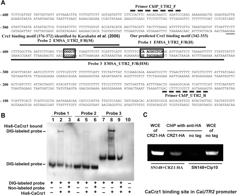
Base on the consensus motif [5′-GGAGGC(G/A)C(T/A)G-3′] from the MEME analysis described above, we found one putative CaCrz1 binding motif, the 5′-TGAGGCGTTG-3′ region in the complementary sequence of the 5′-C(− 342)AACGCCTCA(− 333)-3′ site in the promoter of one of the CaCrz1 target genes, CaUTR2 (Fig. 3a). Next, we tested the roles of this motif and the other putative CaCrz1 binding motif, 5′-G(− 376)GGCT(− 372)-3′, which was identified previously [28].
Fig. 3.
CaCrz1 binds in vitro and in vivo to two motifs in the promoter of UTR2. (a) Locations of two potential Crz1-binding motifs (boxed) in the UTR2 promoter. The 5′-TGAGGCGTTG-3′ region in the complementary sequence of the 5′-C(− 342)AACGCCTCA(− 333)-3′ site is the potential Crz1 binding motif predicted in our study, and the 5′-G(− 376)GGCT(− 372)-3′ region is the putative Crz1 binding motif identified previously (28). Locations of EMSA Probe 1 [EMSA_UTR2_F/R(H)] and Probe 2 [EMSA_UTR2_F/R(M)] are indicated with dark lines above their corresponding sequences, and EMSA Probe 3 [EMSA_UTR2_F/R(HM)] is indicated with a dark line under its corresponding sequence. Locations of the ChIP PCR primer pair [CHIP_UTR2_F和CHIP_UTR2_R] are indicated with broken lines above and under their corresponding sequences, respectively. (b) DIG-labelled probe 1 [EMSA_UTR2_F/R (H)] was added into samples in Lanes 1–3. DIG-labelled probe 2 [EMSA_UTR2_F/R(M)] was added into samples in Lanes 4–6. DIG-labelled probe 3 [EMSA_UTR2_ F/R(HM)] was added into samples in Lanes 7–9. Purified His6-Crz1 protein of 1 μg was added into Lanes 2, 3, 5, 6, 8 and 9. Unlabelled probes 1, 2 and 3 were added into samples in Lanes 3, 6 and 9, respectively. Only purified His6-Crz1 protein, but not probe DNA, were added into the sample in Lane 10. (c) Detection of Crz1 binding to the UTR2 promoter in vivo by ChIP analysis. The wild-type strain expressing Crz1-HA and the control strain integrated with CIp10 vector (no tag control) were exposed to 0.2 M CaCl2 for 1 h, and their cells were treated with formaldehyde. Whole cell extractions were obtained from collected cells, and immunoprecipitation was done with anti-HA monoclonal antibodies. Immunoprecipitated pellets were used as templates for PCR with the primer pair ChIP_UTR2_F/R. PCR products were separated on 1% agarose gel
Different from other eukaryotes, C. albicans does not follow the universal genetic code, by translating the CTG codon into serine instead of leucine [51]. Therefore, we first optimized the codon usage by mutating all five CTG codons in CaCRZ1 to TCT codon (L22S), AGC codon (L24S), TCC codons (L601S, L649S and L686S) (Additional file 1: Figure S2). The codon-optimized and Hisx6 tagged full-length CaCrz1 (His6-CaCrz1) was expressed in bacterial cells and purified (Additional file 1: Figure S3). Electrophoretic mobility shift (EMSA) assay showed that His6-CaCrz1 bound to both the P1 probe containing the putative binding motif identified in our study (Lane 2), the P2 probe containing the putative binding motif identified in the previous study [28] (Lane 5), and the Probe 3 containing two of the motifs (Lanes 8) (Fig. 3b). The binding of His6-CaCrz1 to Probe 1, Probe 2 and Probe 3 was abolished by their specific competitors, unlabelled probes, respectively (Lanes 3, 6 and 9) (Fig. 3b). Taken together, these results demonstrate that CaCrz1 can indeed bind in vitro to both motifs in the CaUTR2 promoter.
To examine if CaCrz1 binds to the CaUTR2 promoter region in vivo, we carried out chromatin immunoprecipitation (ChIP) experiments. We examined the wild-type SN148 strain expressing a chromosomally and C-terminally HA-tagged CaCrz1 (CaCrz1-HA) under the control of the CaCRZ1 promoter (left two lanes in Fig. 3c), and the wild-type SN148 strain with the untagged wild type CaCrz1 and with the CIp10 vector integrated as the control (right two lanes in Fig. 3c). DNA samples isolated from their anti-HA chromatin immunoprecipitates were used in PCR assays to detect CaCrz1-HA target promoters (The second and the third lanes in Fig. 3c). As controls, their whole-cell extracts (WCEs) were used in parallel PCR assays to ensure the equivalence of the IP starting materials (The first and the fourth lanes in Fig. 3c). We found that the promoter region containing two putative binding motifs in the CaUTR2 promoter were enriched in the anti-HA IPs of the CaCrz1-HA strain (The second lane in Fig. 3c), but not in the untagged CaCrz1 strain (The third lane in Fig. 3C). Together, these data demonstrate that CaCrz1 binds in vivo to the promoter region containing the two motifs of CaUTR2.
Mutations of two putative binding motifs in the promoter abolish the CaCrz1-regulated expression of CaUTR2
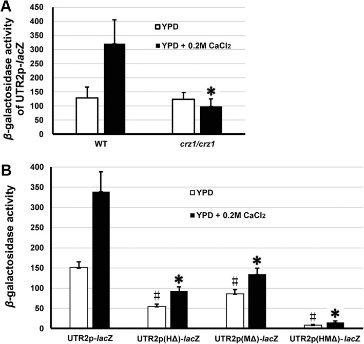
To further characterize the effects of two CaCrz1-binding motifs on the expression of CaUTR2, we generated four plasmids, CIp10-UTR2-lacZ, CIp10-UTR2(HΔ)-lacZ, CIp10-UTR2(MΔ)-lacZ and CIp10-UTR2(HMΔ)-lacZ, containing the wild-type CaUTR2 promoter, the single-motif mutated promoter UTR2(HΔ), the single-motif mutated promoter UTR2(MΔ) and the double-motif mutated promoter UTR2(HMΔ). In the absence of supplemented calcium, a basal expression level was detected for the wild type promoter UTR2-lacZ in the wild type cells (Fig. 4a). As expected, in response to 0.2 M CaCl2, the β-galactosidase activity of the wild type promoter UTR2-lacZ was increased by more than two times in the wild-type cells, but did not change significantly in the crz1/crz1 mutant cells (Fig. 4a). This indicates that the calcium-induced expression of CaUTR2 is dependent on CaCrz1.
Fig. 4.
Two CaCrz1-binding motifs in the promoter play additive roles in the regulation of CaUTR2 expression. (a), β-galactosidase activities of the wild-type promoter UTR2-lacZ in the wild-type SN148 and the crz1/crz1 mutant cells in the absence or presence of 0.2 M CaCl2. The asterisk (*) indicates statistically significant differences (P < 0.05) in the β-galactosidase activity between the wild type strain SN148 and the crz1/crz1 mutant strain in the absence or presence of 0.2 M CaCl2, respectively. (b), β-galactosidase activities of the wild-type promoter UTR2-lacZ, two single mutated promoters UTR2(HΔ)-lacZ and UTR2(MΔ)-lacZ as well as the double mutated promoter UTR2(HMΔ)-lacZ in the wild-type SN148 cells in the absence or presence of 0.2 M CaCl2. The asterisks (#) and (*) indicate statistically significant differences (P < 0.05) in the β-galactosidase activity between the wild type promoter and each of the mutated promoters in the wild-type strain SN148 in the absence or presence of 0.2 M CaCl2, respectively
As compared to the wild-type promoter UTR2(HΔ), the β-galactosidase activities of two single mutated promoters UTR2(HΔ) and UTR2(MΔ) were significantly reduced in the absence or presence of 0.2 M CaCl2 in the wild type cells (Fig. 4b). The β-galactosidase activity of the double mutated promoter UTR2(HMΔ) were even further reduced than those of two single mutated promoters UTR2(HΔ) and UTR2(MΔ) in the absence or presence of 0.2 M CaCl2 in the wild type cells (Fig. 4b). Taken together, these results suggest that two CaCrz1-binding motifs play additive roles in the regulation of CaUTR2 expression.
CaCrz1 binds in vitro and in vivo to its own promoter
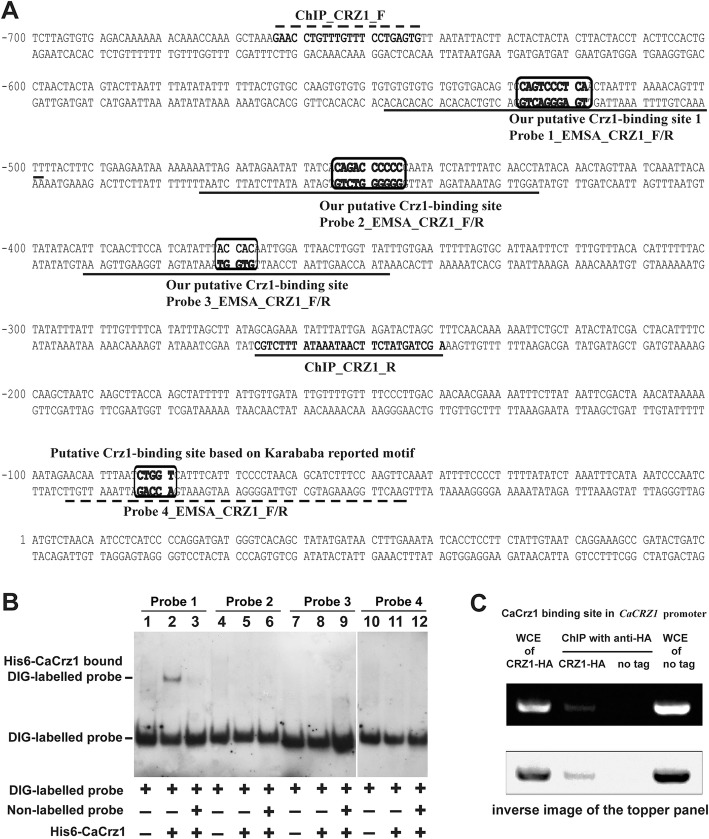
Both a previous study and our current study have observed that CaCRZ1 itself is positively regulated by CaCrz1 (Table 2; 28). Base on the consensus motif [5′-GGAGGC(G/A)C(T/A)G-3′] identified in our study, we identified two putative CaCrz1 binding motif, the 5′-T(− 519)GAGGGACTG(− 528)-3′ site (within the Probe 1 sequence) and the 5′-G(− 446)GGGGGTCTG(− 455)-3′ site (within the Probe 2 sequence) in the complementary sequence, in its own promoter (Fig. 5a). Based on the consensus motif [5′-G(C/T)GGT-3′] identified previously [28], we also identified one putative CaCrz1 binding motif, the 5′-G(− 368)TGGT(− 372)-3′ site (within the Probe 3 sequence), in the complementary sequence of CaCRZ1 promoter (Fig. 5a). The fourth putative CaCrz1 binding motif, the 5′-C(− 84)TGGT(− 80)-3′ site (within the Probe 4 sequence) was identified previously [28].
Fig. 5.
CaCrz1 binds in vitro and in vivo to its own promoter. (a) Locations of three predicated CaCrz1-binding motifs (boxed and within Probe 1, Probe 2 and Probe 3 sequences) based on the consensus motif we discovered in this study and one predicated CaCrz1-binding motif (boxed and within the Probe 4 sequence). Locations of the ChIP PCR primer pair [CHIP_CRZ1_F和CHIP_CRZ1_R] are indicated with broken lines above and under their corresponding sequences, respectively. (b) DIG-labelled Probe 1_EMSA_CRZ1_F/R was added into samples in Lanes 1–3. DIG-labelled Probe 2_EMSA_CRZ1_F/R was added into samples in Lanes 4–6. DIG-labelled Probe 3_EMSA_CRZ1_ F/R was added into samples in Lanes 7–9, and DIG-labelled Probe 4_EMSA_CRZ1_ F/R was added into samples in Lanes 10–12. Unlabelled probes 1, 2, 3 and 4 were added into samples in Lanes 3, 6, 9 and 12, respectively. Purified His6-Crz1 protein of 1 μg was added into Lanes 2, 3, 5, 6, 8, 9, 11 and 12. (C) Detection of CaCrz1 binding to its own promoter in vivo by ChIP analysis. The same pair of strains were treated and their whole cell extracts were immunoprecipitated as in Fig. 3c. PCR reactions were carried out with ChIP primers CHIP_CRZ1_F和CHIP_CRZ1_R. The lower panel is the inverse image of the topper panel, which is for a better view of the PCR band in the second lane
EMSA assay demonstrated that His6-CaCrz1 bound to only the P1 probe (Lane 2), but not to other three probes, Probe 2 (Lane 5), Probe 3 (Lane 8) and Probe 4 (Lane 11) (Fig. 5b). The binding of His6-CaCrz1 to Probe 1 was abolished by its specific competitor, unlabelled Probe 1 (Lane 3) (Fig. 5b). ChIP analysis indicated that the promoter region containing the 5′-T(− 519)GAGGGACTG(− 528)-3′ site (within the Probe 1 sequence) was enriched in the anti-HA IPs of the CaCrz1-HA strain (Lane 2), but not in the untagged CaCrz1 strain (lane 3) (Fig. 5c). These results demonstrate that CaCrz1 regulates its own expression by binding to the motif 5′-T(− 519)GAGGGACTG(− 528)-3′ in its own promoter. The autoregulation phenomenon of this transcription factor gene has also been previously shown in the rice blast pathogen M. oryzae MoCrz1 [49, 50].
Discussion
Microarrays are based on the hybridization of oligonucleotide DNA sequences, representing the entire set of genes of an organism arranged in a grid pattern, with complementary DNA (cDNA) molecules derived from the transcriptome in a cell sample, while cDNA molecules derived from a sample are directly and massively sequenced in the case of RNA-sequencing methodology [52, 53]. As compared to microarrays, RNA sequencing technology offers increased specificity and sensitivity, but the application of multiple transcriptome measurement methods can improve the comprehension of the global gene expression profile of one organism [34, 35]. Through RNA sequencing, we have identified 219 genes positively, and 59 genes negatively, regulated by CaCrz1 in response to calcium stress in C. albicans. A total of 40 out of the 219 genes identified in this study to be positively regulated by CaCrz1 account for the majority of 60 genes identified by DNA microarray analysis in the previous study (Table 2; 28). Therefore, our current study has expanded the global expression profile of genes controlled by CaCrz1 in response to calcium stress in C. albicans. This provides a basis for further understanding the regulation of calcium homeostasis in this important human fungal pathogen.
In addition to the CaCrz1-binding motif (M) identified in the previous study [28], we have revealed a novel CaCrz1-binding motif (H) through the MEME analysis of promoters of 40 common genes identified to be controlled by CzCrz1 through both RNA sequencing and microarray approaches (Fig. 3). Furthermore, we have demonstrated that CaCrz1 binds in vitro and in vivo to these two motifs in the promoter of its target gene CaUTR2, and that these two calcineurin-dependent response elements (CDREs) might play additive roles in the regulation of CaUTR2 expression (Fig. 6). Similarly, two MoCrz1-binding motifs in promoters of target genes have been demonstrated in the rice fungal pathogen M. oryzae [49]. Among the 219 genes positively regulated by CaCrz1, we found that promoters of 79 genes contain both motifs (M and H), promoters of 59 genes contain only motif H, promoters of 45 genes contain only motif M, and promoters of 36 genes contain neither motif H or motif M (Additional file 2). Therefore, expression of target genes seems to be differentially regulated by CaCrz1.
Fig. 6.
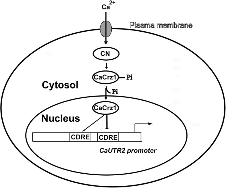
Schematic model for the regulation of CaUTR2 expression by the transcription factor CaCrz1 in response of C. albicans cells to extracellular calcium stress. Influx of extracellular calcium ions to the cytosol leads to the activation of calcineurin, which in turn dephosphorylates and activates CaCrz1. Dephosphorylated CaCrz1 enters to the nucleus to bind to two CaCrz1 binding motifs (calcineurin dependent response element; CDRE) in the promoter of CaUTR2, which results in the activation of CaUTR2 expression
In S. cerevisiae, 125 calcium-specific and calcineurin-dependent genes reported in a previous study [48]. Out of these 125 genes, there are 83 genes that are positively regulated by ScCrz1 (Additional file 3). From the C. albicans database (http://www.candidagenome.org/), we were able to find 38 C. albicans homologs for these ScCrz1-dependent S. cerevisiae genes, but only 9 of these 38 C. albicans homologs are present in the list of genes identified in this study to be CaCrz1-dependent (Table 2; Additional file 3). Therefore, target genes of ScCrz1 and CaCrz1 seem to be very divergent. This is supported by our observation that the amino acid sequences of ScCrz1 and CaCrz1 shares only 31.9 and 24% similarity and identity, respectively, although their predicted structures are very similar (Fig. S4 in Additional file 1). Similar to the homologs in S. cerevisiae, M. oryzae and another human fungal pathogen Aspergillus fumigatus [49], expression of PMC1 (C3_01250W_A) and RCT1 (C3_05710W) is positively controlled by CaCrz1, although expression of RCN1 (C6_01160W_A) is not regulated by CaCrz1 (SuppInfo 1 and 2; GEO Accession number: GSE123122). This is consistent with previous observations on Cryptococcus neoformans CBP1, the homolog of RCN1, that neither is regulated by nor interacts with Crz1 in this human fungal pathogen [54, 55]. In contrast, expression of RCN1 is regulated by Crz1 in S. cerevisiae, M. oryzae and another human fungal pathogen Aspergillus fumigatus, which forms a feedback mechanism for the regulatory role of Rcn1 as an inhibitor of calcineurin [48, 55, 56]. Nevertheless, overexpression of C. albicans RCN1 could inhibit S. cerevisiae calcineurin function [21]. Taken together, these data indicate that regulation of the calcium/calcineurin signaling pathway is diverged in fungal pathogens, although the core calcium signaling machinery (calmodulin, calcineurin and Crz1) is highly conserved across these species. This is consistent with the previous hypothesis [49, 56, 57].
It is interesting to note that the calcium-sensitive phenotype of the CRISPR mutant for CaCRZ1 could only be partially reversed by the introduction of the full-length CaCRZ1 gene back to its genome (Fig. 1). Transcripts of the CRISPR mutant CaCRZ1 from the CaCRZ1 locus might compete with those of the wild-type CaCRZ1 transcripts derived from CIp10-CaCRZ1 at the CaRPS1 locus, which might interfere with the translational efficiency of wild-type CaCRZ1 transcripts. This might explain the partial complementation of calcium sensitivity of the CRISPR mutant for CaCRZ1 by CIp10-CaCRZ1. Furthermore, the full-length 6xHis tagged CaCrz1 protein expresses in bacterial cells as a protein of about 100 kDa (Additional file 1: Figure S3), which is much bigger than its predicted size (= 80 kDa). However, the dephosphorylated form of CaCrz1 expressed in C. albicans cells in response to calcium stress also shows a molecular weight of more than 100 kDa (Fig. 2), which is similar to that of CaCrz1 expressed in bacterial cells. Therefore, this mobility shift could be due to the conformation of CaCrz1 itself, but not the host cell environment or the tag type or tag location (N-terminus or C-terminus).
Conclusions
In this study, through RNA sequencing we have identified 219 genes that are positively, and 59 genes that are negatively, controlled by CaCrz1 in response to calcium stress. We have also revealed and demonstrated experimentally a novel consensus CaCrz1-binding motif [5′-GGAGGC(G/A)C(T/A)G-3′] in promoters of CaCrz1 target genes. In addition, CaCrz1 binds to its own promoter and shows an autoregulatory mechanism for CaCRZ1 expression. These findings would contribute to our further understanding of molecular mechanisms regulating calcium homeostasis.
Supplementary information
Additional file 2: Figure S1. Construction and phenotypes of CRISPR mutants for CaCRZ1. Figure S2. Alignment between the amino acid sequences of the wild type and the codon optimized version of CaCrz1. Figure S3. Expression and purification of the codon optimized and His6-tagged CaCrz1 protein in bacterial cells. Figure S4. Differences between CaCrz1 and ScCrz1. Table S1. Primers used in this study.
Additional file 3. Promoter analysis of 219 genes whose expression is positively regulated by CaCrz1#
Additional file 4. Comparison of calcium-specific and Crz1-dependent genes in Saccharomyces cerevisiae and Candida albicans.
Acknowledgements
We gratefully acknowledge the help of Malcolm Whiteway’ laboratory members for technical support.
Ethical approval and consent to participate
N/A
Abbreviations
- ChIP
Chromatin immunoprecipitation
- CRISPR
Clustered regularly interspaced short palindromic repeat
- Crz1
Calcineurin-responsive zinc finger 1
- EMSA
Gel electrophoretic mobility shift assay
- MEME
Multiple em for motif elicitation
- NFAT
the nuclear factor of activated T cells
- PCR
Polymerase chain reaction
- YPD
Yeast peptone dextron
Authors’ contributions
HX, TF and ORP performed the experiments. LJ designed the study and wrote the manuscript. LJ and MW analyzed the data. All authors read and approved the final manuscript.
Authors’ information
N/A
Funding
This work was funded by the National Natural Science Foundation of China to LJ (No. 81571966 and No. 81371784) and the NSERC Discovery to MW (RGPIN/4799) and the CRC to MW (950–228957).
Availability of data and materials
All data generated or analyzed during this study are included in this published article and deposited in the database Gene Expression Omnibus (GEO) site.
Consent for publication
All authors approved the final manuscript.
Competing interests
The authors declare that they have no competing interests.
Footnotes
Publisher’s Note
Springer Nature remains neutral with regard to jurisdictional claims in published maps and institutional affiliations.
Contributor Information
Huihui Xu, Email: 835728196@qq.com.
Tianshu Fang, Email: 531503280@qq.com.
Raha Parvizi Omran, Email: raha.parvizi.o@gmail.com.
Malcolm Whiteway, Email: malcolm.whiteway@concordia.ca.
Linghuo Jiang, Email: linghuojiang@sdut.edu.cn.
Supplementary information
Supplementary information accompanies this paper at 10.1186/s12964-019-0473-9.
References
- 1.Tang RJ, Luan S. Regulation of calcium and magnesium homeostasis in plants: from transporters to signaling network. Curr Opin Plant Biol. 2017;39:97–105. doi: 10.1016/j.pbi.2017.06.009. [DOI] [PubMed] [Google Scholar]
- 2.Bond R, Ly N, Cyert MS. The unique C terminus of the calcineurin isoform CNAβ1 confers non-canonical regulation of enzyme activity by Ca2+ and calmodulin. J Biol Chem. 2017;292:16709–16721. doi: 10.1074/jbc.M117.795146. [DOI] [PMC free article] [PubMed] [Google Scholar]
- 3.Espeso EA. The CRaZy calcium cycle. Adv Exp Med Biol. 2016;892:169–186. doi: 10.1007/978-3-319-25304-6_7. [DOI] [PubMed] [Google Scholar]
- 4.Plattner H, Verkhratsky A. The ancient roots of calcium signalling evolutionary tree. Cell Calcium. 2015;57:123–132. doi: 10.1016/j.ceca.2014.12.004. [DOI] [PubMed] [Google Scholar]
- 5.Medler KF. Calcium signaling in taste cells: regulation required. Chem Senses. 2010;35:753–765. doi: 10.1093/chemse/bjq082. [DOI] [PMC free article] [PubMed] [Google Scholar]
- 6.Serra-Cardona A, Canadell D, Arino J. Coordinate responses to alkaline pH stress in budding yeast. Microb Cell. 2015;2:182–196. doi: 10.15698/mic2015.06.205. [DOI] [PMC free article] [PubMed] [Google Scholar]
- 7.Cui J, Kaandorp JA, Sloot PM, Lloyd CM, Filatov MV. Calcium homeostasis and signaling in yeast cells and cardiac myocytes. FEMS Yeast Res. 2009;9:1137–1147. doi: 10.1111/j.1567-1364.2009.00552.x. [DOI] [PubMed] [Google Scholar]
- 8.Colinet AS, Thines L, Deschamps A, Flémal G, Demaegd D, Morsomme P. Acidic and uncharged polar residues in the consensus motifs of the yeast Ca2+ transporter Gdt1p are required for calcium transport. Cell Microbiol. 2017;19(7). https://doi.org/10.1111/cmi.%2012729. [DOI] [PubMed]
- 9.Zhao Y, Yan H, Happeck R, Peiter-Volk T, Xu H, Zhang Y, Peiter E, van Oostende TC, Whiteway M, Jiang L. The plasma membrane protein Rch1 is a negative regulator of cytosolic calcium homeostasis and positively regulated by the calcium/calcineurin signaling pathway in budding yeast. Eur J Cell Biol. 2016;95:164–174. doi: 10.1016/j.ejcb.2016.01.001. [DOI] [PubMed] [Google Scholar]
- 10.Cyert MS, Philpott CC. Regulation of cation balance in Saccharomyces cerevisiae. Genetics. 2013;193:677–713. doi: 10.1534/genetics.112.147207. [DOI] [PMC free article] [PubMed] [Google Scholar]
- 11.Zhao Y, Du J, Zhao G, Jiang L. Activation of calcineurin is mainly responsible for the calcium sensitivity of gene deletion mutations in the genome of budding yeast. Genomics. 2013;101:49–56. doi: 10.1016/j.ygeno.2012.09.005. [DOI] [PubMed] [Google Scholar]
- 12.Dadar M, Tiwari R, Karthik K, Chakraborty S, Shahali Y, Dhama K. Candida albicans - Biology, molecular characterization, pathogenicity, and advances in diagnosis and control - An update. Microb Pathog. 2018;117:128–138. doi: 10.1016/j.micpath.2018.02.028. [DOI] [PubMed] [Google Scholar]
- 13.Yu SJ, Chang YL, Chen YL. Calcineurin signaling: lessons from Candida species. FEMS Yeast Res. 2015;15(4). https://doi.org/10.1093/femsyr/fov016. [DOI] [PubMed]
- 14.Chauhan N, Latge JP, Calderone R. Signalling and oxidant adaptation in Candida albicans and Aspergillus fumigatus. Nat Rev Microbiol. 2006;4:435–444. doi: 10.1038/nrmicro1426. [DOI] [PubMed] [Google Scholar]
- 15.Jiang L, Xu D, Hameed A, Fang T. Bakr Ahmad Fazili a, Asghar F. The plasma membrane protein Rch1 and the Golgi/ER calcium pump Pmr1 have an additive effect on filamentation in Candida albicans. Fungal Genet Biol. 2018;115:1–8. doi: 10.1016/j.fgb.2018.04.001. [DOI] [PubMed] [Google Scholar]
- 16.Jiang L, Alber J, Wang J, Du W, Li X, Geyer J. The Candida albicans plasma membrane protein Rch1p a member of the vertebrate SLC10 carrier family, is a novel regulator of cytosolic Ca2+ homoeostasis. Biochem J. 2012;444:497–502. doi: 10.1042/BJ20112166. [DOI] [PubMed] [Google Scholar]
- 17.Liu S, Hou Y, Liu W, Lu C, Wang W, Sun S. Components of the calcium-calcineurin signaling pathway in fungal cells and their potential as antifungal targets. Eukaryot Cell. 2015;14:324–334. doi: 10.1128/EC.00271-14. [DOI] [PMC free article] [PubMed] [Google Scholar]
- 18.Wang Y, Wang J, Cheng J, Xu D, Jiang L. Genetic interactions between the Golgi Ca2+/H+ exchanger Gdt1 and the plasma membrane calcium channel Cch1/Mid1 in the regulation of calcium homeostasis, stress response and virulence in Candida albicans. FEMS Yeast Res. 2015;15:(7). https://doi.org/10.1093/femsyr/fov069. [DOI] [PubMed]
- 19.Xu D, Cheng J, Cao C, Wang L, Jiang L. Genetic interactions between Rch1 and the high-affinity calcium influx system Cch1/Mid1/Ecm7 in the regulation of calcium homeostasis, drug tolerance, hyphal development and virulence in Candida albicans. FEMS Yeast Res. 2015;15:(7). https://doi.org/10.1093/femsyr/fov079. [DOI] [PubMed]
- 20.Alber J, Jiang L, Geyer J. CaRch1p does not functionally interact with the high-affinity Ca2+ influx system (HACS) of Candida albicans. Yeast. 2013;30:449–457. doi: 10.1002/yea.2981. [DOI] [PubMed] [Google Scholar]
- 21.Reedy JL, Filler SG, Heitman J. Elucidating the Candida albicans calcineurin signaling cascade controlling stress response and virulence. Fungal Genet Biol. 2010;47:107–116. doi: 10.1016/j.fgb.2009.09.002. [DOI] [PMC free article] [PubMed] [Google Scholar]
- 22.Li X, Hou Y, Yue L, Liu S, Du J, Sun S. Potential targets for antifungal drug discovery based on growth and virulence in Candida albicans. Antimicrob Agents Chemother. 2015;59:5885–5891. doi: 10.1128/AAC.00726-15. [DOI] [PMC free article] [PubMed] [Google Scholar]
- 23.Thewes S. Calcineurin-Crz1 signaling in lower eukaryotes. Eukaryot Cell. 2014;13:694–705. doi: 10.1128/EC.00038-14. [DOI] [PMC free article] [PubMed] [Google Scholar]
- 24.Gow NA, van de Veerdonk FL, Brown AJ, Netea MG. Candida albicans morphogenesis and host defence: discriminating invasion from colonization. Nat Rev Microbiol. 2011;10:112–122. doi: 10.1038/nrmicro2711. [DOI] [PMC free article] [PubMed] [Google Scholar]
- 25.Sanglard D, Ischer F, Marchetti O, Entenza J, Bille J. Calcineurin a of Candida albicans: involvement in antifungal tolerance, cell morphogenesis and virulence. Mol Microbiol. 2003;48:959–976. doi: 10.1046/j.1365-2958.2003.03495.x. [DOI] [PubMed] [Google Scholar]
- 26.Blankenship JR, Wormley FL, Boyce MK, Schell WA, Filler SG, Perfect JR, Heitman J. Calcineurin is essential for Candida albicans survival in serum and virulence. Eukaryot Cell. 2003;2:422–430. doi: 10.1128/EC.2.3.422-430.2003. [DOI] [PMC free article] [PubMed] [Google Scholar]
- 27.Cruz MC, Goldstein AL, Blankenship JR, del Poeta M, Davis D, Cardenas ME, Perfect JR, McCusker JH, Heitman J. Calcineurin is essential for survival during membrane stress in Candida albicans. EMBO J. 2002;21:546–559. doi: 10.1093/emboj/21.4.546. [DOI] [PMC free article] [PubMed] [Google Scholar]
- 28.Karababa M, Valentino E, Pardini G, Coste AT, Bille J, Sanglard D. CRZ1, a target of the calcineurin pathway in Candida albicans. Mol Microbiol. 2006;59:1429–1451. doi: 10.1111/j.1365-2958.2005.05037.x. [DOI] [PubMed] [Google Scholar]
- 29.Onyewu C, FLJr W, Perfect JR, Heitman J. The calcineurin target, Crz1, functions in azole tolerance but is not required for virulence of Candida albicans. Infect Immun. 2004;72:7330–7333. doi: 10.1128/IAI.72.12.7330-7333.2004. [DOI] [PMC free article] [PubMed] [Google Scholar]
- 30.Xu H, Whiteway M, Jiang L. The tricarboxylic acid cycle, cell wall integrity pathway, cytokinesis and intracellular pH homeostasis are involved in the sensitivity of Candida albicans cells to high levels of extracellular calcium. Genomics. 2018;pii: S0888–7543(18)30307–0. https://doi.org/10.1016/j.ygeno.%202018.08.001. [DOI] [PubMed]
- 31.Chen Y, Mallick J, Maqnas A, Sun Y, Choudhury BI, Cote P, Yan L, Ni TJ, Li Y, Zhang D, Rodríguez-Ortiz R, Lv QZ , Jiang YY, Whiteway M. Chemogenomic profiling of the fungal pathogen Candida albicans. Antimicrob Agents Chemother. 2018;62:pii: e02365–17. https://doi.org/10.1128/AAC.02365-17. [DOI] [PMC free article] [PubMed]
- 32.Roemer T, Jiang B, Davison J, Ketela T, Veillette K, Breton A, Tandia F, Linteau A, Sillaots S, Marta C, Martel N, Veronneau S, Lemieux S, Kauffman S, Becker J, Storms R, Boone C, Bussey H. Large-scale essential gene identification in Candida albicans and applications to antifungal drug discovery. Mol Microbiol. 2003;50:167–181. doi: 10.1046/j.1365-2958.2003.03697.x. [DOI] [PubMed] [Google Scholar]
- 33.Mille C, Janbon G, Delplace F, Ibata-Ombetta S, Gaillardin C, Strecker G, Jouault T, Trinel PA, Poulain D. Inactivation of CaMIT1 inhibits Candida albicans phospholipomannan beta-mannosylation, reduces virulence, and alters cell wall protein beta-mannosylation. J Biol Chem. 2004;279:47952–47960. doi: 10.1074/jbc.M405534200. [DOI] [PubMed] [Google Scholar]
- 34.Zhao W, He X, Hoadley KA, Parker JS, Hayes DN, Perou CM. Comparison of RNA-Seq by poly (A) capture, ribosomal RNA depletion, and DNA microarray for expression profiling. BMC Genomics. 2014;15:419. doi: 10.1186/1471-2164-15-419. [DOI] [PMC free article] [PubMed] [Google Scholar]
- 35.Kogenaru S, Qing Y, Guo Y, Wang N. RNA-seq and microarray complement each other in transcriptome profiling. BMC Genomics. 2012;13:629. doi: 10.1186/1471-2164-13-629. [DOI] [PMC free article] [PubMed] [Google Scholar]
- 36.Wang Z, Gerstein M, Snyder M. RNA-Seq: a revolutionary tool for transcriptomics. Nat Rev Genet. 2009;10:57–63. doi: 10.1038/nrg2484. [DOI] [PMC free article] [PubMed] [Google Scholar]
- 37.Nagalakshmi U, Wang Z, Waern K, Shou C, Raha D, Gerstein M, Snyder M. The transcriptional landscape of the yeast genome defined by RNA sequencing. Science. 2008;320:1344–1349. doi: 10.1126/science.1158441. [DOI] [PMC free article] [PubMed] [Google Scholar]
- 38.Sun Y, Gadoury C, Hirakawa MP, Bennett RJ, Harcus D, Marcil A, Whiteway M. Deletion of a Yci1 domain protein of Candida albicans allows homothallic mating in MTL heterozygous cells. MBio. 2016;7:(2). https://doi:10.1128/mBio.00465-16 [DOI] [PMC free article] [PubMed]
- 39.Vyas VK, Barrasa IM, Fink GR. A Candida albicans CRISPR system permits genetic engineering of essential genes and gene families. Sci Adv. 2015;1:(3). https://doi:10.1126/sciadv.1500248 [DOI] [PMC free article] [PubMed]
- 40.Brand A, Lee K, Veses V, Gow NA. Calcium homeostasis is required for contact-dependent helical and sinusoidal tip growth in Candida albicans hyphae. Mol Microbiol. 2009;71:1155–1164. doi: 10.1111/j.1365-2958.2008.06592.x. [DOI] [PMC free article] [PubMed] [Google Scholar]
- 41.Asghar F, Yan H, Jiang L. The putative transcription factor CaMaf1 controls the sensitivity to lithium and rapamycin and represses RNA polymerase III transcription in Candida albicans. FEMS Yeast Res. 2018;18:(6). https://doi.org/10.1093/femsyr/foy068. [DOI] [PubMed]
- 42.Zhao Y, Feng J, Li J, Jiang L. Mithochondrial type 2C protein phosphatases CaPtc5p, CaPtc6p, and CaPtc7p play vital roles in cellular responses to antifungal drugs and cadmium in Candida albicans. FEMS Yeast Res. 2012;12:897–906. doi: 10.1111/j.1567-1364.2012.00840.x. [DOI] [PubMed] [Google Scholar]
- 43.Yu L, Zhao J, Feng J, Fang J, Feng C, Jiang Y, Cao Y, Jiang L. Candida albicans CaPTC6 is a functional homolog for Saccharomycese cerevisiae ScPTC6 and encodes a type 2C protein phosphatase. Yeast. 2010;27:197–206. doi: 10.1002/yea.1743. [DOI] [PubMed] [Google Scholar]
- 44.Jiang L, Whiteway M, Shen SH. A novel type 2C protein phosphatase from the human fungal pathogen. Candida albicans FEBS Lett. 2001;509:42–144. doi: 10.1016/s0014-5793(01)03125-8. [DOI] [PubMed] [Google Scholar]
- 45.Jiang L, Wang J, Asghar F, Snyder N, Cunningham KW. CaGdt1 plays a compensatory role for the calcium pump CaPmr1 in the regulation of calcium signaling and cell wall integrity signaling in Candida albicans. Cell Commun Signal. 2018;16:33. doi: 10.1186/s12964-018-0246-x. [DOI] [PMC free article] [PubMed] [Google Scholar]
- 46.Yan H, Zhao Y, Jiang L. The putative transcription factor CaRtg3 is involved in tolerance to cations and antifungal drugs as well as serum-induced filamentation in Candida albicans. FEMS Yeast Res. 2014;14:614–623. doi: 10.1111/1567-1364.12148. [DOI] [PubMed] [Google Scholar]
- 47.Zhao Y, Du J, Xiong B, Xu H, Jiang L. ESCRT components regulate the expression of the ER/Golgi calcium pump gene PMR1 through the Rim101/Nrg1 pathway in budding yeast. J Mol Cell Biol. 2013;5:336–344. doi: 10.1093/jmcb/mjt025. [DOI] [PubMed] [Google Scholar]
- 48.Yoshimoto H, Saltsman K, Gasch AP, Li HX, Ogawa N, Botstein D, Brown PO, Cyert MS. Genome-wide analysis of gene expression regulated by the calcineurin/Crz1p signaling pathway in Saccharomyces cerevisiae. J Biol Chem. 2002;277:31079–31088. doi: 10.1074/jbc.M202718200. [DOI] [PubMed] [Google Scholar]
- 49.Kim S, Hu J, Oh Y, Park J, Choi J, Lee YH, Dean RA, Mitchell TK. Combining ChIP-chip and expression profiling to model the MoCRZ1 mediated circuit for Ca/calcineurin signaling in the rice blast fungus. PLoS Pathog. 2010;6:(5). https://doi.org/10.1371/%20journal.ppat.1000909. [DOI] [PMC free article] [PubMed]
- 50.Choi J, Kim Y, Kim S, Park J, Lee YH. MoCRZ1, a gene encoding a calcineurin-responsive transcription factor, regulates fungal growth and pathogenicity of Magnaporthe oryzae. Fungal Genet Biol. 2009;46:243–254. doi: 10.1016/j.fgb.2008.11.010. [DOI] [PubMed] [Google Scholar]
- 51.Miranda I, Rocha R, Santos MC, Mateus DD, Moura GR, Carreto L, Santos MA. A genetic code alteration is a phenotype diversity generator in the human pathogen Candida albicans. PLoS One. 2007;2:(10). https://doi:10.1371/journal.pone.0000996 [DOI] [PMC free article] [PubMed]
- 52.Schena M, Shalon D, Davis RW, Brown PO. Quantitative monitoring of gene expression patterns with a complementary DNA microarray. Science. 1995;270:467–470. doi: 10.1126/science.270.5235.467. [DOI] [PubMed] [Google Scholar]
- 53.Mortazavi A, Williams BA, McCue K, Schaeffer L, Wold B. Mapping and quantifying mammalian transcriptomes by RNA-Seq. Nat Methods. 2008;5:621–628. doi: 10.1038/nmeth.1226. [DOI] [PubMed] [Google Scholar]
- 54.Fu C, Donadio N, Cardenas ME, Heitman J. Dissecting the roles of the calcineurin pathway in unisexual reproduction, stress responses, and virulence in Cryptococcus deneoformans. Genetics. 2018;208:639–653. doi: 10.1534/genetics.117.300422. [DOI] [PMC free article] [PubMed] [Google Scholar]
- 55.Fox DS, Heitman J. Calcineurin-binding protein Cbp1 directs the specificity of calcineurin-dependent hyphal elongation during mating in Cryptococcus neoformans. Eukaryot Cell. 2005;4:1526–1538. doi: 10.1128/EC.4.9.1526-1538.2005. [DOI] [PMC free article] [PubMed] [Google Scholar]
- 56.Kraus PR, Heitman J. Coping with stress: calmodulin and calcineurin in model and pathogenic fungi. Biochem Biophys Res Commun. 2003;311:1151–1157. doi: 10.1016/S0006-291X(03)01528-6. [DOI] [PubMed] [Google Scholar]
- 57.Soriani FM, Mala I, da Silva Ferreira ME, Savoldi M, von Zeska Kress MR, de Souza Goldman MH, Loss O, Bignell E, Goldman GH. Functional characterization of the Aspergillus fumigatus CRZ1 homologue. CrzA Mol Microbiol. 2008;67:1274–1291. doi: 10.1111/j.1365-2958.2008.06122.x. [DOI] [PubMed] [Google Scholar]
- 58.Kingsbury TJ, Cunningham KW. A conserved family of calcineurin regulators. Genes Dev. 2000;14:1595–1604. [PMC free article] [PubMed] [Google Scholar]
Associated Data
This section collects any data citations, data availability statements, or supplementary materials included in this article.
Supplementary Materials
Additional file 2: Figure S1. Construction and phenotypes of CRISPR mutants for CaCRZ1. Figure S2. Alignment between the amino acid sequences of the wild type and the codon optimized version of CaCrz1. Figure S3. Expression and purification of the codon optimized and His6-tagged CaCrz1 protein in bacterial cells. Figure S4. Differences between CaCrz1 and ScCrz1. Table S1. Primers used in this study.
Additional file 3. Promoter analysis of 219 genes whose expression is positively regulated by CaCrz1#
Additional file 4. Comparison of calcium-specific and Crz1-dependent genes in Saccharomyces cerevisiae and Candida albicans.
Data Availability Statement
All data generated or analyzed during this study are included in this published article and deposited in the database Gene Expression Omnibus (GEO) site.


