Abstract
The present study aimed to determine the relationship between leg bone length and running performance in well-trained endurance runners. The lengths of the leg bones in 42 male endurance runners (age: 20.0 ± 1.0 years, body height: 169.6 ± 5.6 cm, body mass: 56.4 ± 5.1 kg, personal best 5000-m race time: 14 min 59 s ± 28 s) were measured using magnetic resonance imaging. The lengths of the femur and tibia were calculated to assess the upper and lower leg lengths, respectively. The total length of the femur + tibia was calculated to assess the overall leg bone length. These lengths of the leg bones were normalized with body height, which was measured using a stadiometer to minimize differences in body size among participants. The relative tibial length was significantly correlated with personal best 5000-m race time (r = -0.328, p = 0.034). Moreover, a trend towards significance was observed in the relative femoral length (r = -0.301, p = 0.053). Furthermore, the relative total lengths of the femur + tibia were significantly correlated with personal best 5000-m race time (r = -0.353, p < 0.05). These findings suggest that although the relationship between the leg bone length and personal best 5000-m race time was relatively minor, the leg bone length, especially of the tibia, may be a potential morphological factor for achieving superior running performance in well-trained endurance runners.
Key words: bone morphology, running economy, Achilles tendon length, step length, magnetic resonance imaging
Introduction
Previous studies have determined that several morphological properties of the muscles and tendons of the lower limb are associated with running performance (Barnes and Kilding, 2015; Saunders et al., 2004). Furthermore, a recent study found that longer forefoot bones, which were measured using magnetic resonance imaging (MRI), were related to better running performance in endurance runners (Ueno et al., 2018a). Thus, in addition to muscle and tendon morphologies, bone morphology may play an important role in achieving superior running performance in endurance runners; however, this is poorly understood.
Previous studies have reported that absolute upper and lower legs in the East African endurance runners, who have superior running performance and running economy, are longer than those in endurance runners of other regions (Kunimasa et al., 2014; Lucia et al., 2006; Sano et al., 2015; Verillo et al., 2013). Additionally, Mooses et al. (2015) determined a positive correlation between absolute upper leg length and running performance in Kenyan endurance runners. They also reported that absolute and relative total lengths (normalized with body height) of the upper and lower legs were positively correlated with running performance. Moreover, Laumets et al. (2017) reported that a longer absolute lower leg was related to better running economy in Caucasian endurance runners. Since leg length affects stride length (Cavanagh and Kram, 1989), longer legs can ensure that endurance runners run more efficiently by devoting less energy to leg acceleration (Rahmani et al., 2004). Thus, leg
length may be one of the important morphological factors for achieving higher running performance in endurance runners. However, considering that previous studies have provided some potential variables for leg length, an optimal variable for assessing running performance is not fully understood.
In general, leg length is measured using an anthropometrical technique, which is performed manually with a tape measure. Thus, previous studies had a technical limitation due to the ambiguity of leg length measurements using this technique. Compared to anthropometrical measurement, MRI is more appropriate for morphological measurements, including bone length measurement, in humans. However, no study has examined the relationship between MRI-measured leg bone lengths and running performance. In this study, we examined whether longer leg bones would be an important morphological factor for achieving better running performance in well-trained endurance runners. Previous studies have determined the relationship between leg length and running performance using anthropometrical measurements of the femoral and tibial lengths, which were measured as markers of the upper and lower leg lengths. Thus, we calculated the lengths of the femur and tibia to assess the upper and lower leg lengths, respectively. Additionally, we calculated the total length of the femur + tibia to assess the overall leg bone length, as in previous studies (Laumets et al., 2017; Mooses et al., 2015).
Methods
Participants
Forty-two Japanese male endurance runners (age: 20.0 ± 1.0 years, body height: 169.6 ± 5.6 cm, body mass: 56.4 ± 5.1 kg) participated in this study. They were all well-trained, being involved in regular training and competition. Their personal best times of the 5000-m race within the past 1 year before the measurement of the present study that were recorded ranged from 13 min 54 s to 15 min 54 s (mean, 14 min 59 s ± 28 s). The runners were informed of the experimental procedures and provided written consent to participate in the study. None of the participants had contraindications to MRI. All procedures were approved by the Ethics Committee of the Ritsumeikan University (BKC-IRB-2016-047).
MRI measurements
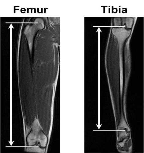
Representative images for calculating the lengths of leg bones on magnetic resonance imaging (MRI) are shown in Figure 1. The MRI measurement was performed using a 1.5-T magnetic resonance system (Signa HDxt; GE Medical Systems, Milwaukee, WI, USA). The participant was positioned supine on the scanner bed with both knees fully extended and both the ankles set at the neutral position (ie, 0°). To measure the lengths of the leg bones, the sagittal T1-weighted MRI scans of the thigh and lower leg were acquired with a standard body coil. The coronal scans were obtained in success slices with a repetition time of 600 ms, echo time of 8.5 ms, slice thickness of 8 mm, field of view of 480 mm, and matrix size of 512 × 256 pixels.
Figure 1.

Representative magnetic resonance imaging scans used for measuring the lengths of the femur and tibia The femoral and tibial lengths were measured using coronal images for the upper and lower legs, respectively. The femoral length was measured as the distance between the tip of the greater trochanter and the distal end of the lateral condyle of the femur. The tibial length was measured as the distance between the proximal end of the lateral condyle and the distal inferior surface of the tibia.
Regarding leg bone length analyses, the femoral and tibial lengths were measured using coronal images for the upper and lower legs, respectively. The femoral length was calculated as the distance between the tip of the greater trochanter and the distal end of the lateral condyle of the femur. The tibial length was calculated as the distance between the proximal end of the lateral condyle and the distal inferior surface of the tibia. Moreover, the total length of the femur + tibia was calculated to assess the overall leg bone length. In addition to absolute bone length, these lengths were normalized with body height to minimize the differences in body size among participants. The body height was measured to the nearest 0.1 cm using a stadiometer under barefoot condition. Furthermore, the ratio of the lengths of tibia/femur was calculated to evaluate interaction between the lengths of thigh and lower leg bones.
The analyses for measuring the lengths of the leg bones were conducted using image analysis software (OsiriX Version 5.6; OsiriX Foundation, Geneva, Switzerland). The measurements of the leg bone lengths were performed twice, and the mean of the two measurements was used. The coefficient of variation for the two measurements in all runners was 0.1 ± 0.2% for femoral length and 0.1 ± 0.1% for tibial length. The intraclass correlation coefficient for the two measurements in all participants was 0.997 (95% CI, 0.995-0.998) for femoral length and 0.999 (95% CI, 0.999-1.000) for tibial length.
To further determine the reproducibility of measurements of the leg bone lengths obtained in this study, we measured femoral and tibial lengths on 2 separate days in 16 healthy young men (age, 20.2 ± 0.9 years; body height, 170. 7 ± 4.8 cm; body mass, 60.5 ± 7.7 kg). The intraclass correlation for the 2 days was 0.998 (95% CI, 0.993-0.999) for the femoral length and 0.997 (95% CI, 0.993- 0.999) for the tibial length.
To ensure the validity of tibial length as the lower leg length, we examined the relationship between tibial and fibular lengths in all participants. The fibular length was measured using sagittal images for the lower leg and calculated as the distance between the proximal end of the head and the distal end of the lateral malleolus of the fibula. Tibial length was very strongly correlated with fibular length (r = 0.979, p < 0.001).
Statistical analysis
The data are presented as the mean ± SD. The relationship between variables was evaluated using a Pearson’s product moment correlation. Statistical significance was defined at p < 0.05. All statistical analyses were conducted using IBM SPSS software (version 19.0; International Business Machines Corp, NY, USA).
Results
The leg bone length variables are summarized in Table 1. The absolute lengths of the femur and tibia were significantly correlated with body height (r = 0.852 and 0.866, respectively; p < 0.001 for both).
Table1.
Leg bone length variables in endurance runners
| Mean ± SD | Range | |
|---|---|---|
| Absolute Leg bone length | ||
| Femur, mm | 420.8 ± 20.2 | 369.1 - 464.0 |
| Tibia, mm | 351.3 ± 18.2 | 309.4 - 389.2 |
| Absolute total leg bone length | ||
| Femur + Tibia, mm | 772.1 ± 37.2 | 678.4 - 852.3 |
| Relative leg bone length | ||
| Femur, % of body height | 24.8 ± 0.6 | 23.4 - 26.0 |
| Tibia, % of body height | 20.7 ± 0.6 | 19.6 - 22.4 |
| Relative total leg length | ||
| Femur + Tibia, % of body height | 45.5 ± 1.1 | 43.0 - 47.8 |
| Ratio of leg bone length | ||
| Tibia/femur | 0.84 ± 0.02 | 0.79 - 0.88 |
The lengths of the leg bones were normalized with body height to calculate relative bone lengths, that were expressed as percentages (i.e., % of body height)
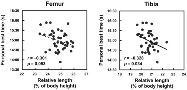
The correlation coefficients between leg bone length variables and running performance are shown in Table 2. There were no significant correlations between the absolute lengths of the femur and tibia and personal best 5000-m race time (r = -0.246 and r = -0.255, respectively; p > 0.050 for both). However, after each leg bone length was normalized with body height, the relative tibial length was significantly correlated with personal best 5000-m race time (Figure 2). Moreover, a trend towards significance was observed in the relative femoral length (r = -0.301, p = 0.053).
Table 2.
Correlation coefficients between leg bone length variables and personal best 5000-m race time
| r | p | |
|---|---|---|
| Absolute leg bone length | ||
| Femur | -0.246 | 0.117 |
| Tibia | -0.255 | 0.104 |
| Absolute total leg bone length | ||
| Femur + Tibia | -0.257 | 0.100 |
| Relative leg bone length | ||
| Femur | -0.301 | 0.053 |
| Tibia | ‐0.328 | 0.034 |
| Relative total leg length | ||
| Femur + Tibia | ‐0.353 | 0.022 |
| Ratio of leg bone length | ||
| Tibia/Femur | -0.045 | 0.778 |
Bold values indicate a significant correlation (p < 0.05) between the leg bone length variable and personal best 5000-m race time
Figure 2.

Relationships between the relative leg bone lengths and personal best 5000-m race time The lengths of the leg bones were normalized with body height to calculate relative bone lengths, that were expressed as percentages (i.e., % of body height).
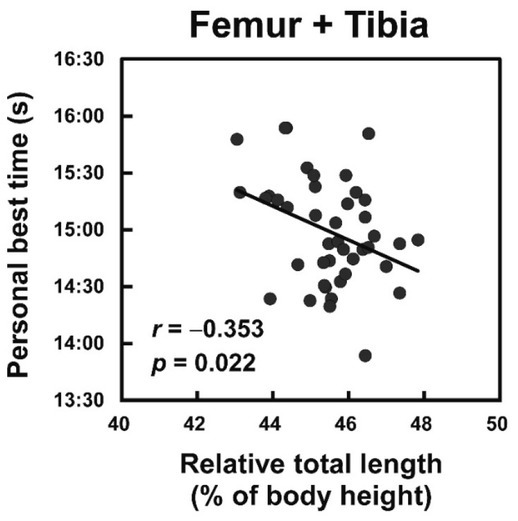
There was no significant correlation between the absolute total length of the femur + tibia and personal best 5000-m race time (r = -0.257, p = 0.100). However, the relative total length of the femur + tibia was significantly correlated with personal best 5000-m race time (Figure 3). In contrast, no such a correlation was noted between the ratio of the length of tibia/femur and personal best 5000-m race time (r = -0.045, p = 0.778).
Figure 3.

Relationship between the relative total length of the leg bones and personal best 5000-m race time The total length of the femur + tibia was normalized with body height to calculate relative bone lengths, that were expressed as percentages (i.e., % of body height).
Discussion
The primary findings of the present study were that the relative tibial length, which was normalized with body height, was significantly correlated with personal best 5000-m race time in endurance runners. Moreover, a trend towards such significance was observed in the relative femoral length. Furthermore, the total length of the femur + tibia was significantly correlated with running performance. Several previous studies have determined that anthropometrical technique-measured leg length was related to running performance in endurance runners (Laumets et al., 2017; Mooses et al., 2015). Thus, the present findings can further provide evidence of the advantage of long legs in achieving superior running performance by showing a positive relationship between MRI-measured leg bone length and running performance in well-trained endurance runners.
Laumets et al. (2017) determined that a longer absolute lower leg was correlated with higher running economy in endurance runners. Running economy is known to be an important factor for superior running performance (Barnes and Kilding, 2015; Saunders et al., 2004). Considering these findings, longer lower leg bones, especially the tibia, may contribute to better running performance due to better running economy. A recent study reported that a longer Achilles tendon was related to better running performance, including greater running economy, in endurance runners (Ueno et al., 2018c). Previous studies have demonstrated that the Achilles tendon was longer in East African endurance runners than in endurance runners of other races, which is in accordance with the results of the lower leg length (Kunimasa et al., 2014; Sano et al., 2015). Furthermore, Morrison et al. (2015) reported that the length of the lower leg was related to that of the Achilles tendon. Considering a positive relationship between lower leg length and Achilles tendon length, a longer tibia may contribute to better running economy. In addition, the present study determined that the total length of the femur + tibia was correlated with better running performance. Previous studies have proposed that, because leg length is related to stride length (Cavanagh and Kram, 1989), longer legs may contribute to more efficient running due to the lower level of internal work compared to that in case of shorter legs (Rahmani et al., 2004). Therefore, the total length of the femoral and tibial bones may play an important role in achieving superior running performance, potentially by improving running economy, in endurance runners.
Mooses et al. (2015) determined that the absolute total length of the upper and lower legs was related to better running performance in Kenyan endurance runners. However, in the results of the present study, the absolute total length of the femur + tibia was not significantly correlated with running performance in Japanese endurance runners. Nevertheless, a study by Mooses et al. (2015) reported that in addition to the absolute length, the relative total leg length, normalized with body height, was related to running performance. Thus, present and other previous findings suggest that leg length relative to body height may be used as a universal factor to predict running performance in Kenyan and Japanese endurance runners. However, Mooses et al. (2015) showed the importance of the upper leg length to running performance by demonstrating that the length of the upper leg, not the lower leg, was significantly correlated with running performance. In contrast, the present study showed that although tibial length was significantly correlated with running performance, only a trend towards such significance was observed in the femoral length. Sano et al. (2015) demonstrated that running in Kenyan runners was performed with a smaller range of knee joint motion than in Japanese runners. When range of knee joint motion is small, range of the swing of the lower limb is naturally limited. Considering the biomechanical theory, the degree of stride length may depend on the length of the thigh rather than the lower leg. Therefore, their findings indicate that compared to the lower leg, longer thighs may play a role in increasing stride length in Kenyan runners. In contrast, because Japanese runners perform the leg swing with more range of knee joint motion during running, longer lower legs may contribute to increased stride length. Thus, these findings suggest that effect of leg length on running performance may be different because of differences in race and/or running kinematics.
The present study demonstrated a positive correlation between leg bone lengths and running performance; however, these correlations were relatively small, and their coefficients of determination were less than 10%. The present study recruited only Japanese endurance runners, and therefore, the participants had similar physical characteristics and leg bone lengths. They also had similar competence levels (within the regional or inter-collegiate levels). Thus, the similar properties among participants may partly explain the relatively small correlation between leg bone length and running performance obtained in the present study.
Physiological (e.g., maximal oxygen uptake) and physiobiological (e.g., fiber type and mitochondrial function) factors are important determinants of running performance (Barnes and Kilding, 2015; Saunders et al., 2004). Moreover, in a recent study, we demonstrated that higher passive planter flexor stiffness, which is a biomechanical factor, was related to better running performance, including greater running economy, in endurance runners (Ueno et al., 2018b). Furthermore, we reported that morphological factors, such as Achilles tendon length and forefoot bone length, were related to running performance in endurance runners (Ueno et al., 2018a, 2018c). Therefore, since running performance is determined by various factors, longer leg bones may be slightly advantageous in achieving better running performance.
The present study has several limitations. First, although we demonstrated positive relationships between leg bone lengths and running performance (e.g. personal best 5000-m race time), we did not ascertain whether longer leg bones would be related to better running economy. Furthermore, we speculated that longer leg bones may contribute to the improvement of running economy, which may be due to increases of stride length. Further studies are needed to examine the relationships of leg bone length with running economy and kinematics. Second, because we recruited only male Japanese endurance runners, whether the present findings can be generalized to other races or female runners remains unclear. Additionally, although all endurance runners recruited in the present study were well-trained, approximately half of them were not the elite levels (i.e., personal best 5000-m race time less than 15 min). Previous studies have reported the importance of leg length to running performance in elite Kenyan and European male endurance runners (Laumets et al., 2017; Mooses et al., 2015). However, to enhance universality of the present findings, further studies are needed to examine the relationships between leg bone lengths and running performance in endurance runners of various races, sexes, and performance levels.
Conclusion
The present findings demonstrated that although the relationship between leg bone length and personal best 5000-m race time was relatively minor, the lengths of the leg bones, especially the tibia, could be related to running performance. Thus, the present study is the first to determine that the MRI-measured lengths of the leg bones may be potential morphological variables for achieving superior running performance in well-trained endurance runners.
Acknowledgements
This study was supported by a Grant-in-Aid for Scientific Research from the Japanese Ministry of Education, Science, Sports and Culture (#15K16497 to T.S; #16H03238 to A.N; #26560361 and #15H03077 to T.I).
References
- Barnes KR, Kilding AE. Running economy: measurement, norms, and determining factors. Sports Med Open. 2015;1(1):1–8. doi: 10.1186/s40798-015-0007-y. [DOI] [PMC free article] [PubMed] [Google Scholar]
- Cavanagh PR, Kram R. Stride length in distance running: velocity, body dimensions, and added mass effects. Med Sci Sports Exerc. 1989;21(4):467–79. [PubMed] [Google Scholar]
- Kunimasa Y, Sano K, Oda T, Nicol C, Komi PV, Ito A, Ishikawa M. Specific muscle-tendon architecture in elite Kenyan distance runners. Scand J Med Sci Sports. 2014;24(4):e269–74. doi: 10.1111/sms.12161. [DOI] [PubMed] [Google Scholar]
- Laumets R, Viigipuu K, Mooses K, Mäestu J, Purge P, Pehme A, Kaasik P, Mooses M. Lower leg length is associated with running economy in high level Caucasian distance runners. J Hum Kinet. 2017;56:229–39. doi: 10.1515/hukin-2017-0040. [DOI] [PMC free article] [PubMed] [Google Scholar]
- Lucia A, Esteve-Lanao J, Oliván J, Gómez-Gallego F, San Juan AF, Santiago C, Pérez M, Chamorro-Viña C, Foster C. Physiological characteristics of the best Eritrean runners-exceptional running economy. Appl Physiol Nutr Metab. 2006;31(5):530–40. doi: 10.1139/h06-029. [DOI] [PubMed] [Google Scholar]
- Mooses M, Mooses K, Haile DW, Durussel J, Kaasik P, Pitsiladis YP. Dissociation between running economy and running performance in elite Kenyan distance runners. J Sports Sci. 2015;33(2):136–44. doi: 10.1080/02640414.2014.926384. [DOI] [PubMed] [Google Scholar]
- Morrison SM, Dick TJ, Wakeling JM. Structural and mechanical properties of the human Achilles tendon: Sex and strength effects. J Biomech. 2015;48(12):3530–3. doi: 10.1016/j.jbiomech.2015.06.009. [DOI] [PMC free article] [PubMed] [Google Scholar]
- Rahmani A, Locatelli E, Lacour JR. Differences in morphology and force/velocity relationship between Senegalese and Italian sprinters. Eur J Appl Physiol. 2004;91(4):399–405. doi: 10.1007/s00421-003-0989-x. [DOI] [PubMed] [Google Scholar]
- Sano K, Nicol C, Akiyama M, Kunimasa Y, Oda T, Ito A, Locateli E, Komi PV, Ishikawa M. Can measures of muscle-tendon interaction improve our understanding of the superiority of Kenyan endurance runners? Eur J Appl Physiol. 2015;115(4):849–59. doi: 10.1007/s00421-014-3067-7. [DOI] [PubMed] [Google Scholar]
- Saunders PU, Pyne DB, Telford RD, Hawley JA. Factors affecting running economy in trained distance runners. Sports Med. 2004;34(7):465–85. doi: 10.2165/00007256-200434070-00005. [DOI] [PubMed] [Google Scholar]
- Ueno H, Suga T, Takao K, Tanaka T, Misaki J, Miyake Y, Nagano A, Isaka T. Association between forefoot bone length and performance in male endurance runners. Int J Sports Med. 2018a;39(4):275–81. doi: 10.1055/s-0043-123646. [DOI] [PubMed] [Google Scholar]
- Ueno H, Suga T, Takao K, Tanaka T, Misaki J, Miyake Y, Nagano A, Isaka T. Potential relationship between passive planter flexor stiffness and running performance. Int J Sports Med. 2018b;39(3):204–9. doi: 10.1055/s-0043-121271. [DOI] [PubMed] [Google Scholar]
- Ueno H, Suga T, Takao K, Tanaka T, Miyake Y, Nagano A, Isaka T. Relationship between Achilles tendon length and running performance in well-trained endurance runners. Scand J Med Sci Sports. 2018c;28(2):446–51. doi: 10.1111/sms.12940. [DOI] [PubMed] [Google Scholar]
- Verillo G, Schena F, Berardelli C, Rosa G, Galvani C, Maggioni M, Agnello L, La Torre A. Anthropometric characteristics of top-class Kenyan marathon runners. J Sports Med Phys Fitness. 2013;53:403–8. [PubMed] [Google Scholar]


