Abstract
The purpose of this study was to determine whether Far-Infrared Emitting Ceramic Materials worn as Bioceramic pants would improve neuromuscular performance, biochemical and perceptual markers in healthy individuals after maximal eccentric exercise. Twenty-two moderately active men were randomized into Bioceramic (n = 11) or Placebo (n = 11) groups. To induce muscle damage, three sets of 30 maximal isokinetic eccentric contractions of the quadriceps were performed at 60°·s-1. Participants wore the bioceramic or placebo pants for 2 hours immediately following the protocol, and then again for 2 hours prior to each subsequent testing session at 24, 48 and 72 hours post. Plasma creatine kinase and lactate dehydrogenase activity, delayed-onset muscle soreness, perceived recovery status, and maximal voluntary contraction were measured pre-exercise and 2, 24, 48, and 72 hours post-exercise. Eccentric exercise induced muscle damage as evident in significant increases in delayed-onset muscle soreness at 24 - 72 hours (p < 0.05) and creatine kinase between Pre to 2, 24, 48 and 72 hours (p < 0.05). Despite the increased delayed-onset muscle soreness and creatine kinase values, no effect of Bioceramic was evident (p > 0.05). Furthermore, decreases in maximal voluntary contraction between Pre and immediately, 2, 24, 48 and 72 hours post (p < 0.05) were reported. However, the standardized difference was moderate lower for lactate dehydrogenase at 24 h (ES = 0.50), but higher at 48 h (ES = -0.58) in the Bioceramic compared to the Placebo group. Despite inducing muscle damage, the daily use of Far-Infrared Emitting Ceramic Materials clothing over 72 hours did not facilitate recovery after maximal eccentric exercise.
Key words: muscle damage, delayed-onset muscle soreness, bioceramic, neuromuscular performance, post-exercise recovery
Introduction
High-intensity and eccentric exercise can induce deleterious effects on skeletal muscle fibers, which might result in muscle soreness, swelling, and reduction of muscle strength and power for several days after exercise (Paulsen et al., 2012; Proske and Morgan, 2001; Gołaś et al., 2017). Exercise-induced muscle damage (EIMD) is typically assessed by indirect markers, including delayed-onset muscle soreness (DOMS), and by the appearance of muscle-specific proteins in the blood, such as creatine kinase (CK) and lactate dehydrogenase (LDH) (Hody et al., 2013; Paulsen et al., 2012; Proske and Morgan, 2001). Considering that high mechanical loads can result in considerable muscle damage, and inadequate recovery may affect subsequent physical performance and increase the risk of injury/illness, it is necessary to investigate strategies to improve the post-exercise recovery process (Duffield et al., 2014; Halson, 2014; Vaile et al., 2013).
In this regard, a host of different strategies
are proposed to aid post-exercise recovery with varying degrees of effectiveness and logistical practicality (Duffield et al., 2014; Halson, 2014; Laurent et al., 2011; Vaile et al., 2013). Strategies that aid skeletal muscle recovery from damage and promote anti-inflammatory responses via practical methods are deemed critical. Hence, recently use of bioceramic materials, also called Far-Infrared Emitting Ceramic Materials (cFIR), has been proposed as a post-exercise recovery method (Hausswirth et al., 2011; Loturco et al., 2016). Previous in vitro and animal model studies report that the emitted heat and radiation from FIR materials can increase blood circulation, facilitate cell growth (Vatansever and Hamblin, 2012) and tissue regeneration (Segovia et al., 2003), leading to calcium-dependent nitric oxide (NO) and calmodulin upregulation in different cell lines (Leung et al., 2009). Given the proposition that such properties can positively influence anti-oxidative, anti-inflammatory and analgesic activities (Vatansever and Hamblin, 2012), it is suggested that cFIR may play a role as a post-exercise recovery tool.
Bioceramics are produced by a combination of oxides (Vatansever and Hamblin, 2012), which reflect/emit high-performance far-infrared rays (FIR) (Segovia et al., 2003). FIR-emitting polymers or ceramic nanoparticles have recently been incorporated into sports apparel to aid the facilitation and practical application of their use (Cian et al., 2015; Loturco et al., 2016). Accordingly, cFIR have become a promising method to reduce pain and induce tissue repair (Ko and Berbrayer, 2002; Leung et al., 2012; Loturco et al., 2016; Vatansever and Hamblin, 2012), however, conflicting results were reported regarding post-exercise recovery improvement (Hausswirth et al., 2011; Leung et al., 2013; Loturco et al., 2016).
For instance, FIR treatments with small far-infrared radiators (e.g., lamps, ceramic disks, plaster, and pads) have shown positive effects in chronic diseased populations (Bagnato et al., 2012; Lai et al., 2014; Silva et al., 2009), though the exact mechanisms of cFIR remain unknown. In exercise models, Hausswirth et al. (2011) reported positive effects on neuromuscular performance after 30 min of FIR (via lamps) in endurance runners, yet no decrease in blood-based markers was observed. In this line, although Loturco et al. (2016) suggested that FIR emitting clothes used for three days during sleep might reduce perceived DOMS after an intense plyometric session in professional soccer players, no effects were shown regarding indirect markers (i.e., CK) and physical performance recovery. The results suggested that FIR clothes might reduce perceived DOMS, despite the absence of improved physical performance in those soccer players. Hence the role of a placebo effect remains present, though further application of cFIR therapy following EIMD remains to be investigated. Nevertheless, to our knowledge, no studies have used the bioceramic material as a recovery strategy during a short period. Furthermore, according to a pilot study, this material used for 2 hours on rats was adequate to reduce inflammation induced by Complete Freud’s Adjuvant (Emer et al., 2014). Thus, the aim of this study was to verify whether the use of cFIR emitting clothes for 2 hours over three days could improve neuromuscular performance as well as biochemical and perceptual markers in healthy individuals after maximal eccentric exercise.
Methods
Participants
A total of twenty-two moderately active, healthy men (mean age: 25.7 ± 3.8 y, body height: 177.3 ± 0.01 cm, body mass: 77.2 ± 9.6 kg) volunteered to participate in this study. Participants had no personal history of lower limb injury and were not involved in regular training programs before the study. The sample size was determined using power analysis software G*Power software (V.3.0.10). Accordingly, each group should comprise 11 participants to provide 80% power at the 0.05 level of significance. The inclusion criteria were: to attend 100% of physical tests and blood sample collections on each occasion, to be free from chronic diseases, not to be taking any medication (e.g. painkillers, anti-inflammatory), nutritional supplements, illegal substances or applying technical recovery strategies (e.g. cold-water immersion, massage, therapeutic exercise) and to wear the bioceramic (BIO) or placebo (PL) impregnated pants for ~2 h during the experimental period. The subjects were also instructed to avoid intense exercise, as well as alcoholic and caffeinated products within 48 h preceding the tests. Each individual signed a written informed consent form after being informed about the purpose, experimental procedures, possible risks, and benefits of the study. The experimental procedures were approved by the University of Santa Catarina Ethics Committee, according to the Helsinki Declaration.
Experimental overview
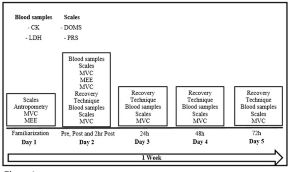
The study was designed as a randomized double-blind placebo-controlled trial (Figure 1). Participants were randomly allocated in the BIO (n = 11) group or the PL (n = 11) group, according the first generator randomizer where each participant was allocated to a single treatment by using the method of randomly permuted blocks (available at http://www.randomization.com) Participants completed testing sessions at a standardised time of day (±2 h) within a 1-week period. The first visit consisted of a familiarization session for all tests and procedures, including a modified (shorter) version of the eccentric exercise protocol. During the second session, participants performed the maximal eccentric exercise protocol to induce skeletal muscle damage. Before and after this session data on maximal voluntary contractions, and perceptual markers were recorded as well as blood samples were drawn at Pre, and 2 h, 24 h, 48 h, and 72 h post exercise. The BIO or PL pants were worn for 2 h immediately post-exercise and then again for 2 h prior to each of the ensuing measurement time points. Although the individuals were instructed about how to adapt their meal calories (approximately 60% carbohydrates, 15% proteins, and 25% lipids), no recalls of foods or diets were performed.
Figure 1.

Experimental protocol. MVC = maximal voluntary contraction; MEE = maximal eccentric exercise; CK = creatine kinase; LDH = lactate dehydrogenase; DOMS = delayed-onset muscle soreness; PRS = perceived recovery status.
Treatments
Both the BIO and PL pants were made of polyester (81%) and spandex (19%) and came in three different sizes (M, L, and XL). The bioceramic material used in this study was composed of microscopic particles including kaolinite, tourmaline, and aluminum oxide. A radiant power emission analysis in the infrared region in the range between 9 and 11 microns was conducted with a Scientech calorimeter (Boulder, CO, USA), Astral model (S AC2500S series), reporting the following value: 1) Fabric with bioceramic emissivity 0.91; 2) Fabric without bioceramic emissivity 0.83. For any given size, BIO and PL pants were identical in size, appearance, and elasticity. The only difference between bioceramic and placebo pants was a garment label indicating 1 or 2. All participants wore the pants for 2 h after the exercise protocol, and then again for 2 h over three successive days at the same time during which they remained seated at rest in the laboratory. To eliminate any potential bias, participants in the placebo group were led to believe that their pants were also a beneficial active treatment for recovery. Of note, the use of cFIR in 2 h interventions was deemed appropriate from an ecological perspective for athletes, though it had been previously demonstrated that acute exposure to FIR (i.e. 30 min) (Hausswith et al., 2011) improved indirect markers of EIMD.
Maximal eccentric exercise (MEE)
The MEE protocol was performed using an isokinetic dynamometer Biodex IV (Shirley Corporation, New York, USA). A standardized warm-up consisted of 10-min cycling on a bicycle ergometer (Lode, Excalibur) at 70–80 rpm and 50 W. Participants were then placed in a supine position to induce maximal lengthening of the rectus femoris (the only biarticular muscle of the knee extensor muscles) as muscle damage mainly occurs at longer muscle lengths (Kanda et al., 2013). Participants were strapped across the femur distal extremity to avoid compensation from the hip joint. Initially two submaximal voluntary isometric knee extensions of 5 s with 30 s recovery were performed as part of the warm up. Then three sets of five submaximal concentric contractions of the extensor muscles at an angular velocity of 120°·s-1 throughout a constant motion range (100° of flexion from the maximal active extension) were performed. The contraction intensity was progressively increased across repetitions until they reached their own maximal performance. Afterwards, the participants performed three sets of five eccentric contractions (at 60°·s-1 angular velocity) progressively intensified until reaching their own maximal performance. After the familiarization, the participants performed the eccentric contraction bout, which consisted of three sets of 30 maximal eccentric contractions of the quadriceps muscle group at 60°·s-1 angular velocity throughout the same range of motion used for their familiarization. Each set was separated by a 30 s rest interval (Hody et al., 2013). The choice of the leg for this unilateral eccentric bout was randomly assigned (Hody et al., 2013). During each contraction, the participants were verbally encouraged to produce their maximal performance, and the examiner checked the temporal evolution of the isokinetic curves on the screen to ensure that the participants worked at their maximal intensity.
Maximal voluntary contraction (MVC)
The MVC was performed using the same isokinetic dynamometer outlined earlier (Biodex IV Shirley Corporation, New York, USA). At Pre, 24 h, 48 h and 72 h post, following an initial warm up on a cycle ergometer, participants were then strapped across the femur distal extremity to avoid compensation from the hip joint. The dynamometer chair backrest inclination was 70°·s- 1, and the evaluated leg knee was positioned in an extension angle of 60°·s-1. The dynamometer rotation axis was aligned to the knee joint rotation axis. Initially two submaximal voluntary isometric knee extensions of 5 s with 30 s of recovery were performed as part of a warm up, followed by three 5 s MVC separated by a 2 min rest interval. During each contraction, the participants were verbally encouraged to produce their maximal performance. The knee extensor peak force output was calculated based on the highest value of 3 maximal isometric contractions held for 5 s each.
Blood sampling CK and LDH activity
Blood samples (5 mL) were drawn from an antecubital vein using a standard venipuncture technique with an ethylene diamine tetra acetic acid (EDTA) tube. Samples were centrifuged for 10 min at 1500 rpm to separate the plasma. Samples were extracted, stored at -80°C until analysis according to specialized laboratory conditions for the following variables: Creatine Kinase (CK) and Lactate Dehydrogenase (LDH). CK and LDH activity was determined spectrophotometrically using commercial kits (Cobas Mira Plus, Roche, Basel, Switzerland and Sigma Diagnostics, St. Louis, MO for CK and LHL, respectively). The CK and LDH intra- and inter-assay CVs were 3.8% and 2.8%, and 3.2% and 1.6%, respectively.
Delayed-onset muscle soreness (DOMS)
Each participant was asked to complete a questionnaire about the perceived leg muscle soreness on a scale from 0 (absence of soreness) to 10 (very intense soreness). This method has been previously used as a non-invasive way to monitor changes in perceived pain following muscle damage protocols (Vaile et al., 2007). Prior to reporting their DOMS ranking, the participants were required to perform a standardized half squat to ensure that all of them were experiencing the same movement/sensation.
Perceived recovery status scale (PRS)
The Perceived Recovery Status scale (PRS) consists of scores ranging between 0 and 10, in which 0–2 means very poorly recovered and with anticipated declines in performance, 4–6 means low to moderately recovered and with similar performance expected, and 8–10 represents high perceived recovery with increases in performance expected (Laurent et al., 2011). The participants were asked to draw a vertical line that intersected the horizontal descriptor scale at the position that best described their perceived recovery level.
Statistical Analysis
The Shapiro-Wilk and Levene’s tests were used to verify normality and data homogeneity, respectively. The isometric peak torque, blood variables, and perceptual marker data were analyzed using a two-way repeated measures ANOVA (group × time), followed by the Bonferroni post-hoc test. The statistical analyses were performed using SPSS 21.0 for Windows (SPSS Inc., Chicago, USA). The effect sizes (ES) were calculated and classified following the scale proposed by Cohen (1988): small (ES from 0.2 to 0.5), moderate (ES from 0.5 to 0.8), and large (ES ≥ 0.8). Values are reported as mean values and standard deviation (SD).
Results
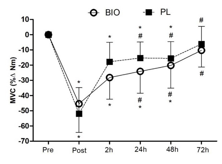
The %ΔMVC for BIO and PL groups are shown in Figure 2. There were no significant main effects of group or interaction (p > 0.05). Nevertheless, a time main effect was found for %ΔMVC between Pre compared to Post, 2 h, 24 h, 48 h and 72 h and immediately Post compared to 24 h, 48 h and 72 h conditions (F = 18.087; p < 0.05).
Figure 2.

Perceptual delta changes of maximal voluntary contraction (MVC) between interventions (Bioceramic [BIO] and Placebo [PL]) throughout 72 hours post maximal eccentric exercise (MEE). * Statistically significant differences in relation to Pre (p < 0.05); # Statistically significant differences in relation to Post (p < 0.05).
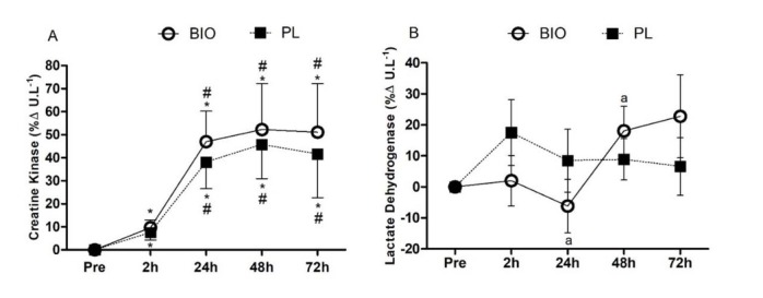
The %ΔCK and %ΔLDH in BIO and PL groups at different time points are presented in Figures 3A and 3B. There was no between-group significant effect (p > 0.05), although the main effect for time was evident for %ΔCK between Pre compared to 2 h, 24 h, 48 h and 72 h, and 2 h compared to 24 h, 48 h and 72 h (F = 13.046; p<0.05). There were no significant main effects in the groups, time, nor group × time interaction at %ΔLDH. However, the standardized difference was moderate lower in 24 h (ES = 0.50), but higher when compared to the PL group in 48 h (ES = -0.58) (Figure 3B).
Figure 3.

Perceptual delta changes of creatine kinase (CK) (A) and lactate dehydrogenase (LDH) (B) between interventions (Bioceramic [BIO] and Placebo [PL]) throughout 72 hours post maximal eccentric exercise (MEE). * Statistically significant differences in relation to Pre (p < 0.05); # Statistically significant differences in relation to 2 h (p < 0.05). a Moderate effect size compared with the Placebo group.
There were no significant main effects for group or interaction (p < 0.05) for either DOMS or PRS (Figures 4A and 4B, respectively). That said, DOMS was significantly increased at 24 h, 48 h, 72 h compared to Pre, and 48 h compared to 2 h (F = 29.821; p < 0.05). Similarly, a significant decrease was reported in the PRS scale at 24 and 48 h compared to Pre (F = 6.765; p < 0.05).
Figure 4.

Quadriceps delayed-onset muscle soreness (DOMS) (A) and perceived recovery status scale (PRS) (B) between interventions (Bioceramic [BIO] and Placebo [PL]) throughout 72 hours post maximal eccentric exercise (MEE). * Statistically significant differences in relation to Pre (p < 0.05); # Statistically significant differences in relation to 2 h post (p < 0.05).
Discussion
The present study was designed to evaluate the effect of using cFIR immediately after and on the days following completion of the eccentric exercise training program on neuromuscular performance, muscle damage and perceived recovery. The MEE bout was effective at inducing significant reductions in MVC and increases in CK and DOMS. However, neither treatment nor time x treatment interactions were observed in isometric peak torque (MVC), biochemical measures (CK and LDH), or perceptual markers (DOMS and PRS). Consequently, the use of BIO was not effective in improving recovery following muscle damaging exercise.
Although cFIR treatment has not been extensively explored, previous studies have suggested it as a post-exercise recovery method (Hausswirth et al., 2011; Leung et al., 2013; Loturco et al., 2016). Hausswirth et al. (2011) performed a running protocol to induce muscle damage in runners, and applied FIR (via lamps) immediately, 24 h and 48 h post-exercise. The results showed that trained runners exposed to 30 min of FIR in the whole body were able to recover knee extensor MVC by 48 h post. Conversely, Loturco et al. (2016) did not observe any positive effects of cFIR clothing in male soccer players who performed damaging drop jumps and used FIR for ten hours sleeping over three successive nights. Nevertheless, the authors reported that athletes appeared to be well-trained, adapted, and hence resilient to the damaging effects of plyometric exercise, which might have attenuated possible cFIR effects.
While no differences were observed for post-exercise CK or LDH responses between groups, the effect size in LDH was lower at 24 h, but higher at 48 h in the BIO group compared to the PL group. This finding is in agreement with previous studies that demonstrated that FIR sauna and clothes applied during three consecutive days did not decrease CK levels in highly-trained endurance runners (Hausswirth et al., 2011) and in elite soccer players (Loturco et al., 2016). One of the proposed effects of FIR is to improve local microcirculation, increasing the blood flow development of leukocyte migration into tissues (Yang et al., 2010) and irradiation of endothelial cells (Park et al., 2013), resulting in vasodilatation and a rise in tissue temperature to facilitate neutrophil migration into the muscle (Kanda et al., 2013). While such mechanisms were not investigated in the present study, the lack of difference in CK or LDH responses following BIO suggests insufficient emissivity to reflect/emit FIR ray by the clothing into human tissues, or was insufficient to promote biological effects and improve recovery in the current population.
Although scientific evidence has been reported regarding the biological and hyperthermic effects of cFIR, its mechanisms affecting muscle performance are still unclear. The loss of maximal voluntary contraction force is considered one of the best clinical methods for quantifying muscle damage (Miles et al., 2008). Thus, EIMD assessed by indirect markers (e.g. inflammatory mediators) may concur with the inhibition of force development in skeletal muscles (Wilcox et al., 1992) and partially explain the muscle strength loss. Therefore, it has been suggested that cFIR therapy may modulate cytokine expression during the muscle repair process, inducing a decrease in pro-inflammatory responses (Lin et al., 2007). However, our results showed no significant difference between the two groups.
Previous studies in clinical trials have also demonstrated that FIR reduced pain (Bagnato et al., 2012; Ko and Berbrayer, 2002; Lin et al., 2007; Silva et al., 2009), intolerance to cold (Silva et al., 2009), muscle stiffness (Lai et al., 2014) and serum IL-6 and endothelin-1 concentrations in individuals following total knee arthroplasty (Wong et al., 2012). However, in the sports field few studies have investigated the cFIR effect on perceptual markers, such as DOMS and PRS. Loturco et al. (2016) were the first to report that FIR emitting clothes over three successive nights might have contributed to reduced DOMS within 72 h (moderate and large effect sizes, respectively) after the plyometric exercise bout. The authors suggested FIR clothes might be used to accelerate muscle pain recovery after eccentrically-biased exercises in soccer players. Hausswirth et al. (2011) observed that the perception of muscle pain was significantly reduced after 48 hours in relation to the non-treatment condition. In the present study, DOMS and PRS did not differ between conditions, either due to insufficient exposure or lack of perceptual or biological effect of the Biocermic pants given the placebo effect was masked by the PL condition.
Despite these findings, certain limitations should be acknowledged. These include the short time and possible insufficient microscopic particles emissivity to emit cFIR from clothing into human tissues. Consequently, insufficient biological effects and acceleration of the recovery of skeletal muscle function with decreasing pain were evident. Furthermore, a longer time lapse between the end of the exercise protocol and the time the participants started to wear the cFIR pants could prevent cFIR acute effects upon neutrophil migration (Loturco et al., 2016). Thus, it is suggested that the use of cFIR during sleep, under more controlled exposure time and ambient conditions would make the treatment more effective (Loturco et al., 2016). Finally, our study was also limited by the sole use of scales to quantify DOMS.
In conclusion, the selected eccentric exercise stimulus used in our study induced muscle damage in the quadriceps muscles. However, the use of cFIR emitting clothes for 2 hours over three days did not contribute to improved MVC, perceptual scales ratings and blood-based markers (although lower ES in 24 h in the BIO compared to the PL group for LDH was observed) after a maximal eccentric exercise. Despite scarce research regarding cFIR in exercise models, the present findings suggest that the use of this material for 2 hours on a daily basis does not facilitate recovery in healthy individuals. Although the use of recovery clothes could be practical and applicable, longer exposure time has to be considered, since this method does not affect the daily routines of individuals. Future studies should investigate the effects of FIR-emitting clothes on other blood markers in order to understand their biological mechanisms after exercise. Additionally, further studies should be conducted to evaluate the required time for cFIR use, different protocols and study populations to induce beneficial effects on human tissues and its possible effects when associated to different recovery strategies.
Acknowledgements
We acknowledge support from Conselho Nacional de Desenvolvimento Científico e Tecnológico (CNPq), Fundação de Amparo à Pesquisa e Inovação do Estado de Santa Catarina (FAPESC), Coordenação de Aperfeiçoamento de Pessoal de Nível Superior (CAPES) and, Universidade Federal de Santa Catarina (UFSC) and Universidade do Sul de Santa Catarina (UNISUL).
References
- Bagnato GL, Miceli G, Atteritano M, Marino N, Bagnato GF. Far infrared emitting plaster in knee osteoarthritis: a single blinded, randomised clinical trial. Reumatismo. 2012;64:388–394. doi: 10.4081/reumatismo.2012.388. [DOI] [PubMed] [Google Scholar]
- Cian C, Gianocca V, Barraud PA, Guerraz M, Bresciani JP. Bioceramic fabrics improve quiet standing posture and handstand stability in expert gymnasts. Gait & Posture. 2015;42:419–423. doi: 10.1016/j.gaitpost.2015.07.008. [DOI] [PubMed] [Google Scholar]
- Cohen J. Statistical Power Analysis for the Behavioral Sciences. Hillsdale (NJ): Lawrence Erlbaum Associates; 1988. 2nd ed. [Google Scholar]
- Duffield R, Murphy A, Kellett A, Reid M. Recovery from repeated on-court tennis sessions: combining cold-water immersion, compression, and sleep interventions. Int J Sports Physiol Perform. 2014;9:273–282. doi: 10.1123/ijspp.2012-0359. [DOI] [PubMed] [Google Scholar]
- Emer AA, Cidral-Filho FJ, Madeira F, Martins DF. Effect of Far Infrared Emitted by Bioceramics on Parameters of Physical Performance in Mice. J Altern Complement Med. 2014;20(6):A34. [Google Scholar]
- Gołaś A, Maszczyk A, Pietraszewski P, Stastny P, Tufano JJ, Zając A. Effects of Pre-exhaustion on the Patterns of Muscular Activity in the Flat Bench Press. J Strength Cond Res. 2017;31(7):1919–1924. doi: 10.1519/JSC.0000000000001755. [DOI] [PubMed] [Google Scholar]
- Halson SL. Sleep in elite athletes and nutritional interventions to enhance sleep. Sports Med. 2014;44(Suppl 1):S13–S23. doi: 10.1007/s40279-014-0147-0. [DOI] [PMC free article] [PubMed] [Google Scholar]
- Hausswirth C, Louis J, Bieuzen F, Pournot H, Fournier J, Filliard JR, Brisswalter J. Effects of whole-body cryotherapy vs. far-infrared vs. passive modalities on recovery from exercise induced muscle damage in highly-trained runners. PLoS One. 2011;6(12):e27749. doi: 10.1371/journal.pone.0027749. [DOI] [PMC free article] [PubMed] [Google Scholar]
- Hody S, Rogister B, Leprince P, Wang F, Croisier JL. Muscle fatigue experienced during maximal eccentric exercise is predictive of the plasma creatine kinase (CK) response. Scand J Med Sci Sports. 2013;23:501–507. doi: 10.1111/j.1600-0838.2011.01413.x. [DOI] [PubMed] [Google Scholar]
- Kanda K, Sugama K, Hayashida H, Sakuma J, Kawakami Y, Miura S, Yoshioka H, Mori Y, Suzuki K. Eccentric exercise-induced delayed-onset muscle soreness and changes in markers of muscle damage and inflammation. Exerc Immunol. 2013;19:72–85. [PubMed] [Google Scholar]
- Kimura T, Takahashi K, Suzuki Y, Konishi Y, Ota Y, Mori C, Ikenaga T, Takanami T, Saito R, Ichiishi E, Awaji S, Watanabe K, Higashitani A. The effect of high strength static magnetic fields and ionizing radiation on gene expression and DNA damage in Caenorhabditis elegans. Bioelectromagnetics. 2008;29:605–614. doi: 10.1002/bem.20425. [DOI] [PubMed] [Google Scholar]
- Ko GD, Berbrayer D. Effect of ceramic-impregnated thermoflow gloves on patients with Raynaud’s syndrome: randomized, placebo-controlled study. Altern Med. 2002;7:328–335. [PubMed] [Google Scholar]
- Lai CH, Leung TK, Peng CW, Chang KH, Lai MJ, Lai WF, Chen SC. Effects of far-infrared irradiation on myofascial neck pain: a randomized, double-blind, placebo-controlled pilot study. J Altern Complement Med. 2014;20:123–129. doi: 10.1089/acm.2013.0122. [DOI] [PubMed] [Google Scholar]
- Laurent CM, Green JM, Bishop PA, Sjokvist J, Schumacker RE, Richardson MT, Curtner-Smith M. A practical approach to monitoring recovery: development of a perceived recovery status scale. J Strength Cond Res. 2011;25:620–628. doi: 10.1519/JSC.0b013e3181c69ec6. [DOI] [PubMed] [Google Scholar]
- Leung TK, Kuo CH, Lee CM, Kan NW, Hou CW. Physiological effects of bioceramic material: harvard step, resting metabolic rate and treadmill running assessments. Chin J Physiol. 2013;56:334–40. doi: 10.4077/CJP.2013.BAB132. [DOI] [PubMed] [Google Scholar]
- Leung TK, Lin YS, Chen YC, Shang HF, Lee YH, Su CH, Liao HC. Immunomodulatory effects of far infrared ray irradiation via increasing calmodulin and nitric oxide production in RAW 264.7 macrophages. Biomed Eng Appl Basis. 2009;21:317–323. [Google Scholar]
- Leung TK, Lin JM, Chien HS, Day TC. Biological effects of melt spinning fabrics composed of 1% bioceramic material. Textile Res J. 2012;82(11):1121–1130. [Google Scholar]
- Lin YS, Lin MY, Leung TK, Liao CH, Huang TT, Huang HS, Pan HC. Properties and biological effects of high performance ceramic powder emitting far-infrared irradiation. Instrum Today. 2007;6:60–66. [Google Scholar]
- Loturco I, Abad CCC, Nakamura FY, Ramos SP, Kobal R, Gil S, Pereira LA, Burini FHP, Roschel H, Ugrinowitsch C, Tricoli V. Effects of far infrared rays emitting clothing on recovery after an intense plyometric exercise bout applied to elite soccer players: a randomized double-blind placebo-controlled trial. Biol Sport. 2016;33:277–283. doi: 10.5604/20831862.1208479. [DOI] [PMC free article] [PubMed] [Google Scholar]
- Miles MP, Andring JM, Pearson SD, Gordon LK, Kasper C, Depner CM, Kidd JR. Diurnal variation, response to eccentric exercise, and association of inflammatory mediators with muscle damage variables. J Appl Physiol. 2008;104:451–458. doi: 10.1152/japplphysiol.00572.2007. [DOI] [PubMed] [Google Scholar]
- Park JH, Lee S, Cho DH, Park YM, Kang DH, Jo I. Far-infrared radiation acutely increases nitric oxide production by increasing Ca2+ mobilization and Ca2+/calmodulin-dependent protein kinase II-mediated phosphorylation of endothelial nitric oxide synthase at serine 1179. Biochem Biophys Res Commun. 2013;436:601–606. doi: 10.1016/j.bbrc.2013.06.003. [DOI] [PubMed] [Google Scholar]
- Paulsen G, Mikkelsen UR, Raastad T, Peake JM. Leucocytes, cytokines and satellite cells: what role do they play in muscle damage and regeneration following eccentric exercise? Exerc Immunol. 2012;18:42–97. [PubMed] [Google Scholar]
- Proske U, Morgan DL. Muscle damage from eccentric exercise: mechanism, mechanical signs, adaptation and clinical applications. J Physiol. 2001;537:333–345. doi: 10.1111/j.1469-7793.2001.00333.x. [DOI] [PMC free article] [PubMed] [Google Scholar]
- Segovia M, Gordillo FJ, Figueroa FL. Cyclic-AMP levels in the lichen Evernia prunastri are modulated by light quantity and quality. J Photochem Photobiol Biol. 2003;70:145–151. doi: 10.1016/s1011-1344(03)00074-5. [DOI] [PubMed] [Google Scholar]
- Silva TM, Moreira GA, Quadros AA, Pradella-Hallinan M, Tufik S, Oliveira AS. Effects of the use of MIG3 bioceramics fabrics use-long infrared emitter-in pain, intolerance to cold and periodic limb movements in post-polio syndrome. Arq Neuropsiquiatr. 2009;67:1049–1053. doi: 10.1590/s0004-282x2009000600016. [DOI] [PubMed] [Google Scholar]
- Vaile J, Gill N, Blazevich AJ. The effect of contrast water therapy on symptoms of delayed onset muscle soreness (DOMS) and explosive athletic performance. J Strength Cond. 2007;21:697–702. doi: 10.1519/R-19355.1. [DOI] [PubMed] [Google Scholar]
- Vaile J, Halson S, Gill N, Dawson B. Effect of hydrotherapy on the signs and symptoms of delayed onset muscle soreness. Eur J Appl Physiol. 2008;102:447–455. doi: 10.1007/s00421-007-0605-6. [DOI] [PubMed] [Google Scholar]
- Vatansever F, Hamblin MR. Far infrared radiation (FIR): its biological effects and medical applications. Photonics Lasers Med. 2012;4:255–266. doi: 10.1515/plm-2012-0034. [DOI] [PMC free article] [PubMed] [Google Scholar]
- Wilcox P, Osborne S, Bressler B. Monocyte inflammatory mediators impair in vitro hamster diaphragm contractility. Am Ver Respir Dis. 1992;146:462–466. doi: 10.1164/ajrccm/146.2.462. [DOI] [PubMed] [Google Scholar]
- Wong CH, Lin LC, Lee HH, Liu CF. The analgesic effect of thermal therapy after total knee arthroplasty. J Altern Complement Med. 2012;18:175–179. doi: 10.1089/acm.2010.0815. [DOI] [PubMed] [Google Scholar]
- Yang CS, Yeh CH, Tung CL, Chen MY, Jiang CH, Yeh ML. Impact of far-infrared ray exposure on the mechanical properties of unwounded skin of rats. Exp Biol Med. 2010;235:952–956. doi: 10.1258/ebm.2010.009372. [DOI] [PubMed] [Google Scholar]


