Abstract
Herein, we describe an alkyl thiolate-ligated iron complex that reacts with dioxygen to form an unprecedented example of an iron superoxo (O2•−) intermediate, [FeIII(S2Me2N3(Pr,Pr))(O2)] (4), which is capable of cleaving strong C–H bonds. A cysteinate-ligated iron superoxo intermediate is proposed to play a key role in the biosynthesis of β-lactam antibiotics by isopenicillin N-synthase (IPNS). Superoxo 4 converts to a metastable putative Fe(III)–OOH intermediate, at rates that are dependent on the C–H bond strength of the H atom donor, with a kinetic isotope effect (kH/kD = 4.8) comparable to that of IPNS (kH/kD = 5.6). The bond dissociation energy of the C–H bonds cleaved by 4 (92 kcal/mol) is comparable to C–H bonds cleaved by IPNS (93 kcal/mol). Both the calculated and experimental electronic absorption spectra of 4 are comparable to those of the putative IPNS superoxo intermediate, and are shown to involve RS− → Fe–O2•− and O2•− → Fe charge transfer transitions. The π-back-donation by the electronrich alkyl thiolate presumably facilitates this reactivity by increasing the basicity of the distal oxygen. The frontier orbitals of 4 are shown to consist of two strongly coupled unpaired electrons of opposite spin, one in a superoxo π*(O–O) orbital, and the other in an Fe(dxy) orbital.
Isopenicillin N synthase (IPNS)1–4 and cysteine dioxygenase (CDO)5–10 are nonheme Fe enzymes that catalyze the O2-promoted oxidation of cysteinates (RS−). Although O2 oxidation reactions are thermodynamically favored, they are kinetically slow in the absence of a transition-metal catalyst, because they are spin-forbidden.11 The electron donor properties of cysteinate and high covalency of FeIII–SR bonds12,13 lower the activation barrier to O2 binding4 to iron, promote O–O bond cleavage,4,14 and increase the reactivity of high-valent Fe-oxo intermediates.15,16 This helps to facilitate the oxidative bicyclization reaction involved in the biosynthesis of β-lactam antibiotics (e.g., penicillin, cephalosporins) by IPNS,1,3 as well as the CDO-catalyzed regulation of cysteine concentration, toxic levels of which can lead to neurological disorders,17 or the metastasis of cancerous tumors.18,19 The proposed mechanism of both IPNS2,3 and CDO5,6 involves the initial formation of a cis thiolate-ligated iron superoxo intermediate (cis-RS-Fe-O2•−). With the former, this intermediate abstracts a H atom from substrate, and with the latter it is proposed to attack the adjacent sulfur to form a transient peroxythiolate species. The putative IPNS Fe–O2•− is spectroscopically detected in small amounts (14%) via transient absorption (λmax = 630 nm; t = 2–10 ms) and Mössbauer spectroscopies if the cysteinate β-hydrogens are deuterated.2 A CDO intermediate, proposed to be an Fe(III)-peroxythiolate, is also observed by transient absorption spectroscopy (λmax = 500 nm, 640 nm).5 Vibrational data is not available to support these assignments, however. Two strong C–H bonds are cleaved during the proposed IPNS mechanism (Figure S1): a β-hydrogen from cysteine (93 kcal/mol) and a β-hydrogen from valine (96 kcal/mol).20 The former is proposed to involve the putative Fe(III)-superoxo, and the latter an Fe(IV)-oxo intermediate.2 There are few well-characterized examples of Fe(III)-superoxo compounds,21–24 however, and none of these cleave strong C–H bonds. An aryl thiolate-ligated Fe–O2•− was recently reported; however, the sulfur lone pair is tied up in π-bonding to the aryl carbon in one of its resonance forms making it less reactive.23 Although it has yet to be demonstrated with a superoxo, π-back-donation by an electron rich alkyl thiolate has been shown to facilitate the cleavage of strong C–H bonds by increasing the basicity of an iron oxo.25 Herein, we report the synthesis and structure of an alkyl thiolate-ligated iron complex that reacts with O2 to afford a spectroscopically observable reactive intermediate.
Reduced [FeII(S2Me2N3(Pr,Pr))] (1) was synthesized and structurally characterized according to the method outlined in the Supporting Information, and was shown to contain Fe2+ in a distorted trigonal bipyramidal coordination environment (τ = 0.78; Figure 1, Tables S2–S6). In solution, 1 has a magnetic moment of μ = 2.63 μB at 298 K in MeCN consistent with an S = 1 spin-state, and has a characteristic electronic absorption band at λmax = 420 (εM 1600) nm (Figure S2). Previously, we showed that, like IPNS and CDO,3,10,26 the oxidized derivative of 1, [FeIII(S2Me2N3(Pr,Pr))]+ (2), binds small molecules (azide and NO),27–29 cis with respect to one of the thiolate sulfurs. The latter are frequently used to probe enzymatic O2 binding sites.10,14
Figure 1.
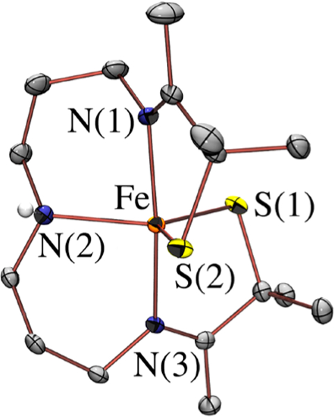
ORTEP of 1 with thermal ellipsoids at the 50% probability level. With the exception of the secondary amine proton, hydrogens have been removed for clarity.
The addition of dry O2 to 1 in THF at 25 °C causes an immediate color change from pale yellow to watermelon pink, with an associated shift in λmax to 510(1500) nm (Figure S3), and the growth of a signal (g = 2.17, 2.11, 1.98) in the electron paramagnetic resonance (EPR) spectrum (Figure S4) consistent with the formation (in 93% yield) of low-spin (S = ½) [FeIII(η2-SMe2O)(SMe2N3(Pr,Pr))]+ (3).30 Electrospray mass spectroscopy (ESI-MS) of isotopically labeled samples shows that the oxo of 3 is derived from 18O2 (Figure S5). Azide inhibits this reaction (Figures S6) indicating that O2 must bind to the metal ion in order for oxo atom transfer to occur. At low temperatures (−73 °C), a new metastable cranberry red O2-derived intermediate, 4 (Figure 2), is observed en route to singly oxygenated 3,30 the low energy band (~700 nm) of which is characteristic of six-coordinate, bis-thiolate-ligated Fe(III).12,31 When this reaction is monitored by 1H NMR, the paramagnetic signals of 1 collapse to diamagnetic (S = 0) signals upon the addition of O2 (Figure S7). The ESI-MS of 4 contains an M + 32 peak at m/z = 417.3 (Figure S8), consistent with the addition of two oxygen atoms to the parent ion, 1 (m/z = 385.4). An identical intermediate can be generated via the low temperature (−73 °C) addition of excess (50 equiv) potassium superoxide (KO2) to oxidized [FeIII(S2Me2N3(Pr,Pr))]+ (2) (Figure S9). The resonance Raman (rR) spectrum of 4 reproducibly shows isotopically sensitive features (a Fermi doublet) at 1093 and 1122 cm−1 (Figure 3) that shift to 1022 cm−1 when generated from 18O2 (Figure S10), and disappear after 30 min, demonstrating its transient nature. All of this data would be consistent with the formation of a metastable ferric superoxo species. The calibrated (vide infra) density functional theory (DFT) calculated structure of [FeIII(S2Me2N3(Pr,Pr))(O2)] (4) contains an O2 moiety cis to one of the thiolate sulfurs (Figure S11), with bond lengths (O–O = 1.289 Å, Table S1), and a calculated νO–O stretch (Figure S12, 1154 cm−1), consistent with a ferric superoxo (FeIII-O2•−), analogous to the prop osed IPNS and CDO intermediates. The frontier orbitals of 4 (Figure 4) contain two unpaired electrons of opposite spin, one in a superoxo π*(O–O) orbital, and the other in a Fe(dxy) orbital. The calculated overlap parameter of T=0.17, and coupling constant Jcalc = − 450 cm−1 indicate that the two unpaired spins are strongly coupled antiferromagnetically, consistent with the absence of paramagnetically shifted peaks in the 1H NMR and EPR silence of 4. The time-dependent DFT (TD-DFT) calculated electronic absorption spectrum of 4 (Figures S13 and S14) reproduces the experimental spectrum (Figure 2), and shows that superoxo π*(O–O) → dxy(Fe) charge transfer transitions are responsible for the higher energy bands, and a RS− → Fe–O2•− charge transfer transition for the lower energy band. Both the calculated and experimental spectrum of 4 are similar to that of the putative IPNS superoxo intermediate,2 supporting its assignment as a superoxo species. The reported CDO intermediate spectrum5 is also similar to that of 4, suggesting that it too is a ferric superoxo.
Figure 2.
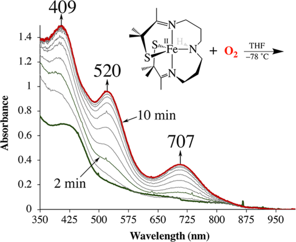
Monitoring the low temperature (−73 °C) reaction between 1 (0.48 mM) and excess O2 in THF by electronic absorption spectroscopy.
Figure 3.
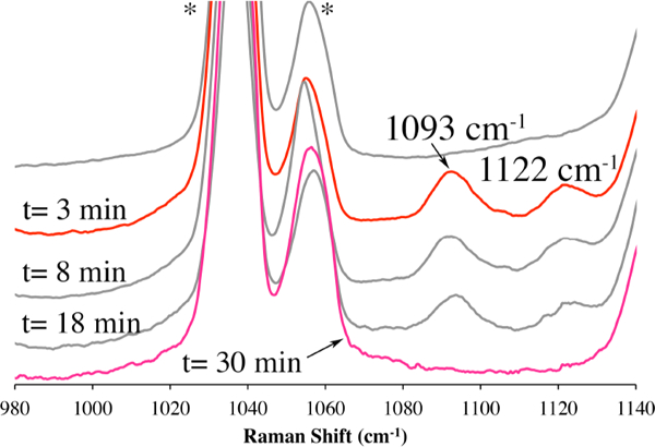
Monitoring the reaction between 1 (5 mM) and O2 in THF at −73 °C by resonance Raman spectroscopy. Samples were frozen in liquid N2 (77 K) at the time-intervals indicated. Excitation wavelength λex = 527 nm; 4.0 mW power; * = solvent peak.
Figure 4.
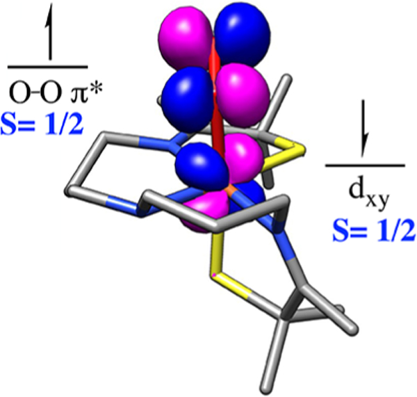
Singly occupied molecular orbitals (SOMO) of 4 contain strongly coupled electrons of opposite spin, one on the superoxo (O2•−) and the other on the metal ion.
Ferric superoxo (FeIII−O2•−) 4 converts to a second metastable intermediate, 5 (λmax = 696 nm), at −73 °C in THF (Figure S15), en route to 3 (Figure S16), at a rate that is dependent on the C–H bond strength of the solvent or H atom donor. Reaction rates decrease in deuterated THF (Figure 5), and increase upon the addition of a sacrificial H atom donor (100 equiv of 1,4-cyclohexadiene (CHD), BDE = 76 kcal/mol). The observed deuterium isotope effect, kH/kD = 4.8, is comparable to that of IPNS (kH/kD = 5.6),32 and indicates that superoxo 4 is capable of abstracting hydrogen atoms from strong C–H bonds (BDE(THF) = 92 kcal/mol).33 A likely product of this reaction would be a ferric hydroperoxo, [FeIII(S2 Me2N3(Pr,Pr))(OOH)] (5). Consistent with this, a new rhombic signal grows in when the reaction between 1 and O2 is monitored by EPR (Figure 6). Spin-quantitation using double integration (Figure S18) indicates that the EPR signal of 5 represents 87% of the sample (Figures S4 and S17). The remaining 13% can be attributed to 1 and/or 4, both of which are EPR-silent in ⊥-mode. Together, these results show that in contrast to the few reported Fe(III)-superoxo complexes,21–24 alkylthiolate-ligated 4 is capable of abstracting H atoms from strong C–H bonds, on par with that of the β C–H bonds of cysteine (93 kcal/mol).33 It is plausible that π-back-donation by the electron-rich alkyl thiolate facilitates this reactivity by increasing the basicity of the distal oxygen. Spectroscopic characterization of 4, along with calibrated DFT calculations, provides additional evidence to support the assignment of the IPNS and CDO intermediates detected via transient absorption spectroscopy,2,5 as cis RS-FeIII-O2•− species.
Figure 5.
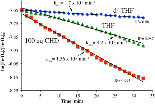
Pseudo-first-order kinetic plots associated with the reaction between 4 (0.48 mM) and THF (12 M), or CHD (48 mM) in THF at −73 °C.
Figure 6.
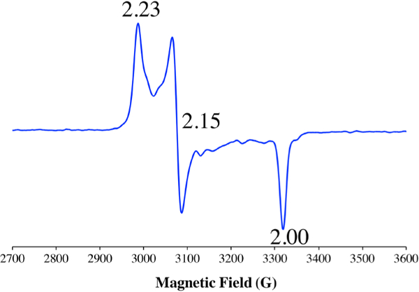
X-band EPR spectrum (⊥-mode) of putative hydroperoxo FeIII–OOH (5), formed from superoxo FeIII–O2•− (4) via H atom abstraction.
Supplementary Material
ACKNOWLEDGMENTS
Funding from the NIH (RO1-GM123062) is gratefully acknowledged. We thank Elizabeth Canarie and Donald Mannikko for assistance with EPR spin quantitation.
Footnotes
ASSOCIATED CONTENT
Supporting Information
The Supporting Information is available free of charge on the ACS Publications website at DOI: 10.1021/jacs.8b12670.
Experimental Section, crystallographic tables for 1; quantitative UV/vis of 1; ESI-MS of 3 (generated from 1+18O2), and 4; LT 1H NMR of the reaction 1+O2; rR spectrum of 1, 3, and 4 generated from 16O2 and 18O2; electronic absorption spectrum of 2+KO2, putative FeIII–OOH 5, and its conversion to 3; DFT optimized structure of 4; TD-DFT calculated vs experimental electronic absorption spectrum of 4; calibration curve used for spin quantitation of EPR spectra (PDF)
The authors declare no competing financial interest.
REFERENCES
- (1).Burzlaff NI; Rutledge PJ; Clifton IJ; Hensgens CMH; Pickford M; Adlington RM; Roach PL; Baldwin JE The reaction cycle of isopenicillin N synthase observed by X-ray diffraction. Nature 1999, 401, 721–724. [DOI] [PubMed] [Google Scholar]
- (2).Tamanaha EY; Zhang B; Guo Y; Chang W-C; Barr EW; Xing G; St. Clair J; Ye S; Neese F; Bollinger JM Jr; Krebs C Spectroscopic evidence for the two C-H-cleaving intermediates of Aspergillus nidulans isopenicillin N synthase. J. Am. Chem. Soc. 2016, 138, 8862–8874. [DOI] [PMC free article] [PubMed] [Google Scholar]
- (3).Roach PL; Clifton IJ; Hensgens CMH; Shibata N; Schofield CJ; Hajdu J; Baldwin JE Structure of isopenicillin N synthase complexed with substrate and the mechanism of penicillin formation. Nature 1997, 387, 827–830. [DOI] [PubMed] [Google Scholar]
- (4).Brown CD; Neidig ML; Neibergall MB; Lipscomb JD; Solomon EI VTVH-MCD and DFT Studies of Thiolate Bonding to {FeNO}7/{FeO2}8 Complexes of Isopenicillin N Synthase: Substrate Determination of Oxidase versus Oxygenase Activity in Nonheme Fe Enzymes. J. Am. Chem. Soc. 2007, 129 (23), 7427–7438. [DOI] [PMC free article] [PubMed] [Google Scholar]
- (5).Tchesnokov EP; Faponle AS; Davies CG; Quesne MG; Turner R; Fellner M; Souness RJ; Wilbanks SM; deVisser SP; Jameson GNL An iron–oxygen intermediate formed during the catalytic cycle of cysteine dioxygenase. Chem. Commun. 2016, 52, 8814–8817. [DOI] [PMC free article] [PubMed] [Google Scholar]
- (6).Kumar D; Thiel W; de Visser SP Theoretical Study on the Mechanism of the Oxygen Activation Process in Cysteine Dioxygenase Enzymes. J. Am. Chem. Soc. 2011, 133, 3869–3882. [DOI] [PubMed] [Google Scholar]
- (7).Crawford JA; Li W; Pierce BS Single Turnover of Substrate-Bound Ferric Cysteine Dioxygenase with Superoxide Anion: Enzymatic Reactivation, Product Formation, and a Transient Intermediate. Biochemistry 2011, 50, 10241–10253. [DOI] [PubMed] [Google Scholar]
- (8).Aluri S; de Visser SP The Mechanism of Cysteine Oxygenation by Cysteine Dioxygenase Enzymes. J. Am. Chem. Soc. 2007, 129, 14846–14847. [DOI] [PubMed] [Google Scholar]
- (9).Joseph CA; Maroney MJ Cysteine dioxygenase: structure and mechanism. Chem. Commun. 2007, 3338–3349. [DOI] [PubMed] [Google Scholar]
- (10).Pierce BS; Gardner JD; Bailey LJ; Brunold TC; Fox BG Characterization of the nitrosyl adduct of substrate-bound mouse cysteine dioxygenase by electron paramagnetic resonance: electronic structure of the active site and mechanistic implications. Biochemistry 2007, 46, 8569–8578. [DOI] [PubMed] [Google Scholar]
- (11).Kovacs JA How Iron Activates O2. Science 2003, 299, 1024–1025. [DOI] [PubMed] [Google Scholar]
- (12).Kennepohl P; Neese F; Schweitzer D; Jackson HL; Kovacs JA; Solomon EI Spectroscopy of Non-Heme Iron Thiolate Complexes: Insight into the Electronic Structure of the Low-Spin Active Site of Nitrile Hydratase. Inorg. Chem. 2005, 44, 1826–1836. [DOI] [PMC free article] [PubMed] [Google Scholar]
- (13).Kovacs JA; Brines LM Understanding How the Thiolate Sulfur Contributes to the Function of the Non-Heme Iron Enzyme Superoxide Reductase. Acc. Chem. Res. 2007, 40 (7), 501–9. [DOI] [PMC free article] [PubMed] [Google Scholar]
- (14).Villar-Acevedo G; Nam E; Fitch S; Benedict J; Freudenthal J; Kaminsky W; Kovacs JA Influence of Thiolate Ligands on Reductive N–O Bond Activation. Oxidative Addition of NO to a Biomimetic SOR Analogue, and its Proton-Dependent Reduction of Nitrite. J. Am. Chem. Soc. 2011, 133, 1419–1427. [DOI] [PMC free article] [PubMed] [Google Scholar]
- (15).Yosca TH; Rittle J; Krest CM; Onderko EL; Silakov A; Calixto JC; Behan RK; Green MT Iron(IV)hydroxide pKa and the Role of Thiolate Ligation in C–H Bond Activation by Cytochrome P450. Science 2013, 342, 825–829. [DOI] [PMC free article] [PubMed] [Google Scholar]
- (16).Green MT C–H bond activation in heme proteins: the role of thiolate ligation in cytochrome P450. Curr. Opin. Chem. Biol. 2009, 13, 84–88. [DOI] [PubMed] [Google Scholar]
- (17).Heafield MT; Fearn S; Steventon GB; Waring RH; Williams AC; Sturman SG Plasma cysteine and sulphate levels in patients with motor neurone, Parkinson’s and Alzheimer’s disease. Neurosci. Lett. 1990, 110, 216–220. [DOI] [PubMed] [Google Scholar]
- (18).Dietrich D; Krispin M; Dietrich J; Fassbender A; Lewin J; Harbeck N; Schmitt M; Eppenberger-Castori S; Vuaroqueaux V; Spyratos F; Foekens JA; Lesche R; Martens JWM CDO1 promoter methylation is a biomarker for outcome prediction of anthracycline treated, estrogen receptor-positive, lymph node-positive breast cancer patients. BMC Cancer 2010, 10, 247. [DOI] [PMC free article] [PubMed] [Google Scholar]
- (19).Jeschke J; O’Hagan HM; Zhang W; Vatapalli R; Freitas Calmon M; Danilova L; Nelkenbrecher C; Van Neste L; Bijsmans ITGW; Van Engeland M; Gabrielson E; Schuebel KE; Winterpacht A; Baylin SB; Herman JG; Ahuja N Frequent Inactivation of Cysteine Dioxygenase Type 1 Contributes to Survival of Breast Cancer Cells and Resistance to Anthracyclines. Clin. Cancer Res. 2013, 19, 3201–3211. [DOI] [PMC free article] [PubMed] [Google Scholar]
- (20).Rauk A; Yu D; Armstrong DA Oxidative Damage to and by Cysteine in Proteins: An ab Initio Study of the Radical Structures, C-H, S-H, and C-C Bond Dissociation Energies, and Transition Structures for H Abstraction by Thiyl Radicals. J. Am. Chem. Soc. 1998, 120, 8848–8855. [Google Scholar]
- (21).Hong S; Sutherlin KD; Park J; Kwon E; Siegler MA; Solomon EI; Nam W Crystallographic and spectroscopic characterization and reactivities of a mononuclear non-haem iron-(III)-superoxo complex. Nat. Commun. 2014, 5, 5440–5447. [DOI] [PMC free article] [PubMed] [Google Scholar]
- (22).Chiang C-W; Kleespies ST; Stout HD; Meier KK; Li P-Y; Bominaar EL; Que L Jr.; Munck E; Lee W-Z Characterization of a Paramagnetic Mononuclear Nonheme Iron-Superoxo Complex. J. Am. Chem. Soc. 2014, 136, 10846–10849. [DOI] [PMC free article] [PubMed] [Google Scholar]
- (23).Fischer AA; Lindeman SV; Fiedler AT A synthetic model of the nonheme iron–superoxo intermediate of cysteine dioxygenase. Chem. Commun. 2018, 54, 11344–11347. [DOI] [PMC free article] [PubMed] [Google Scholar]
- (24).Oddon F; Chiba Y; Nakazawa J; Ohta T; Ogura T; Hikichi S Characterization of Mononuclear Non-heme Iron(III)-Superoxo Complex with a Five-Azole Ligand Set. Angew. Chem., Int. Ed. 2015, 54, 7336–7339. [DOI] [PubMed] [Google Scholar]
- (25).Krest CM; Silakov A; Rittle J; Yosca TH; Onderko EL; Calixto JC; Green MT Significantly shorter Fe–S bond in cytochrome P450-I is consistent with greater reactivity relative to chloroperoxidase. Nat. Chem. 2015, 7, 696–702. [DOI] [PMC free article] [PubMed] [Google Scholar]
- (26).Blaesi EJ; Gardner JD; Fox BG; Brunold TC Spectroscopic and Computational Characterization of the NO Adduct of Substrate-Bound Fe(II) Cysteine Dioxygenase: Insights into the Mechanism of O2 Activation. Biochemistry 2013, 52, 6040–6051. [DOI] [PMC free article] [PubMed] [Google Scholar]
- (27).Ellison JJ; Nienstedt A; Shoner SC; Barnhart D; Cowen JA; Kovacs JA Reactivity of Five-Coordinate Models for the Thiolate-Ligated Fe Site of Nitrile Hydratase. J. Am. Chem. Soc. 1998, 120 (23), 5691–5700. [Google Scholar]
- (28).Schweitzer D; Ellison JJ; Shoner SC; Lovell S; Kovacs JA A Synthetic Model for the NO-Inactivated Form of Nitrile Hydratase. J. Am. Chem. Soc. 1998, 120 (42), 10996–10997. [Google Scholar]
- (29).Scarrow RC; Strickler B; Ellison JJ; Shoner SC; Kovacs JA; Cummings JG; Nelson MJ X-Ray Spectroscopy of Nitric Oxide Binding to Iron in Inactive Nitrile Hydratase and a Synthetic Model Compound. J. Am. Chem. Soc. 1998, 120, 9237–9245. [Google Scholar]
- (30).Villar-Acevedo G; Lugo-Mas P; Blakely MN; Rees JA; Ganas AS; Hanada EM; Kaminsky W; Kovacs JA Metal-Assisted Oxo Atom Addition to an Fe(III)-Thiolate. J. Am. Chem. Soc. 2017, 139, 119–129. [DOI] [PMC free article] [PubMed] [Google Scholar]
- (31).Shearer J; Jackson HL; Schweitzer D; Rittenberg DK; Leavy TM; Kaminsky W; Scarrow RC; Kovacs JA The first example of a nitrile hydratase model complex that reversibly binds nitriles. J. Am. Chem. Soc. 2002, 124, 11417–11428. [DOI] [PMC free article] [PubMed] [Google Scholar]
- (32).Baldwin JE; Abraham E The Biosynthesis of Penicillins and Cephalosporins. Nat. Prod. Rep. 1988, 5, 129–145. [DOI] [PubMed] [Google Scholar]
- (33).Luo Y-R Comprehensive Handbook of Chemical Bond Energies; Taylor and Francis Group: Boca Raton, FL, 2007. [Google Scholar]
Associated Data
This section collects any data citations, data availability statements, or supplementary materials included in this article.


