Abstract
KRAS mutations are detected in numerous human cancers, but there are few effective drugs for KRAS‐mutated cancers. Transporters for amino acids and glucose are highly expressed on cancer cells, possibly to maintain rapid cell growth and metabolism. Alanine‐serine‐cysteine transporter 2 (ASCT2) is a primary transporter for glutamine in cancer cells. In this study, we developed a novel monoclonal antibody (mAb) recognizing the extracellular domain of human ASCT2, and investigated whether ASCT2 can be a therapeutic target for KRAS‐mutated cancers. Rats were immunized with RH7777 rat hepatoma cells expressing human ASCT2 fused to green fluorescent protein (GFP). Splenocytes from the immunized rats were fused with P3X63Ag8.653 mouse myeloma cells, and selected and cloned hybridoma cells secreting Ab3‐8 mAb were established. This mAb reacted with RH7777 transfectants expressing ASCT2‐GFP proteins in a GFP intensity‐dependent manner. Ab3‐8 reacted with various human cancer cells, but not with non‐cancer breast epithelial cells or ASCT2‐knocked out HEK293 and SW1116 cells. In SW1116 and HCT116 human colon cancer cells with KRAS mutations, treatment with Ab3‐8 reduced intracellular glutamine transport, phosphorylation of AKT and ERK, and inhibited in vivo tumor growth of these cells in athymic mice. Inhibition of in vivo tumor growth by Ab3‐8 was not observed in HT29 colon and HeLa uterus cancer cells with wild‐type KRAS. These results suggest that ASCT2 is an excellent therapeutic target for KRAS‐mutated cancers.
Keywords: ASCT2, CRC, glutamine, KRAS, mAb
ASCT2 is a major glutamine transporter in various cancer cells, and higher expression of ASCT2 is correlated with poor prognosis of cancer patients. This study strongly suggests that ASCT2 is an excellent target molecule for the treatment of malignancies, especially KRAS‐mutated cancers.

1. INTRODUCTION
Colon or rectal (colorectal) cancer (CRC) is one of the leading causes of cancer‐related mortality,1 and the worldwide incidence and death rates have increased, especially in developing countries.2 Multiple gene mutations have been reported in patients with CRC, especially, KRAS mutations are detected in approximately 40% of patients,3 whereas NRAS and BRAF mutations are less common.4, 5 In addition to surgical resection and radiotherapy, chemotherapy and molecular target therapy are used in CRC. Cetuximab and panitumumab, therapeutic monoclonal antibodies (mAb) against epidermal growth factor receptor (HER1/EGFR), are frequently used, but are less effective in a subset of cancers with KRAS mutations.6 In clinical trials in patients with KRAS‐mutated CRC, multiple combination therapies such as AKT inhibitor‐MEK inhibitor,7 MEK inhibitor‐FOLFIRI,8 and cetuximab‐FOLFOX9 have shown disappointing results. Similar to strategies to suppress KRAS signaling pathway and function, developing attempts at direct targeting KRAS is anticipated to be long and difficult way.10 As mentioned above, there are no effective therapies for KRAS‐mutated CRC, and furthermore, the exact molecular pathology of CRC with KRAS mutations is still unsolved.
Glutamine is considered as a conditional essential amino acid for proliferation and survival of cancer cells.11, 12, 13 In particular, KRAS‐mutated cancer cells are reported to be prone to increase the demand of glutamine.14 Indeed, several studies demonstrated the effects of oncogenic KRAS on glutamine metabolism. Utilization of glutamine for anabolic synthesis and the expression of genes associated with glutamine metabolism are upregulated in NIH3T3 cells expressing KRAS‐mutated proteins and in KRAS‐mutated human breast cancers.15 In pancreatic cancers, oncogenic KRAS plays an important role in the reprogramming of glutamine metabolism.16 Furthermore, glutamine catabolism in the TCA cycle is necessary for KRAS‐induced anchorage‐independent growth.17 Taken together, glutamine plays an essential role in the growth of KRAS‐mutated cells.
ASCT2 (SLC1A5) is a multi‐pass transmembrane protein that transports neutral amino acids. In many cancer cells, ASCT2 is highly expressed and regarded as a primary transporter for glutamine.18 Several studies have reported that treatment with ASCT2 inhibitor or knockdown of ASCT2 causes marked decreases in intracellular glutamine concentration and cell viability in various human cancer cells.19, 20, 21, 22, 23, 24 In human tumor tissues harboring KRAS mutation, ASCT2 proteins were highly expressed as compared with KRAS wild‐type tumors.25 Although the relationship between KRAS mutations and ASCT2 remains unclear, inhibition of ASCT2 function may be a promising method to treat KRAS‐mutated cancers considering the importance of glutamine in cells with KRAS mutation. In this context, we developed specific mAb recognizing the extracellular domain of human ASCT2 and examined the effects of mAb on in vitro and in vivo growth of KRAS‐mutated human colon cancer cells.
2. MATERIALS AND METHODS
2.1. Cell culture
Following cell lines were purchased from American Type Cell Collection (ATCC): HT29 (#HTB‐38), SW1116 (#CCL‐233), HCT15 (#CCL‐225), DLD‐1 (#CCL‐221), HCT116 (#CCL‐247), BT20 (#HTB‐19), T47D (#HTB‐133), AU565 (#CRL‐2351), MDA‐436 (#HTB‐130), H2170 (#CRL‐5928), H1975 (#CRL‐5908), HeLa (#CRM‐CCL‐2), T24 (#HTB‐4), HME1 (#CRL‐4010), and MCF10A (#CRL‐10317). ABC‐1 (#JCRB0815) was purchased from Japanese Collection of Research Bioresources (JCRB). Tagawa was provided by Dr Ohuchi N, Second Department of Surgery, School of Medicine, Tohoku University. Cells were cultured in equal volumes of RPMI‐1640 medium and Dulbecco's modified Eagle's medium (RD medium; #05918 and #05919, Nissui Pharmaceutical Co., Ltd) supplemented with 7% fetal bovine serum (FBS; #10270‐106, Thermo Fisher Scientific Inc) in humidified CO2 incubators. RH7777 rat hepatoma (#CRL‐1601; provided by Dr Chiba, Tanabe Mitsubishi Pharm) and HEK293F human embryonic kidney cells (#R79007, Invitrogen) expressing human ASCT2 fused to green fluorescent protein (GFP) (ASCT2‐GFP) were cultured in RD medium with 7% FBS and 0.4 mg/mL of G418 (#09380‐44, Nacalai Tesque).
2.2. Establishment of ASCT2‐knocked out (KO) cells
pX330‐U6‐Chimeric_BB‐CBh‐hSpCas9 (#42230)26 and pCAG‐EGxxFP (#50716)27 plasmids were purchased from Addgene. For CRISPR/Cas9‐based ASCT2 gene disruption, guide (g) RNA sequences (5′‐GCGGAGCCCACCGCCAACGG‐3′) corresponding to ASCT2 gene (43 bp‐62 bp from the initiation ATG site) were designed using CRISPR direct (https://crispr.dbcls.jp/). The efficiency of KO by pX330 plasmids expressing codon‐optimized SpCas9 and chimeric gRNA was confirmed by double‐strand break‐mediated enhanced GFP reconstitution with co‐transfection of pX330 and pCAG‐EGxxFP plasmids into HEK293F cells. Cells were seeded into 35‐mm dishes in 1 mL of RD medium with 7% FBS, grown to 80% confluency, and plasmid DNA (5 μg) was introduced into these cells using Xfect transfection reagent (#631317, Takara Bio Inc).
2.3. Animals
Six‐week‐old female F344/N rats and 6‐week‐old male KSN athymic (nude) mice were purchased from SLC Inc (Hamamatsu, Japan). They were housed in specific pathogen‐free conditions, kept individually in cages under a standard light/dark cycle (12‐hr light cycle starting at 7:00) at a constant temperature of 23 ± 1°C, and had ad libitum access to food and water. Animal experiments in this study were approved by the Committee for the Care and Use of Laboratory Animals at Kindai University, and performed following the institutional guidelines and the United States National Institutes of Health Guide for the Care and Use of Laboratory Animals.
2.4. Rat mAb against human ASCT2
Production of the anti‐human ASCT2 mAb was performed according to previous reports.28, 29 In brief, RH7777 cells expressing human ASCT2‐GFP were injected three times into F344/N rats every 2 weeks. Three days after the final immunization, cell fusion was carried out by mixing the splenocytes of immunized rats with P3X63Ag8.653 mouse myeloma cells (#CRL‐1580, ATCC) using 50% polyethylene glycol (#10783641001, Roche). Hybridomas were chosen for their binding capacity of antibodies in culture supernatant to transfectants expressing ASCT2. Selected and cloned hybridoma cells were injected into athymic mice pretreated with 2,6,10,14‐tetramethylpentadecane (Pristane; #161‐27483, Wako). Ascites was harvested and purified using Protein G sepharose (#17061801, GE Healthcare). The isotype of mAb was determined with the Rapid Monoclonal Antibody Isotyping Kit (#ISO‐M6‐20, Antagen Pharmaceuticals, Inc).
2.5. Flow cytometry (FCM)
FCM was performed as previously described.28, 29 For the screening of hybridomas, RH7777 or HEK293 (1 × 10⁶ cells) expressing ASCT2‐GFP were reacted with undiluted hybridoma culture supernatants, followed by the incubation with PE‐conjugated donkey anti‐rat IgG (1:300; #712‐116‐153, Jackson ImmunoResearch Inc). For measurement of ASCT2 proteins on the cell surface, cells (1 × 10⁶) were stained with 10 μg/mL of Ab3‐8, followed by incubation with PE‐conjugated above secondary antibody. Between the incubation steps, cells were washed with Dulbecco's phosphate‐buffered saline (PBS) containing 0.2% bovine serum albumin (#01281‐84, BSA; Nacalai Tesque). For two‐color immunostaining, cells (1 × 10⁶) were fixed with 4% paraformaldehyde (PFA; #162‐16065, Wako) in PBS for 15 minutes, and incubated in 90% methanol at 4°C for 30 minutes for permeabilization. The cells were reacted with a combination of Ab3‐8 (10 μg/mL) and anti‐ASCT2 rabbit mAb (1:200; #8057, Cell Signaling Technology, Inc) at room temperature for 1 hour, and reacted with Alexa Fluor 488‐labeled anti‐rat IgG (1:200; #712‐545‐153) and Alexa Fluor 647‐labeled anti‐rabbit IgG (1:200; #711‐605‐152) (Jackson ImmunoResearch) at 4°C for 45 minutes. The fluorescence intensity of each cell was measured by a flow cytometer (BD LSR Fortessa; Becton‐Dickinson) and analyzed using FlowJo software (TreeStar). Cells stained with only fluorochrome‐conjugated secondary antibodies, but not Ab3‐8 or anti‐ASCT2 rabbit mAb, were used as the control.
2.6. Immunoprecipitation
Immunoprecipitation was performed according to our previous report.28 Briefly, cells were lysed in the buffer containing 50 mmol/L Tris‐HCl (pH 7.4), 150 mmol/L NaCl, 1% Nonidet P‐40 (#18551‐95, Nacalai Tesque), and protease inhibitor cocktail (#04080‐24, Nacalai Tesque). Cell lysates were incubated with Ab3‐8 (20 μg) at 4°C overnight, and Protein G sepharose was added. The precipitated proteins were separated by SDS‐PAGE. The following procedure was described in “Western blotting” in the materials and methods section with anti‐ASCT2 rabbit mAb.
2.7. Immunocytochemistry
Cells (1 × 10⁵) were plated on Geltrex (#A1413202, Thermo Fisher Scientific)‐coated eight‐well chamber slides (#192‐008, WATSON Bio Lab). The cells were fixed with 4% PFA‐PBS for 15 minutes and coated with Block Ace (#UKB40, Dainippon Pharmaceutical Co.) containing 0.3% Triton X‐100 (#12969‐25, Nacalai Tesque) for 1 hour at room temperature. Subsequently, the cells were reacted with Ab3‐8 (10 μg/mL) and anti‐ASCT2 rabbit mAb (1:200) overnight at 4°C. The cells were incubated with Alexa Fluor 488‐labeled anti‐rat IgG (1:200) and Alexa Fluor 647‐labeled anti‐rabbit IgG (1:200) for 2 hours at room temperature. The confocal fluorescence images were obtained by confocal laser fluorescence microscopy using FV10C‐O (Olympus, Tokyo, Japan) with a 60 × oil immersion lens. We observed at least 30 cells in each cell line.
2.8. Internalization of ASCT2 proteins
Microscopy study of direct internalization: HEK293 and RH7777 cells overexpressing human ASCT2‐GFP were used. Cells were incubated with Ab3‐8 (10 μg/mL) for 1 hour at 37°C. Fluorescence images were obtained using a BioZero microscope (Keyence) with a 20 × Plan Fluor objective lens. We observed at least 20 cells in each cell line. Quantification analysis using FCM: Cells (2 × 10⁶) were reacted with Ab3‐8 (10 μg/mL) at 4°C for 30 minutes. After that, the cells were divided into two groups; one was incubated at 4°C, and the other did at 37°C for 1 hour. Cells were reacted with PE‐labeled anti‐rat IgG at 4°C for 1 hour, followed by FCM analysis.
2.9. Glutamine‐dependent cell proliferation assay
Cells (2 × 10³) were seeded onto each well of 96‐well plates in serum‐ and glutamine‐free DMEM medium (#A1443001, Thermo Fisher Scientific) containing 7% dialyzed FBS, 1 mg/mL of D‐glucose, and 0.11 mg/mL of sodium pyruvate. The cells were cultured for 3 days in the absence or presence of 2 mmol/L glutamine, and were measured for cell proliferation every 24 hours. For the evaluation of cell proliferation, WST‐8‐based Cell Count Reagent SF (#07553‐44, Nacalai Tesque) was added to each well, and incubated for 3 hours at 37°C. After the incubation, the absorbance was read at 450 nm using a FilterMax F3 microplate reader (Molecular Devices, LLC). The assay was performed in duplicate.
2.10. Glutamine uptake
Cells (2 × 10⁶) were cultured in 35‐mm culture dishes overnight. After cells were incubated for 2 hours with Ab3‐8 (30 μg/mL) in PBS containing 1 g/L of D‐glucose and 0.11 mg/mL of sodium pyruvate, L‐glutamine (2 mmol/L) was added for 10 minutes. Concentrations of intracellular glutamine were measured in cell lysates using the glutamine/glutamate‐Glo Assay kit (#J8021, Promega Co.) according to the manufacturer's instructions. Luminescence was measured using a FilterMax F3 microplate reader. The data were normalized to total protein levels and this assay was performed in duplicate.
2.11. Western blotting
Cells (3 × 10⁶) were starved for 24 hours in serum‐ and glutamine‐free DMEM medium adding 1 g/L of D‐glucose and 0.11 mg/mL of sodium pyruvate in the presence of Ab3‐8. Fifteen min after the addition of glutamine, cells were lysed in lysis buffer containing 50 mmol/L Tris‐HCl (pH 7.4), 150 mmol/L NaCl, 1% Nonidet P‐40, 0.1% SDS, protease inhibitor cocktail, and phosphatase inhibitor cocktail (#07574‐61, Nacalai Tesque) for 15 minutes at 4°C. The cell lysates were centrifuged for 10 minutes at 15 000 × g at 4°C. Protein concentrations were measured using a BCA Protein Assay Kit (#T9300A, Takara Bio). Proteins (15 μg) were boiled for 5 minutes, and separated by SDS‐polyacrylamide gel electrophoresis (reduced; 8%). The samples were loaded in duplicate. The proteins were transferred electrophoretically to a hydrophobic PVDF membrane (#IPVH00010, pore size 0.45 μm, Merck Millipore). The blotted membranes were blocked for 1 hour at room temperature in 3% skim milk/Tris‐buffered saline (20 mmol/L Tris‐HCl, 137 mmol/L NaCl and 0.1% Tween 20, pH 7.6) and incubated overnight at 4°C with the following antibodies: phospho‐Akt (Ser473; #4060), phospho‐p44/42 MAPK (Erk1/2) (Thr202/Tyr204; #9101), Akt (#4691), p44/42 MAPK (Erk1/2) (#9102), and GAPDH (#2118). Membranes were incubated with horseradish peroxidase (HRP)‐conjugated anti‐rabbit IgG (1:5000; #711‐035‐152, Jackson ImmunoResearch) for 2 hours at room temperature. The immune complexes were visualized using Chemi‐Lumi One Super (#02230, Nacalai Tesque) and an ImageQuant RT ECL Imager (GE Healthcare). All primary antibodies were purchased from Cell Signaling and diluted 1:2000 (except for GAPDH 1:5000) in 3% skim milk/TBS‐T. The band densitometry was quantified using Image J Software (US National Institutes of Health).
2.12. In vivo tumor formation
The in vivo tumor study was performed as previously described.28, 29 Tumor‐bearing mice were prepared by subcutaneous inoculation of 5 × 10⁶ of human colon cancer cells or 1.4 × 10⁶ of HeLa cells in the left or right flank. The day on which the tumor was engrafted was defined as day 1. Tumor volumes were measured every third day using digital calipers, and were quantified using the formula: volume [mm3] = (length [mm]) × (width [mm])2 × 0.5. Ab3‐8 (100 μg/mouse) was administered intraperitoneally on days 1 and 9.
2.13. Immunohistochemistry
Immunohistochemistry was performed as previously described.28 In brief, serial 10‐μm‐thick sections were fixed with 4% PFA‐PBS for 10 minutes and blocked with Block Ace for 1 hour. The sections were incubated overnight with the following antibodies: phospho‐Akt (Ser473), phospho‐ERK (Thr202/Tyr204), and biotinylated Ki‐67 (#13‐5699, Thermo Fisher Scientific). They were reacted with biotinylated anti‐rabbit IgG (1:1000; #111‐065‐144, Jackson ImmunoResearch), except for anti‐Ki‐67 antibody‐treated sections, for 2 hours, followed by incubation with Elite ABC solution (#PK‐6100, Vector Laboratories) for 1 hour. After that, the sections were colored with 3,3′‐diaminobenzidine (DAB, #347‐00904, Dojin Chemicals, Kumamoto, Japan). Images were obtained using a BioZero microscope with a 4 × and 20 × Plan Apo objective lens.
2.14. Statistical analysis
Data are presented as mean ± standard deviation (SD). The significance of the differences was determined by the Mann‐Whitney U test using GraphPad Prism 7 for Windows (GraphPad Software). The criterion for significance was P < .05.
3. RESULTS
3.1. Production of a novel anti‐human ASCT2 mAb
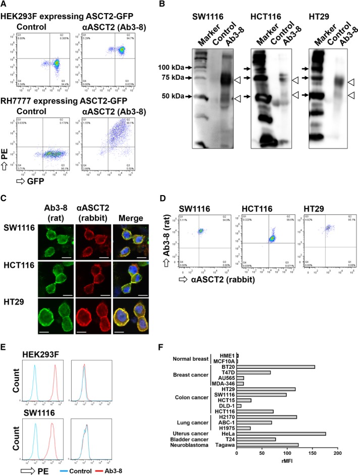
We have previously established a screening method for mAbs using flow cytometry based on the reactivity with transfectants stably expressing GFP‐tagged target protein.30, 31 We examined the reactivity of rat antibodies in supernatants of hybridoma cultures with cells expressing human ASCT2‐GFP. The Ab3‐8 (IgG2a/κ) rat mAb definitely reacted with HEK293 cells expressing human ASCT2‐GFP, and with RH7777 cells expressing ASCT2‐GFP in a GFP expression level‐dependent manner (Figure 1A). Furthermore, immunoprecipitation analysis revealed that Ab3‐8 immunoprecipitated glycosylated 75‐ and non‐glycosylated 50‐kDa proteins in extracts prepared from three human colon cancer cell lines (Figure 1B). As anti‐ASCT2 rabbit mAb (D7C12) is available for biochemical experiments, we evaluated the specificity of Ab3‐8 by comparing staining pattern of Ab3‐8 with that of D7C12. On two‐color immunostaining of human colon cancer cell lines (SW1116, HCT116, and HT29), the components stained by Ab3‐8 were merged with components stained by the anti‐ASCT2 rabbit mAb (Figure 1C). In FCM, the Ab3‐8‐positive populations in these colon cancer cells were also stained by the anti‐ASCT2 rabbit mAb (Figure 1D). Next, we examined the reactivity of Ab3‐8 with ASCT2‐KO cells using the CRISPR‐Cas9 system. Ab3‐8 did not react with ASCT2‐KO‐HEK293 and SW1116 cells (Figure 1E). Furthermore, Ab3‐8 reacted with various human cancer cells, but its reactivity with non‐cancer cells was weak (Figure 1F). Among these cell lines, SW1116, HCT116, DLD‐1, and HCT15 cells harbor a KRAS mutation, whereas other cell lines have wild‐type KRAS.32, 33, 34, 35, 36, 37
Figure 1.

Specificity of the Ab3‐8 anti‐human ASCT2 rat mAb. A, FCM analysis of Ab3‐8 against HEK293 (upper) and RH7777 (lower) cells expressing ASCT2‐GFP. PE, phycoerythrin. B, Immunoprecipitation. Lysates of SW1116, HCT116 and HT29 cells were immunoprecipitated with Ab3‐8, and then detected using a commercial anti‐ASCT2 rabbit mAb. Arrowhead, position of human ASCT2 proteins. C, PFA‐fixed cells were stained with a combination of Ab3‐8 and anti‐ASCT2 rabbit mAb, followed by Alexa Fluor 488‐conjugated anti‐rat IgG and Alexa Fluor 647‐conjugated anti‐rabbit IgG. Nuclei were counterstained with 4′,6‐diamidino‐2‐phenylindole (DAPI). Images were taken at 60 × magnification. Scale bar, 10 μm. D, PFA‐fixed cells were stained with the same antibodies as described above. E, ASCT2‐KO HEK293 and SW1116 cells were reacted with Ab3‐8, followed by PE‐conjugated anti‐rat IgG. The reactivity of Ab3‐8 with these cells was analyzed by flow cytometry. F, FCM analysis of cell surface expression of ASCT2 proteins in various human cell lines. The indicated cells were stained with Ab3‐8, followed by PE‐conjugated anti‐rat IgG. From values of the mean fluorescence intensity (MFI), the ratio (+mAb/ −mAb) of MFI (rMFI) was calculated.
3.2. Functional activity of Ab3‐8
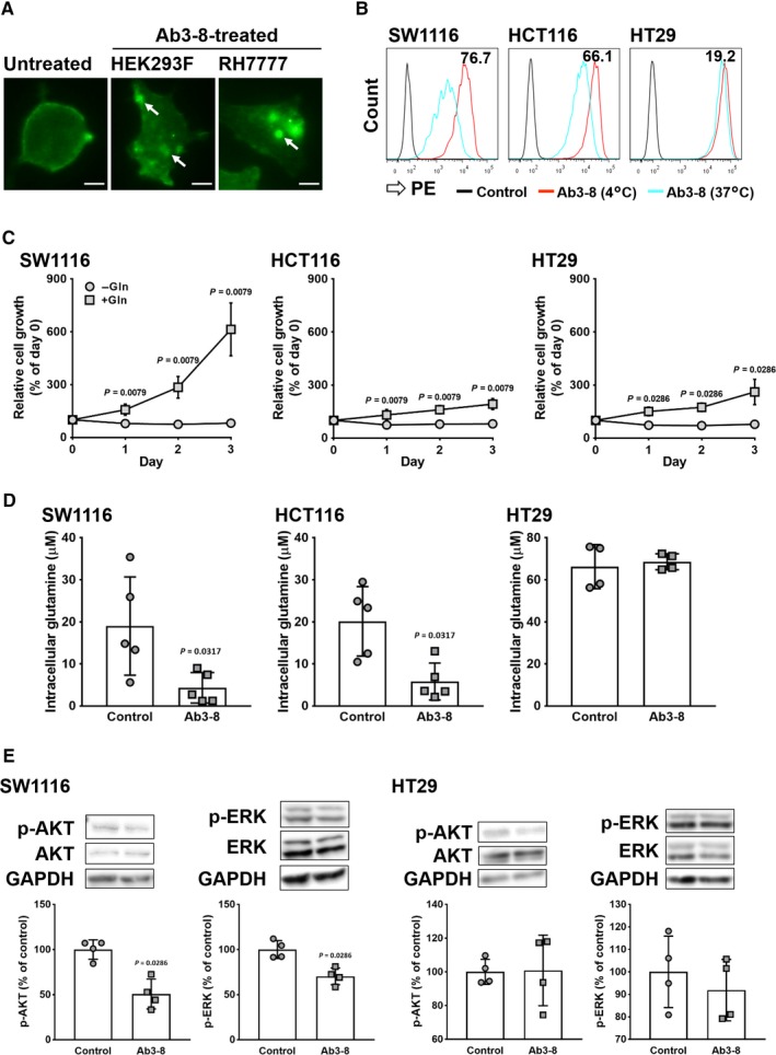
We investigated the functional activity of Ab3‐8. One of the anti‐cancer mechanisms mediated by mAb is internalization; that is, cell surface targets are transferred into the intracellular part after the binding of antibodies.38, 39, 40 Membrane localization of green fluorescence was observed in untreated HEK293 (Figure 2A, left) and RH7777 (data not shown) cells expressing ASCT2‐GFP, whereas intracellular localization of green fluorescence was observed in cells treated with Ab3‐8 (Figure 2A, middle and right). Furthermore, we carried out FCM analysis to quantitate cell surface expression of ASCT2 proteins. In SW1116 and HCT116 cells, which harbor KRAS mutation, treatment with Ab3‐8 at 37°C reduced surface expression of ASCT2 compared with at 4°C. On the other hand, treatment with Ab3‐8 had little effect on ASCT2 surface expression levels in HT29 with no KRAS mutation (Figure 2B).
Figure 2.

Inhibitory effects of Ab3‐8 on ASCT2 functions. A, Microscopy study of ASCT2 internalization. HEK293 and RH7777 cells overexpressing ASCT2‐GFP were reacted with Ab3‐8 at 37°C for 1 h. Images were taken at 20 × magnification. Arrows indicate internalized ASCT proteins. Scale bar, 10 μm. B, Quantitative analysis of ASCT2 internalization using FCM. Human CRC cells were incubated with Ab3‐8 at 4°C or 37°C for 1 h, followed by PE‐conjugated secondary antibody. Numeric data in each panel indicate internalization (%) calculated from the reactivity (MFI) of Ab3‐8 with CRC cells at 37°C and 4°C. C, Glutamine‐dependent cell growth. Cells were cultured with or without glutamine (2 mmol/L) for 3 d. Relative cell growth was measured every 24 h. Each dot represents the mean of four (HT29) or five (SW1116 and HCT116) independent experiments. D, Intracellular glutamine uptake. Cells were incubated in PBS in the presence of Ab3‐8 (30 μg/mL) for 2 h, and then treated with 2 mmol/L glutamine for 10 min. Each dot represents value of individual experiments. E,Cells were incubated in serum‐ and glutamine‐free media in the presence of Ab3‐8 for 24 h, and then treated with 2 mmol/L glutamine for 15 min. Total and phosphorylated (p‐) AKT and ERK protein levels were measured by western blotting. GAPDH was used as a loading control. Each dot represents value of individual experiments. Data are expressed as mean ± SD of at least four independent experiments
Next, we examined whether growth of KRAS‐mutated and wild‐type KRAS colon cancer cells is dependent on glutamine (Figure 2C). All cell lines did not grow under glutamine‐depleted conditions. However, SW1116 cells markedly proliferated in the presence of glutamine. Although HCT116 and HT29 cells proliferated in the presence of glutamine, the proliferation rate of these cells was very small as compared with that of SW1116 cells.
Since ASCT2 is a major glutamine transporter in cancer cells,18 we examined whether Ab3‐8 inhibited intracellular glutamine transport. As shown in Figure 2D, Ab3‐8 markedly inhibited glutamine uptake into SW1116 and HCT116 cells. On the other hand, treatment with Ab3‐8 had no effect on intracellular glutamine concentration in HT29 cells.
AKT and ERK signaling pathways have been reported to be involved in growth and survival of cancer cells.41, 42, 43 As shown in Figure 2E, treatment with Ab3‐8 suppressed the phosphor‐ (p‐) AKT and p‐ERK levels in SW1116 cells. However, consistent with the glutamine uptake assay, Ab3‐8 did not change the p‐AKT and p‐ERK levels in HT29 cells.
3.3. Effects of Ab3‐8 on in vivo tumor growth of colon cancer cells
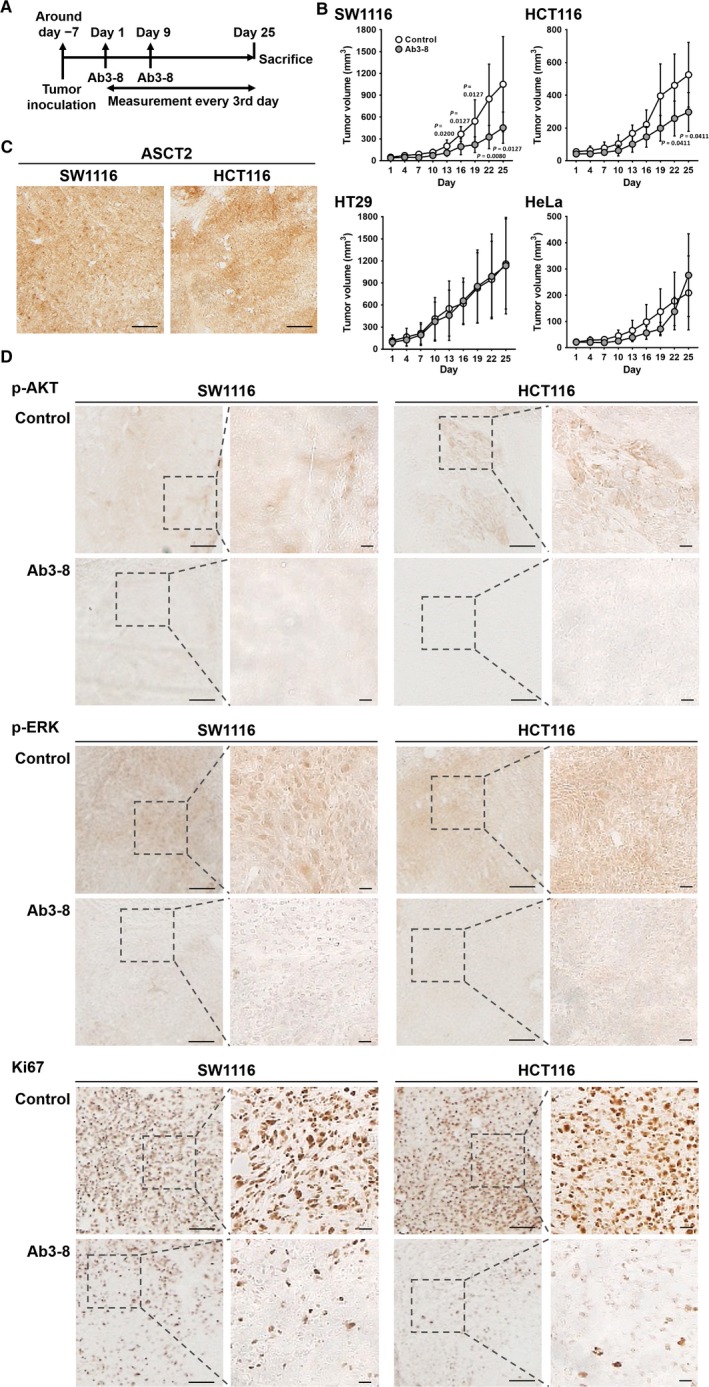
We have next evaluated whether Ab3‐8 inhibits tumor growth of KRAS‐mutated colon cancer cells (Figure 3A). Treatment with Ab3‐8 slowed tumor growth of both SW1116 and HCT116 (Figure 3B). Similar to the in vitro cultured colon cancer cells (Figure 1F), ASCT2 proteins were highly expressed in these in vivo colon cancer tumors (Figure 3C). In addition to inhibitory effects on tumor growth, Ab3‐8 suppressed the expression of p‐AKT, p‐ERK, and Ki67 proteins (Figure 3D). However, Ab3‐8 had no effect on the tumor growth of HT29 and HeLa cells, both of which do not have a KRAS mutation (Figure 3B).
Figure 3.

Anti‐tumor effects of Ab3‐8. Cells were injected subcutaneously into the left/right flanks of male nude mice. A, Time course of the in vivo anti‐tumor study. B, Tumor volumes of CRC and HeLa cells were measured every 3 d using digital calipers, and were quantified using the formula: volume [mm3] = (length [mm]) × (width [mm])2 × 0.5. Ab3‐8 (100 μg/mouse) was intraperitoneally administered on days 1 and 9. Each dot represents the mean of 4‐9 mice. Data are expressed as the mean ± SD. Tumor volume of individual mice shows in Figure S3. C,Tumor sections from control mice were subjected to immunoperoxidase staining using Ab3‐8. Scale bar, 20 μm. D, Tumor sections were subjected to immunoperoxidase staining using the indicated antibodies. Representative images of the SW1116‐ and HCT116‐derived tumor sections, which immune labeled with p‐AKT, p‐ERK and Ki67. Images were obtained at 4 × magnification. Scale bar, 20 μm. Insets, higher magnification of the boxes. Images were obtained at 20 × magnification. Scale bar, 20 μm
4. DISCUSSION
KRAS mutations are observed in many human malignancies, such as non‐small‐cell lung cancer,44, 45 CRC,3, 46 and pancreatic ductal adenocarcinoma47 and are related to their oncogenesis. However, there are no effective methods to treat KRAS‐mutated cancers.
Cancer cells increase the demand for amino acids compared with normal cells because of their rapid metabolism and proliferation. To fulfill this demand, cancer cells upregulate the expression of amino acid transporters on the cell membrane.11 In particular, glutamine is one of the most important nutrients in cancer cells because of enhanced glutaminolysis.11, 12, 13 In this context, glutamine is estimated as a conditional essential amino acid. Previous studies have revealed that ASCT2 is a major glutamine transporter, and is upregulated in multiple cancer cell types such as breast, gastric, prostate, and CRC.18, 48 Furthermore, expression of ASCT2 is correlated with malignant features.18 Regarding CRC, several studies have suggested that glutamine metabolism plays an essential role in cellular growth.49 Pharmacological blockade and genetic silencing of ASCT2 can suppress cancer cell proliferation and tumor growth through the inhibition of glutamine influx.19, 20, 21, 22, 23, 24, 50 Taken together, these findings suggest that ASCT2 is a promising target for cancer therapy.
Recently, production of anti‐human ASCT2 mAbs was reported, and these mAbs suppressed glutamine‐dependent cancer cell growth.51 However, it remains unclear whether these mAbs have in vivo anti‐tumor activity against KRAS‐mutated cancers. Here, we successfully established a novel hybridoma clone secreting the Ab3‐8 mAb against human ASCT2. Ab3‐8 reacted with transfectants expressing human ASCT2‐GFP in a GFP‐expression‐dependent manner. Furthermore, immunoprecipitation assay revealed that Ab3‐8 detected unglycosylated 50‐kDa and glycosylated mature 75‐kDa ASCT2 proteins in human colon cancer cells. These results indicate that Ab3‐8 specifically reacts with human ASCT2 proteins.
We attempted to directly observe Ab3‐8‐mediated internalization of ASCT2 proteins by means of a fluorescent microscopy. Treatment with Ab3‐8 resulted in increased green fluorescent dots in the intracellular region. The dots are assumed to be endocytic vesicles. However, it remains unclear whether Ab3‐8 is released into the cytoplasm or is degrade in the lysosome after the internalization. To confirm this, it needs to further stain lysosome using antibody against the lysosome marker or dye as other groups have done.52, 53
We found that Ab3‐8 inhibited the tumor growth of SW1116 and HCT116 colon cancer cells. Furthermore, treatment with Ab3‐8 reduced intracellular glutamine concentrations in both cell lines. On the other hand, Ab3‐8 had no effects on intracellular glutamine concentration and in vivo tumor growth in HT29 cells. We assume that cause of these results would be attributed to difference in Ab3‐8‐mediated internalization of ASCT2 proteins. FCM analysis revealed that treatment with Ab3‐8‐induced internalization of ASCT2 in both SW1116 and HCT116 cells, but not in HT29 cells. Ma et al54 had reported that treatment with L‐γ‐glutamyl‐p‐nitroanilide, an ASCT2 inhibitor, decreased glutamine consumptions in wild‐type KRAS Caco‐2 cells as well as in KRAS‐mutated SW480 cells. Taken together, Ab3‐8 does not directly inhibit function of ASCT2, but suppresses glutamine uptake through internalization of ASCT2 expressing on cell surface. Although Ab3‐8 binds to ASCT2 proteins expressed on membrane of HT29 cells, the reason why Ab3‐8 did not induce internalization in HT29 cells remains unclear.
HT29 cell line does not harbor KRAS mutation, whereas the cell line harbors BRAF and PIK3CA, the most commonly altered genes in human CRC, mutations.32 For this reason, we cannot accurately judge the effects of Ab3‐8 on tumor growth of KRAS‐mutated cancer. Therefore, we used HeLa cells because the cell line harbors wild‐type KRAS, BRAF, and PIK3CA genes.35, 55, 56 Similar to HT29 cells, treatment with Ab3‐8 did not affect tumor growth of HeLa cells, although we found that the expression of ASCT2 proteins in HT29 and HeLa cells was higher than that in SW1116 and HCT116 cells. Many studies have reported the importance of glutamine metabolism in KRAS‐mutated cancers.15, 16, 17 For this reason, glutamine is likely more important for the survival of SW1116 and HCT116 cells than for HT29 and HeLa cells. In fact, we found that SW1116 cells effectively grew under the glutamine‐enriched conditions as compared with HT29 cells. Accordingly, we speculate that inhibition of ASCT2 function is more effective for KRAS‐mutated cancer than for wild‐type KRAS cancer regardless of the ASCT2 expression level. However, we found unexpectedly that glutamine‐dependent growth rate of HCT116 cells was nearly identical to that of HT29 cells. It would be of great interest to elucidate why Ab3‐8 had no effect on KRAS wild‐type tumors, and this could lead to the development of novel drugs for KRAS‐mutated cancers.
ASCT2 is also expressed in normal mouse tissues such as lung, skeletal muscle, and large intestine.57 However, Ab3‐8 did not recognize mouse ASCT2 protein (data not shown); therefore, we speculated that Ab3‐8 specifically bound to the inoculated tumor expressing human ASCT2 proteins. It was recently reported that Slc1a5 knockout mice exhibit normal growth, survival, and development of adaptive immune cells.58, 59 This suggests that ASCT2‐targeted cancer therapy will be well tolerated with few adverse effects.
Although the importance of glutamine in KRAS‐mutated cancers has been investigated, the exact effects of the KRAS mutation status on ASCT2 function are unclear. KRAS regulates the expression of amino acid transporters including ASCT2, and glutamine uptake in KRAS‐mutated lung cancer cells.60 Furthermore, ASCT2 was upregulated in human CRC cells expressing mutant KRAS as compared with cells expressing wild‐type KRAS.61 Analysis of human CRC samples revealed a significant positive correlation between the KRAS mutation status and ASCT2 protein expression, and an association between tumor depth and ASCT2 expression.25 These findings suggest that regulation of the expression and function of ASCT2 is closely related to the KRAS mutation status. Further studies are needed to clarify the precise relationship between KRAS mutation and ASCT2, particularly how KRAS regulates ASCT2 expression.
ASCT2 is often expressed together with L‐type amino acid transporter 1 (LAT1)/CD98hc (SLC7A5/SLC3A2), a heterodimeric antiporter that exchanges large neutral amino acids.48, 62, 63 Both transporters have been implicated in cancer growth and mTOR signaling in many studies.63, 64 ASCT2 was proposed to take up glutamine, which then acts as an exchange substrate to accumulate leucine via LAT1/CD98hc. We have recently reported the anti‐tumor effects of a mAb against LAT1, which preferentially transports many essential amino acids.29 As tumor growth highly depends on the nutritional condition, maintaining a low‐nutrient environment is likely to induce tumor regression. Taken together, simultaneous inhibition of ASCT2 and LAT1 may be more effective than their individual inhibition for treating cancer.
In conclusion, this study demonstrated that the Ab3‐8 anti‐ASCT2 mAb inhibits the tumor growth of colon cancer cells with KRAS mutation by inhibiting glutamine uptake. Together with the finding that the growth of KRAS‐mutated cancers, including pancreatic and lung cancers, is strongly dependent on glutamine metabolism,16, 65 this study suggests inhibition of glutamine uptake as a potential therapeutic target in KRAS‐mutated various human malignancies.
CONFLICT OF INTEREST
The authors declare no potential conflicts of interest.
AUTHORS’ CONTRIBUTIONS
YH contributed to the study design, carried out almost all experiments, analyzed and interpreted the data, compiled all the figures, and wrote the manuscript. YM, SY, AY, NH, and SU carried out parts of the experiments. KM performed cDNA cloning. TM conceived and supervised the study, participated in its design and coordination, and contributed to the data analysis and interpretation. All authors have read and approved the final version of the manuscript.
Supporting information
ACKNOWLEDGMENTS
The authors gratefully acknowledge the research grants (K18012 and K17030, Kindai University) sponsored by Mr Hina K (PIERAS Co., Ltd). Corresponding author (TM) is deeply grateful to Dr Yamamoto K (Osaka hospital, Japan) for the complete endoscopic submucosal dissection of a colon tumor of TM, which was detected in the process of this manuscript preparation. This study was supported in part by the MEXT‐Supported Program for the Strategic Research Foundation at Private Universities, 2014–2018 (S1411037), JSPS KAKENHI [JP19K16876 (YH)].
Hara Y, Minami Y, Yoshimoto S, et al. Anti‐tumor effects of an antagonistic mAb against the ASCT2 amino acid transporter on KRAS‐mutated human colorectal cancer cells. Cancer Med. 2020;9:302–312. 10.1002/cam4.2689
REFERENCES
- 1. Ferlay J, Soerjomataram I, Dikshit R, et al. Cancer incidence and mortality worldwide: sources, methods and major patterns in GLOBOCAN 2012. Int J Cancer. 2015;136(5):E359‐386. [DOI] [PubMed] [Google Scholar]
- 2. Arnold M, Sierra MS, Laversanne M, Soerjomataram I, Jemal A, Bray F. Global patterns and trends in CRC cancer incidence and mortality. Gut. 2017;66(4):683‐691. [DOI] [PubMed] [Google Scholar]
- 3. Normanno N, Tejpar S, Morgillo F, De Luca A, Van Cutsem E, Ciardiello F. Implications for KRAS status and EGFR‐targeted therapies in metastatic CRC. Nat Rev Clin Oncol. 2009;6(9):519‐527. [DOI] [PubMed] [Google Scholar]
- 4. Irahara N, Baba Y, Nosho K, et al. NRAS mutations are rare in colorectal cancer. Diagn Mol Pathol. 2010;19(3):157‐163. [DOI] [PMC free article] [PubMed] [Google Scholar]
- 5. Chang Y‐Y, Lin P‐C, Lin H‐H, et al. Mutation spectra of RAS gene family in colorectal cancer. Am J Surg. 2016;212(3):537‐544. [DOI] [PubMed] [Google Scholar]
- 6. Misale S, Yaeger R, Hobor S, et al. Emergence of KRAS mutations and acquired resistance to anti‐EGFR therapy in colorectal cancer. Nature. 2012;486(7404):532‐536. [DOI] [PMC free article] [PubMed] [Google Scholar]
- 7. Do K, Speranza G, Bishop R, et al. Biomarker‐driven phase 2 study of MK‐2206 and selumetinib (AZD6244, ARRY‐142886) in patients with colorectal cancer. Invest New Drugs. 2015;33(3):720‐728. [DOI] [PMC free article] [PubMed] [Google Scholar]
- 8. Macarulla T, Cervantes A, Tabernero J, et al. Phase I study of FOLFIRI plus pimasertib as second‐line treatment for KRAS‐mutated metastatic colorectal cancer. Br J Cancer. 2015;112(12):1874‐1881. [DOI] [PMC free article] [PubMed] [Google Scholar]
- 9. Ciardiello F, Normanno N, Martinelli E, et al. Cetuximab continuation after first progression in metastatic colorectal cancer (CAPRI‐GOIM): a randomized phase II trial of FOLFOX plus cetuximab versus FOLFOX. Ann Oncol. 2016;27(6):1055‐1061. [DOI] [PubMed] [Google Scholar]
- 10. Porru M, Pompili L, Caruso C, Biroccio A, Leonetti C. Targeting KRAS in metastatic colorectal cancer: current strategies and emerging opportunities. J Exp Clin Cancer Res. 2018;37(1):57. [DOI] [PMC free article] [PubMed] [Google Scholar]
- 11. Bhutia YD, Babu E, Ramachandran S, Ganapathy V. Amino Acid transporters in cancer and their relevance to "glutamine addiction": novel targets for the design of a new class of anticancer drugs. Cancer Res. 2015;75(9):1782‐1788. [DOI] [PubMed] [Google Scholar]
- 12. Jin L, Alesi GN, Kang S. Glutaminolysis as a target for cancer therapy. Oncogene. 2016;35(28):3619‐3625. [DOI] [PMC free article] [PubMed] [Google Scholar]
- 13. Zhang J, Pavlova NN, Thompson CB. Cancer cell metabolism: the essential role of the nonessential amino acid, glutamine. EMBO J. 2017;36(10):1302‐1315. [DOI] [PMC free article] [PubMed] [Google Scholar]
- 14. Bernfeld E, Foster DA. Glutamine as an essential amino acid for KRas‐driven cancer cells. Trends Endocrinol Metab. 2019;30(6):357‐368. [DOI] [PubMed] [Google Scholar]
- 15. Gaglio D, Metallo CM, Gameiro PA, et al. Oncogenic K‐Ras decouples glucose and glutamine metabolism to support cancer cell growth. Mol Syst Biol. 2011;7:523. [DOI] [PMC free article] [PubMed] [Google Scholar]
- 16. Son J, Lyssiotis CA, Ying H, et al. Glutamine supports pancreatic cancer growth through a KRAS‐regulated metabolic pathway. Nature. 2013;496:101‐105. [DOI] [PMC free article] [PubMed] [Google Scholar]
- 17. Weinberg F, Hamanaka R, Wheaton WW, et al. Mitochondrial metabolism and ROS generation are essential for Kras‐mediated tumorigenicity. Proc Natl Acad Sci USA. 2010;107(19):8788‐8793. [DOI] [PMC free article] [PubMed] [Google Scholar]
- 18. Liu Y, Zhao T, Li Z, Wang L, Yuan S, Sun L. The role of ASCT2 in cancer: a review. Eur J Pharmacol. 2018;837:81‐87. [DOI] [PubMed] [Google Scholar]
- 19. Wang Q, Beaumont KA, Otte NJ, et al. Targeting glutamine transport to suppress melanoma cell growth. Int J Cancer. 2014;135(5):1060‐1071. [DOI] [PubMed] [Google Scholar]
- 20. Wang Q, Hardie R‐A, Hoy AJ, et al. Targeting ASCT2‐mediated glutamine uptake blocks prostate cancer growth and tumour development. J Pathol. 2015;236(3):278‐289. [DOI] [PMC free article] [PubMed] [Google Scholar]
- 21. van Geldermalsen M, Wang Q, Nagarajah R, et al. ASCT2/SLC1A5 controls glutamine uptake and tumour growth in triple‐negative basal‐like breast cancer. Oncogene. 2016;35(24):3201‐3208. [DOI] [PMC free article] [PubMed] [Google Scholar]
- 22. Lu J, Chen M, Tao Z, et al. Effects of targeting SLC1A5 on inhibiting gastric cancer growth and tumor development in vitro and in vivo . Oncotarget. 2017;8(44):76458‐76467. [DOI] [PMC free article] [PubMed] [Google Scholar]
- 23. Marshall AD, van Geldermalsen M, Otte NJ, et al. ASCT2 regulates glutamine uptake and cell growth in endometrial carcinoma. Oncogenesis. 2017;6(7):e367. [DOI] [PMC free article] [PubMed] [Google Scholar]
- 24. van Geldermalsen M, Quek L‐E, Turner N, et al. Benzylserine inhibits breast cancer cell growth by disrupting intracellular amino acid homeostasis and triggering amino acid response pathways. BMC Cancer. 2018;18(1):689. [DOI] [PMC free article] [PubMed] [Google Scholar]
- 25. Toda K, Nishikawa G, Iwamoto M, et al. Clinical role of ASCT2 (SLC1A5) in KRAS‐mutated colorectal cancer. Int J Mol Sci. 2017;18:E1632. [DOI] [PMC free article] [PubMed] [Google Scholar]
- 26. Cong L, Ran FA, Cox D, et al. Multiplex genome engineering using CRISPR/Cas systems. Science. 2013;339(6121):819‐823. [DOI] [PMC free article] [PubMed] [Google Scholar]
- 27. Mashiko D, Fujihara Y, Satouh Y, Miyata H, Isotani A, Ikawa M. Generation of mutant mice by pronuclear injection of circular plasmid expressing Cas9 and single guided RNA. Sci Rep. 2013;3:3355. [DOI] [PMC free article] [PubMed] [Google Scholar]
- 28. Hara Y, Torii R, Ueda S, et al. Inhibition of tumor formation and metastasis by a monoclonal antibody against lymphatic vessel endothelial hyaluronan receptor 1. Cancer Sci. 2018;109(10):3171‐3182. [DOI] [PMC free article] [PubMed] [Google Scholar]
- 29. Ueda S, Hayashi H, Miyamoto T, et al. Anti‐tumor effects of mAb against l‐type amino acid transporter 1 (LAT1) bound to human and monkey LAT1 with dual avidity modes. Cancer Sci. 2019;110(2):674‐685. [DOI] [PMC free article] [PubMed] [Google Scholar]
- 30. Ohno Y, Suda K, Masuko K, Yagi H, Hashimoto Y, Masuko T. Production and characterization of highly tumor‐specific rat monoclonal antibodies recognizing the extracellular domain of human L‐type amino‐acid transporter 1. Cancer Sci. 2008;99(5):1000‐1007. [DOI] [PMC free article] [PubMed] [Google Scholar]
- 31. Masuko T, Ohno Y, Masuko K, et al. Towards therapeutic antibodies to membrane oncoproteins by a robust strategy using rats immunized with transfectants expressing target molecules fused to green fluorescent protein. Cancer Sci. 2011;102(1):25‐35. [DOI] [PubMed] [Google Scholar]
- 32. Ahmed D, Eide PW, Eilertsen IA, et al. Epigenetic and genetic features of 24 colon cancer cell lines. Oncogenesis. 2013;2:e71. [DOI] [PMC free article] [PubMed] [Google Scholar]
- 33. Wong CC, Qian Y, Li X, et al. SLC25A22 promotes proliferation and survival of colorectal cancer cells with KRAS mutations and xenograft tumor progression in mice via intracellular synthesis of aspartate. Gastroenterology. 2016;151(5):945‐960. [DOI] [PubMed] [Google Scholar]
- 34. Hollestelle A, Elstrodt F, Nagel JH, Kallemeijn WW, Schutte M. Phosphatidylinositol‐3‐OH kinase or RAS pathway mutations in human breast cancer cell lines. Mol Cancer Res. 2007;5(2):195‐201. [DOI] [PubMed] [Google Scholar]
- 35. Perera D, Venkitaraman AR. Oncogenic KRAS triggers MAPK‐dependent errors in mitosis and MYC‐dependent sensitivity to anti‐mitotic agents. Sci Rep. 2016;6:29741. [DOI] [PMC free article] [PubMed] [Google Scholar]
- 36. Sumimoto H, Takano A, Teramoto K, Daigo Y. RAS‐mitogen‐activated protein kinase signal is required for enhanced PD‐L1 expression in human lung cancers. PLoS ONE. 2016;11(11):e0166626. [DOI] [PMC free article] [PubMed] [Google Scholar]
- 37. Smakman N, Kranenburg O, Vogten JM, Bloemendaal AL, van Diest P, Borel Rinkes IH. Cyclooxygenase‐2 is a target of KRASD12, which facilitates the outgrowth of murine C26 colorectal liver metastases. Clin Cancer Res. 2005;11(1):41‐48. [PubMed] [Google Scholar]
- 38. Vu T, Claret FX. Trastuzumab: updated mechanisms of action and resistance in breast cancer. Front Oncol. 2012;2:62. [DOI] [PMC free article] [PubMed] [Google Scholar]
- 39. Terp MG, Olesen KA, Arnspang EC, et al. Anti‐human CD73 monoclonal antibody inhibits metastasis formation in human breast cancer by inducing clustering and internalization of CD73 expressed on the surface of cancer cells. J Immunol. 2013;191(8):4165‐4173. [DOI] [PubMed] [Google Scholar]
- 40. Okada Y, Kimura T, Nakagawa T, et al. EGFR downregulation after anti‐EGFR therapy predicts the antitumor effect in colorectal cancer. Mol Cancer Res. 2017;15(10):1445‐1454. [DOI] [PubMed] [Google Scholar]
- 41. Aguirre‐Ghiso JA, Estrada Y, Liu D, Ossowski L. ERKMAPK activity as a determinant of tumor growth and dormancy; regulation by p38SAPK. Cancer Res. 2003;63(7):1684‐1695. [PubMed] [Google Scholar]
- 42. Papa S, Choy PM, Bubici C. The ERK and JNK pathways in the regulation of metabolic reprogramming. Oncogene. 2019;38:2223‐2240. [DOI] [PMC free article] [PubMed] [Google Scholar]
- 43. Song M, Bode AM, Dong Z, Lee MH. AKT as a therapeutic target for cancer. Cancer Res. 2019;79(6):1019‐1031. [DOI] [PubMed] [Google Scholar]
- 44. Karachaliou N, Mayo C, Costa C, et al. KRAS mutations in lung cancer. Clin Lung Cancer. 2013;14(3):205‐214. [DOI] [PubMed] [Google Scholar]
- 45. Maus M, Grimminger PP, Mack PC, et al. KRAS mutations in non‐small‐cell lung cancer and colorectal cancer: implications for EGFR‐targeted therapies. Lung Cancer. 2014;83(2):163‐167. [DOI] [PubMed] [Google Scholar]
- 46. Network CGA. Comprehensive molecular characterization of human colon and rectal cancer. Nature. 2012;487(7407):330‐337. [DOI] [PMC free article] [PubMed] [Google Scholar]
- 47. Bryant KL, Mancias JD, Kimmelman AC, Der CJ. KRAS: feeding pancreatic cancer proliferation. Trends Biochem Sci. 2014;39(2):91‐100. [DOI] [PMC free article] [PubMed] [Google Scholar]
- 48. Fuchs BC, Bode BP. Amino acid transporters ASCT2 and LAT1 in cancer: partners in crime? Semin Cancer Biol. 2005;15(4):254‐266. [DOI] [PubMed] [Google Scholar]
- 49. Miyo M, Konno M, Nishida N, et al. Metabolic adaptation to nutritional stress in human colorectal cancer. Sci Rep. 2016;6:38415. [DOI] [PMC free article] [PubMed] [Google Scholar]
- 50. Schulte ML, Fu A, Zhao P, et al. Pharmacological blockade of ASCT2‐dependent glutamine transport leads to antitumor efficacy in preclinical models. Nat Med. 2018;24(2):194‐202. [DOI] [PMC free article] [PubMed] [Google Scholar]
- 51. Suzuki M, Toki H, Furuya A, Ando H. Establishment of monoclonal antibodies against cell surface domains of ASCT2/SLC1A5 and their inhibition of glutamine‐dependent tumor cell growth. Biochem Biophys Res Commun. 2017;482(4):651‐657. [DOI] [PubMed] [Google Scholar]
- 52. Shukla RS, Jain A, Zhao Z, Cheng K. Intracellular trafficking and exocytosis of a multi‐component siRNA nanocomplex. Nanomedicine. 2016;12(5):1323‐1334. [DOI] [PMC free article] [PubMed] [Google Scholar]
- 53. Jain A, Barve A, Zhao Z, et al. Targeted delivery of an siRNA/PNA hybrid nanocomplex reverses carbon tetrachloride‐induced liver fibrosis. Adv Ther. 2019;2:1900046. [DOI] [PMC free article] [PubMed] [Google Scholar]
- 54. Ma H, Wu Z, Peng J, et al. Inhibition of SLC1A5 sensitizes colorectal cancer to cetuximab. Int J Cancer. 2018;142(12):2578‐2588. [DOI] [PubMed] [Google Scholar]
- 55. Arjumand W, Merry CD, Wang C, et al. Phosphatidyl inositol‐3 kinase (PIK3CA) E545K mutation confers cisplatin resistance and a migratory phenotype in cervical cancer cells. Oncotarget. 2016;7(50):82424‐82439. [DOI] [PMC free article] [PubMed] [Google Scholar]
- 56. Pédeboscq S, Gravier D, Casadebaig F, et al. Synthesis and evaluation of apoptosis induction of thienopyrimidine compounds on KRAS and BRAF mutated colorectal cancer cell lines. Bioorg Med Chem. 2012;20(22):6724‐6731. [DOI] [PubMed] [Google Scholar]
- 57. Utsunomiya‐Tate N, Endou H, Kanai Y. Cloning and functional characterization of a system ASC‐like Na+‐dependent neutral amino acid transporter. J Biol Chem. 1996;271(25):14883‐14890. [DOI] [PubMed] [Google Scholar]
- 58. Nakaya M, Xiao Y, Zhou X, et al. Inflammatory T cell responses rely on amino acid transporter ASCT2 facilitation of glutamine uptake and mTORC1 kinase activation. Immunity. 2014;40(5):692‐705. [DOI] [PMC free article] [PubMed] [Google Scholar]
- 59. Masle‐Farquhar E, Bröer A, Yabas M, Enders A, Bröer S. ASCT2 (SLC1A5)‐deficient mice have normal B‐cell development, proliferation, and antibody production. Front Immunol. 2017;8:549. [DOI] [PMC free article] [PubMed] [Google Scholar]
- 60. Gwinn DM, Lee AG, Briones‐Martin‐del‐Campo M, et al. Oncogenic KRAS regulates amino acid homeostasis and asparagine biosynthesis via ATF4 and alters sensitivity to L‐asparaginase. Cancer Cell. 2018;33(1):91‐107. [DOI] [PMC free article] [PubMed] [Google Scholar]
- 61. Hutton JE, Wang X, Zimmerman LJ, et al. Oncogenic KRAS and BRAF drive metabolic reprogramming in colorectal cancer. Mol Cell Proteomics. 2016;15(9):2924‐2938. [DOI] [PMC free article] [PubMed] [Google Scholar]
- 62. Broer A, Rahimi F, Broer S. Deletion of amino acid transporter ASCT2 (SLC1A5) reveals an essential role for transporters SNAT1 (SLC38A1) and SNAT2 (SLC38A2) to sustain glutaminolysis in cancer Cells. J Biol Chem. 2016;291(25):13194‐13205. [DOI] [PMC free article] [PubMed] [Google Scholar]
- 63. Nicklin P, Bergman P, Zhang B, et al. Bidirectional transport of amino acids regulates mTOR and autophagy. Cell. 2009;136(3):521‐534. [DOI] [PMC free article] [PubMed] [Google Scholar]
- 64. Kandasamy P, Gyimesi G, Kanai Y, Hediger MA. Amino acid transporters revisited: new views in health and desease. Trends Biochem Sci. 2018;43(10):752‐789. [DOI] [PubMed] [Google Scholar]
- 65. Romero R, Sayin VI, Davidson SM, et al. Keap1 loss promotes Kras‐driven lung cancer and results in dependence on glutaminolysis. Nat Med. 2017;23(11):1362‐1368. [DOI] [PMC free article] [PubMed] [Google Scholar]
Associated Data
This section collects any data citations, data availability statements, or supplementary materials included in this article.
Supplementary Materials


