Abstract
We describe a new class of fluorescence polarization immunoassays based on the luminescence from a Re(I) metal–ligand complex. Re(I) complexes are extremely photostable and possess useful photophysical properties including long lifetimes, high quantum yields, and high emission polarization in the absence of rotational diffusion. In the present study, a conjugatable, highly luminescent Re(I) metal-ligand complex, [Re(bcp)(CO)3(4-COOHPy)](ClO4), where bcp is 2,9-dimethyl-4,7-diphenyl-1,10-phenanthroline and 4-COOHPy is isonicotinic acid, has been evaluated for use in fluorescence polarization immunoassays (FPIs) with high-molecular-weight antigens. This Re(I) complex (Re) displays highly polarized emission (with a maximum anisotropy near 0.3) in the absence of rotational diffusion and a long average lifetime (2.7 μs) when bound to human serum albumin (HSA) in oxygenated aqueous solution. The emission polarization of the Re–HSA conjugate is sensitive to the binding of anti-HSA, resulting in a significant increase in anisotropy. The labeled HSA was also used in a competition immunoassay where unlabeled HSA was also used as an antigen. These experimental results, combined with theoretical predictions, demonstrate the potential of this Re(I) metal–ligand complex as a luminescence probe in FPIs of high-molecular-weight analytes (105–108 Da).
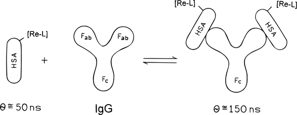
Fluorescence polarization (FP) was first theoretically described by Perrin1 in 1926; this description was subsequently expanded and measured by Weber.2,3 Dandliker and co-workers adapted FP for use in analytical biochemistry, including antigen (Ag)–antibody (Ab) interactions4–6 and hormone–receptor interactions.7 Since establishment of the theory and method by Dandliker et al.,4–6 the use of fluorescence polarization immunoassays (FPIs) for the quantitative and qualitative measurement of various types of molecules and bioconjugates has been reported. Such uses include therapeutic drug monitoring and determination of hormones, drugs of abuse, proteins and peptides, proteases, and inhibitors, as well as DNA binding interactions.7–25 In fact, FPI technology is presently in widespread commercial use in several instruments. A generic representation of a FPI as it applies to the present study is shown in Scheme 1.
Scheme 1.
Intuitive Description of a Fluorescence Polarization Immunoassay (Re-L is [Re(bcp)(CO)3(4-COOHPy)]+, and θ is the Rotational Correlation Time)
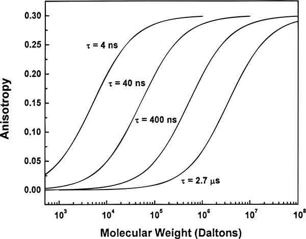
A serious limitation of the present immunoassays is that they are limited to low-molecular-weight antigens. This limitation is a result of the use of fluorophores, such as fluorescein, which display lifetimes near 4 ns. A FPI requires that emission from the unbound labeled antigen be depolarized, so that an increase in polarization may be observed upon antigen binding to antibody. For depolarization to occur, the antigen must display a rotational correlation time much shorter than the lifetime of the probe (in the case of fluorescein, less than 4 ns), which limits the dynamic range of the FPI to antigens with low molecular weights (Figure 1). Some long-lifetime fluorophores, such as chelates of Eu3+ and Tb3+, have been used in time-resolved immunoassays,16–18 but they do not display polarized emission and are thus not useful in FPIs.
Figure 1.
Molecular-weight-dependent anisotropy for a protein-bound luminophore with luminescence lifetimes of 4, 40, 400, and 2700 ns. The curves are based on eqs 3 and 8 assuming an aqueous solution at 20 °C with a viscosity of 1 cP and .
More recent studies have shown that [Ru(bpy)2(dcb)]2+, where bpy is 2,2′-bipyridine and dcb is 4,4′-dicarboxy-2,2′-bipyridine, displays high polarization in the absence of rotational diffusion (~0.25), as well as a long lifetime (~400 ns).19 The experimental results demonstrated that the steady-state polarization of [Ru(bpy)2(dcb)]2+ labeled to HSA was sensitive to the binding of anti-HSA, which resulted in a 200% increase in polarization. Another metal-ligand complex, [Os(bpy)2(dcb)]2+, was also used in a FPI to detect a high-molecular-weight bioconjugate using red excitation and emission wavelengths.20
In the present report, we describe a new FPI probe for high-molecular-weight antigens. A highly luminescent Re(I) metal–ligand complex, [Re(bcp)(CO)3(4-COOHPy)](ClO4), where bcp is 2,9-dimethyl-4,7-diphenyl-1,10-phenanthroline and 4-COOHPy is isonicotinic acid, was synthesized and used in a FPI. This Re(I) complex displays highly polarized emission (with a maximum polarization near 0.4 and maximum anisotropy near 0.3) in the absence of rotational diffusion and a long average lifetime (2.7 μs) when bound to proteins in air-equilibrated aqueous solution. We found that the steady-state polarization of the Re(I) complexlabeled HSA conjugate (Re–HSA) was sensitive to the binding of anti-HSA, resulting in a significant increase in luminescence polarization. The labeled HSA was also used in a competitive format, with unlabeled HSA acting as an antigen. More importantly, the lifetime of this probe when covalently labeled to HSA in air-equilibrated aqueous solution is near 3 μs, which theoretically allows immunoassays of antigens with molecular masses to 108 Da (Figure 1).
The theory of fluorescence polarization and its application to immunoassays has appeared previously,4–7,19 however, it is quite useful to present it here for clarity. The fluorescence polarization (P) of a labeled macromolecule depends on the fluorescence lifetime (τ) and the rotational correlation time (θ):
| (1) |
where P0 is the polarization observed in the absence of rotational diffusion. The effect of molecular weight on the polarization values can be seen from an alternative form of eq 1:
| (2) |
where k is the Boltzmann constant, T is the absolute temperature in kelvin, h is the viscosity of the solution, and V is the molecular volume. The molecular volume of the protein is related to the molecular weight (Mr) and the rotational correlation time by
| (3) |
where R is the ideal gas constant, is the specific volume of the protein, and h is the hydration, typically 0.2 g of H2O/g of protein. Generally, the observed correlation times are about 2-fold longer than those calculated for an anhydrous sphere (eq 3 with h = 0) due to the effects of hydration and the nonspherical shapes of most proteins. Therefore, in aqueous solution at 20 °C (η = 1 cP), one can expect a protein such as HSA (Mr ≈ 65 000, with ) to display a rotational correlation time near 50 ns.
The advantage of using a luminophore with a long lifetime is illustrated by comparing the expected polarization values of HSA when free and bound to IgG (Mr ≈ 160 000, Scheme 1). It is convenient to use the anisotropy (r) in this calculation. The anisotropy and polarization are related by
| (4) |
| (5) |
where I∥ and I⊥ are the vertically and horizontally polarized components of the emission. The values of P and r can be interchanged using
| (6) |
| (7) |
The parameters P and r are both commonly used to describe rotational diffusion processes of fluorophores in solution. The values of P are more often used in FPI because they are entrenched by tradition and are slightly larger than the anisotropy values. The parameter r is preferred on the basis of theory. The anisotropy of a labeled macromolecule is
| (8) |
where r0 is the anisotropy in the absence of rotational diffusion and is typically near 0.3 for most fluorophores, although the theoretical limit given collinear transition dipoles for absorption and emission is 0.4.
We simulated the expected anisotropy values for a range of photoluminescence lifetimes. These calculations were based on eqs 3 and 8, established on the assumptions that the limiting anisotropy (r0) was 0.3 in the absence of rotational diffusion, the solution viscosity was 1 cP, and for the protein. These simulations demonstrate how the lifetime of the luminophore determines the range of molecular weights which can be resolved by the luminophore in an immunoassay. Presently, most immunoassays rely on fluorescein and rhodamine derivatives as fluorescent probes (τ ≈ 4 ns). If one considers that most low-molecular-weight antigens are in the range of Mr < 1000, the expected anisotropy of the labeled antigen can be estimated from Figure 1 to be in the range of 0.05. Upon antigen association with antibody, the molecular weight increases (Mr ≈ 160 000), and the anisotropy of the bioconjugate approaches 0.30. Hence, a large change in anisotropy is found upon binding of Ag to Ab for low-molecular-weight antigens when utilizing a 4 ns lifetime fluorophore.
However, if the molecular weight of the labeled antigen is larger, above Mr ≈ 20 000, then the anisotropy changes only slightly upon binding to antibody if the same fluorophore is used. For instance, suppose the molecular weight of the labeled antigen is 160 000, with a rotational correlation time of 125 ns, and that of the antibody-bound form is 600 000, with a rotational correlation time of 470 ns. In this particular case, the anisotropy values will differ by less than 2% between the two forms when using a short-lifetime fluorophore. This small change is attributed to the large discrepancy between the lifetime of the fluorophore and the rotational correlation time of the labeled macromolecular complex. It is for this reason that FPIs are performed only in the low-molecular-weight range with conventional short-lifetime fluorophores.
Suppose the lifetime of the luminophore is in the range of 3 μs, as is the case with [Re(bcp)(CO)3(4-COOHPy)]+. For the example described above, the binding assay would now be detectable using luminescence polarization (Figure 1). Theoretically, a luminophore with a lifetime of 3 μs could allow the analysis of biological systems with molecular weights up to 100 million and correlation times up to 80 μs, thereby greatly expanding the capabilities of FPIs to include the study of entire cells, viruses, and other large biomolecules and biomolecular complexes.
EXPERIMENTAL SECTION
Human serum albumin (HSA), human immunoglobulin G (IgG), and monoclonal IgG specific for HSA (anti-HSA) from mouse ascites were obtained from Sigma Chemical Co. and were used without further purification. All other reagents and all solvents used were reagent grade. The synthesis of [Re(bcp)(CO)3(4-COOHPy)](ClO4) was described in a previous report.21
Synthesis of the NHS Ester of [Re(bcp)(CO)3(4-COOHPy)](ClO4) and Protein Labeling
Five milligrams of N,N′-dicyclohexylcarbodiimide (DCC) and 3 mg of N-hydroxysuccinimide (NHS) were dissolved in 0.15 mL of DMF with stirring. [Re(bcp)(CO)3(4-COOHPy)](ClO4) (10 mg in 0.15 mL of DMF) was added, and the mixture was stirred for 20 h. The formed precipitate was removed by filtration through a syringe filter, and the filtrate containing the activated Re complex was used for labeling the substrates.
The protein HSA (10 mg) was labeled by adding a 15-fold molar excess of the activated Re complex in 50 μL of DMF to 1 mL of stirring protein solution (0.2 M carbonate buffer, pH 8.5), followed by a 5 h incubation. The conjugate was purified by gel filtration chromatography on Sephadex G-25, using 0.1 M PBS, pH 7.0, as eluent. The dye:protein ratio of the Re–HSA conjugate was determined to be 2:1. The concentration of the protein was determined by the Coomassie Plus protein assay. The concentration of the Re(I) complex was determined by its absorbance at 400 nm, assuming the extinction coefficient was the same as that of the free dye (ϵ400 = 5040 M−1cm−1).21 The equilibrium association constants of the bioconjugates were determined from luminescence anisotropy data as described in the literature.6
Photoluminescence Measurements
Uncorrected emission spectra were recorded on an SLM AB2 spectrofluorometer. The frequency domain lifetime measurements were performed on an ISS K2 fluorometer, using a high-intensity Panasonic blue lightemitting diode (LED) configured to provide amplitude-modulated light centered at 390 nm.21,22 An Andover 500 nm long-pass filter (500FH90–50S) was used to isolate the emission.
The frequency domain intensity decay data were fit by a nonlinear least-squares procedure, generally to a sum of three single-exponential decays. The intensity decays were described by
| (9) |
where I(t) is the luminescence intensity at time t, and αi and the τi are the pre-exponential weighting factors and the excited-state lifetimes, respectively. The subscripts denote each component. Mean lifetimes were calculated using
| (10) |
The excitation anisotropy spectrum is defined by
| (11) |
where I∥ and I⊥ are the emission intensities measured with vertically polarized excitation and the emission polarization parallel (I∥) or perpendicular (I⊥) to the excitation. The values of the polarized intensities were corrected for the transmission efficiency of the polarized components by the detection optics.
RESULTS
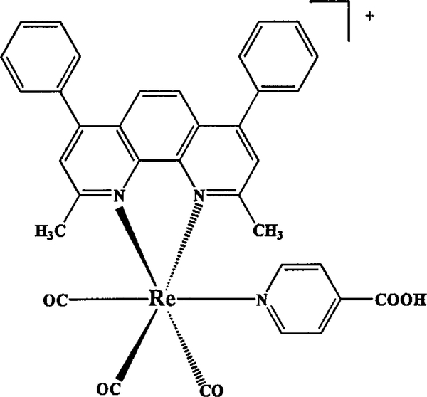
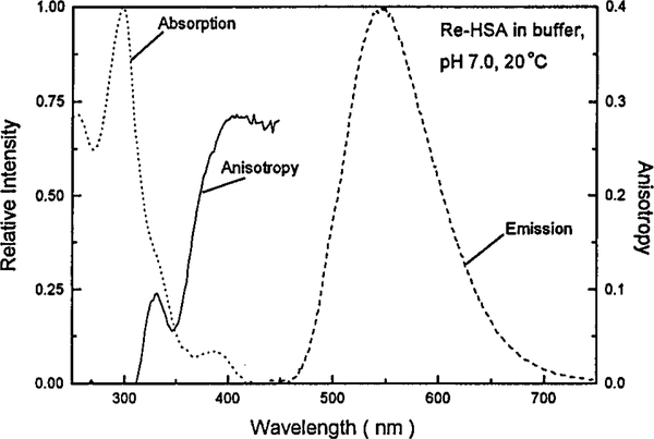
The molecular structure of [Re(bcp)(CO)3(4-COOHPy)]+ is shown in Figure 2. The absorption and emission spectra of [Re(bcp)(CO)3(4-COOHPy)]+ labeled to HSA are shown in Figure 3. The spectra are normalized to unity for comparative purposes. The absorption profile in the low-energy region (340–425 nm) and the more intense higher energy band at 290 nm are characteristic metal-to-ligand charge transfer (MLCT) and π–π* transitions, respectively. The emission spectrum is broad and has a maximum near 550 nm. These photophysical characteristics are similar to that observed with the parent complex [Re(bcp)(CO)3(Py)]+, where Py is pyridine.29b The large Stokes shift of MLCT complexes in general can be exploited in biological media, where multiple labeling of close proximity residues will not result in self-quenching processes.
Figure 2.
Molecular structure of [Re(bcp)(CO)3(4-COOHPy)]+.
Figure 3.
Room temperature absorption and emission spectra of [Re(bcp)(CO)3(4-COOHPy)]+ conjugated to HSA in 0.1 M PBS buffer, pH 7.0. The emission spectrum was obtained with 400 ± 4 nm excitation. The solid line shows the excitation anisotropy spectrum measured in 100% glycerol at −60 °C, with an emission wavelength of 550 ± 8 nm.
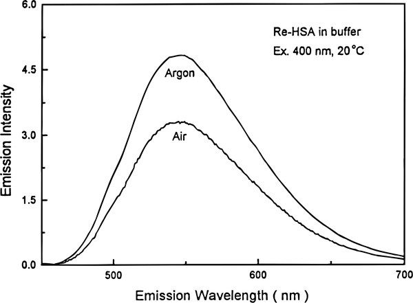
O2 quenching is commonplace from MLCT excited states of Re(I) complexes.21,29 In the case of [Re(bcp)(CO)3(4-COOHPy)]+, the oxygen quenching is modest when the probe is bound to HSA in air-equilibrated aqueous solution. Compared to a deoxygenated buffer solution (1.0), the relative photoluminescence intensity of Re–HSA in air-equilibrated buffer solution is 0.69 (Figure 4). Complete photophysical characterization of [Re(bcp)(CO)3(4-COOHPy)]+ has recently appeared.21
Figure 4.
Emission spectra of [Re(bcp)(CO)3(4-COOHPy)]+ conjugated to HSA in 0.1 M PBS buffer, pH 7.0, equilibrated with argon or air at 20 °C. The excitation wavelength was 400 ± 4 nm.
We examined the steady-state excitation anisotropy spectrum of [Re(bcp)(CO)3(4-COOHPy)]+ in vitrified solution (glycerol, −60 °C), where rotational diffusion does not occur during the excited state lifetime (Figure 3). This complex shows a maximum anisotropy near 0.3, whose values are constant from 390 to 450 nm.
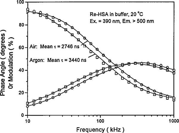
Frequency domain intensity decays of Re–HSA in air-equilibrated and argon-equilibrated 0.1 M PBS buffer solutions are shown in Figure 5. The analysis of the frequency domain intensity decays are summarized in Table 1. The decays were best fit to a sum of three-exponential decay laws. The mean lifetimes are 2.75 μs in air-equilibrated and 3.44 μs in argon-equilibrated buffer solutions, respectively. The elimination of oxygen is, therefore, not required for use in fluorescence polarization immunoassays of high-molecular-weight analytes.
Figure 5.
Frequency domain intensity decays of [Re(bcp)(CO)3(4-COOHPy)]+ conjugated to HSA. Sample was the under same conditions as in Figure 4. The modulated excitation was centered at 390 nm (Panasonic blue LED) and a 500 nm long-pass filter was used to isolate the emission.
Table 1.
Recovered Intensity Decay Parameters of [Re(bcp)(CO)3(4-COOHPy)]+ Conjugated to HSA, Measured in 0.1 M PBS at 20 °C
| condition | τi (μs) | αi | mean τ (μs) |
|---|---|---|---|
| air | 5.76 | 0.02 | |
| 1.27 | 0.13 | ||
| 0.053 | 0.85 | 2.75 | |
| argon | 6.23 | 0.02 | |
| 1.39 | 0.11 | ||
| 0.051 | 0.87 | 3.44 |
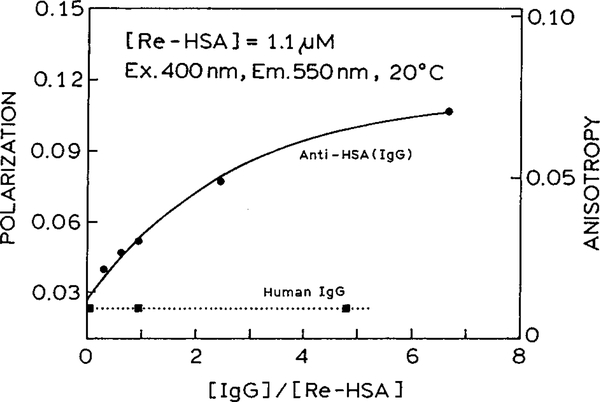
To evaluate the feasibility of using [Re(bcp)(CO)3(4-COOHPy)]+ in a polarization immunoassay, Re–HSA was used as an antigen. We examined the changes in anisotropy of Re-labeled HSA in the presence of increasing amounts of anti-HSA. The polarization increased about 4-fold from 0.023 to 0.108, which corresponds to anisotropy values ranging between 0.017 and 0.075 (Figure 6). Similar results were obtained when using two different batches of anti-HSA with antibody (Ab) concentrations ranging from 0 to 8 times that of Re–HSA (Ag). An association constant was calculated from the data in Figure 6 and found to be 3.3 μM−1. We used nonspecific human IgG as a control, and no detectable changes in polarization of Re–HSA were observed in that experiment (Figure 6).
Figure 6.
Steady-state fluorescence polarization of Re–HSA at various concentrations of IgG specific for HSA (anti-HSA, ●) or nonspecific IgG (■) measured at 20 °C. Anisotropy is also displayed on this plot for comparative purposes. The excitation and observation wavelengths were 400 and 550 nm, respectively, with a bandpass of 8 nm.
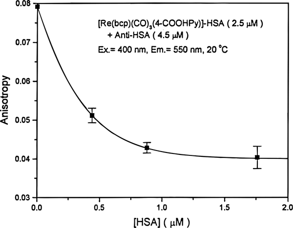
We attempted to develop a competitive assay for HSA wherein labeled and unlabeled antigens are allowed to simultaneously compete for the binding sites on the antibody. The simultaneous exposure of the labeled and unlabeled HSA to anti-HSA resulted in a constant anisotropy at all concentrations. This may reflect a higher affinity of anti-HSA for labeled HSA or the formation of aggregates around the labeled antigen. However, preincubation of the unlabeled HSA with anti-HSA for 30 min, followed by the addition of the Re-labeled antigen, resulted in measurable changes in anisotropy. In this sequential assay, the anisotropy was found to decrease with increasing amounts of unlabeled HSA (Figure 7). The concentrations of Re-labeled HSA and anti-HSA were 2.5 and 4.5 μM, respectively. At high concentrations of unlabeled HSA, the anisotropy could not be reversed to the value for unbound Re–HSA (r = 0.017, p = 0.023), which should be observed on total replacement of Re–HSA with unlabeled HSA. This effect could be explained by nonspecific binding of Re–HSA to other proteins present in the solution. However, the polarization of Re–HSA was not influenced by the presence of nonspecific proteins in the IgG ascites fluid (Figure 6). Another reason for this behavior may be a result of a higher binding affinity for Re–HSA than for free HSA or, possibly, irreversible interactions between the Ab and Ag.
Figure 7.
Steady-state fluorescence anisotropy of Re–HSA added to preincubated mixtures of anti-HSA with various concentrations of unlabeled HSA measured at 20 °C. The excitation and observation wavelengths were 400 and 550 nm, respectively, with a bandpass of 8 nm. Error bars represent the standard deviations of three anisotropy readings.
DISCUSSION
The results described above, combined with our early reports,19,20 demonstrate the utility of fluorescence polarization immunoassays of high-molecular-weight analytes using luminescent metal–ligand complexes which display highly polarized emission and long lifetimes. Many different approaches have been used to circumvent the present limitation of FPIs to low-molecularweight substances.10,23–25 An early attempt to develop FPIs for high-molecular-weight antigens was reported by Grossman.23 The dansyl (dimethylaminonaphthalenesulfonic acid) fluorophore was used because of its 20 ns lifetime. Tsuruoka and co-workers attempted to develop a FPI with IgG by increasing the molecular weight of the antibody.24 This was accomplished by immobilizing the antibody with latex beads or colloidal silver. Urio and Cittanova10 decreased the size of the labeled antibody by using Fab fragments in place of complete IgG molecules. Another approach to enable the measurement of high-molecular-weight antigens was introduced by Wei and Herron.25 They used a tetramethylrhodamine-labeled synthetic peptide, which has a high binding affinity for the Ab of hCG (human chorionic gonadotrophin), as the tracer antigen in their FPI for hCG. In this assay, the tracer antigen, which has a low molecular weight, is replaced by hCG (high molecular weight), thus reducing the amount of polarization.
In our opinion, a superior approach for the direct measurement of high-molecular-weight analytes in an immunoassay is to develop luminescence probes with lifetimes that are comparable to the rotational correlation times of the antibody, the antigen, and the bioconjugates they form. The use of the photoluminescence from MLCT excited states in this regard is the proper direction of this research. As mentioned above, the sensitivity and dynamic range of a generic immunoassay can be correlated well to the lifetime of the probe used and the hydrodynamic volumes (molecular weight) of the bound and free tracer antigen (Figure 1). To observe anisotropy values for a 2.7 μs probe comparable with those obtained for a 4 ns probe, the molecular weight range can be at least 3 orders of magnitude larger in the former case.
Two disadvantages of MLCT complexes are their low extinction coefficients and quantum yields when compared to those of a probe like fluorescein. The extinction coefficients of MLCT compounds are generally 2–5-fold lower than that of fluorescein. There is generally about a 10-fold difference in quantum yield between fluorescein and MLCT compounds as well. These photophysical differences result in a lower sensitivity in the present assay. However, we did not attempt to optimize our conditions to obtain the highest possible sensitivity. Under these conditions, our assay is sensitive in the ~100 nM range, whereas typical fluorescein-based assays are 1–2 orders of magnitude more sensitive. However, these disadvantages are offset by the fact that the photostability of MLCT complexes is remarkable compared to that of fluorescein.19,27,30–32 Demas and co-workers have recently stated that certain Ru(II) MLCT compounds are stable in solution for periods of years,32 which is in agreement with observations in this laboratory.19 MLCT compounds based on Re(I) and Os(II) show higher photochemical stability than their Ru(II) analogues, owing to reduced accessibility of dissociating ligand field states.27 Therefore, immunochemical reagents based on Re(I) complexes can be stored in solution for extended periods of time and will show little or no photochemical decomposition. In addition, MLCT complexes do not display any probe–probe interactions, quite unlike fluorescein, which allows for a much larger dye:protein ratio when labeling macromolecules. The long lifetimes of MLCT complexes permits the off-gating of the autofluorescence from biological samples which takes place on the 1–10 ns time scale, which is not possible with fluorescein. The use of off-gating in time-resolved anisotropy measurements may increase our detection limits as in the case of lanthanide-based time-resolved immunoassays.16–18
MLCT probes are presently known to display lifetimes that range from sub-nanosecond to >100 μs.29–31 This leads us to believe that MLCT compounds can be specifically tailored to be used in any immunoassay. MLCT compounds can be systematically engineered to alter their spectroscopic, photophysical, and chemical properties.26–31 The spectral and chemical versatility of MLCT complexes allows the design of probes displaying lifetimes that respond to specific molecular weights. Compared to the previously reported [Ru(bpy)2(dcb)]2+,19 [Re(bcp)(CO)3(4-COOHPy)]+ displays a higher quantum efficiency, higher anisotropy, and longer lifetime. The quantum yields of [Ru(bpy)2(dcb)]2+ and [Re(bcp)(CO)3(4-COOHPy)]+ are about 0.05 and 0.12, respectively, when bound to protein. It seems probable that further studies will reveal other structures with even more favorable luminescence spectral properties and, possibly, higher initial anisotropy values. It should also be noted that this new Re(I) probe can be of value in biophysical studies of macromolecules, such as for studies of membrane-bound proteins or domain-to-domain motions in proteins.
ACKNOWLEDGMENT
This work was supported by a grant from the National Institutes of Health, (RR-08119), with support for instrumentation from the NIH (RR-07510–01). F.N.C. was supported by the NIH with a postdoctoral fellowship (1F32-GM-18653). This work was also supported by the National Natural Science Foundation of China. X.-Q.G. also expresses appreciation for support from the Department of Chemistry at Xiamen University, China. We express thanks to Jonathan D. Dattelbaum for helpful discussions.
References
- (1).Perrin FJ Phys. Radium 1926, 7, 390. [Google Scholar]
- (2).Weber G Adv. Protein Chem 1953, 8, 415. [DOI] [PubMed] [Google Scholar]
- (3).Weber GJ Opt. Soc. Am 1956, 46, 962. [Google Scholar]
- (4).Dandliker WB; Feigen GA Biochem. Biophys. Res. Commun 1961, 5, 299. [DOI] [PubMed] [Google Scholar]
- (5).Dandliker WB; deSaussure VA Immunochemistry 1970, 7, 799. [DOI] [PubMed] [Google Scholar]
- (6).Dandliker WB; Kelly RJ; Dandliker J; Farquhar J; Levin J Immunochemistry 1973, 10, 219–227. [DOI] [PubMed] [Google Scholar]
- (7).Levison SA; Dandliker WB Endocrinology 1976, 99, 1129. [DOI] [PubMed] [Google Scholar]
- (8).Hemmila IA In Applications of Fluorescence in Immunoassays; Winfordner JD, Kolthoff IM, Eds.; John Wiley & Sons: New York, 1991. [Google Scholar]
- (9).Ozinskas AJ In Topics in Fluorescence Spectroscopy: Vol. 4, Probe Design and Chemical Sensing; Lakowicz JR, Ed.; Plenum Press: New York, 1994; pp 449–496. [Google Scholar]
- (10).Urio P; Cittanova N Anal. Biochem 1990, 185, 308–312. [DOI] [PubMed] [Google Scholar]
- (11).Uematsu T; Sato R; Mizuno A; Nishimoto M; Nagashima S; Nakashima M Clin. Chem 1988, 34 (9), 1880–1882. [PubMed] [Google Scholar]
- (12).Jolley ME J. Biomol. Screening 1996, 1, 33. [Google Scholar]
- (13).Jolley ME J. Anal. Toxicol 1981, 5, 236. [DOI] [PubMed] [Google Scholar]
- (14).Li TM; Parrish RF In Topics in Fluorescence Spectroscopy: Vol. 3, Biochemical Applications; Lakowicz JR, Ed.; Plenum Press: New York, 1992; pp 273–287. [Google Scholar]
- (15).Devlin R Clin. Chem 1993, 39, 1939. [PubMed] [Google Scholar]
- (16).Hemmila I Anal. Chem 1985, 57, 1676–1681. [Google Scholar]
- (17).Bailey MP; Rocks BF; Riley C In Nonisotopic Immunoassays; Ngo T, Ed.; Plenum Press: New York, 1988; pp 187–197. [Google Scholar]
- (18).Hemmila IA; Dakubu S; Mukkala V-M; Siitari H; Lovgren T Anal. Biochem 1984, 137, 335–343. [DOI] [PubMed] [Google Scholar]
- (19).Terpetschnig E; Szmacinski H; Lakowicz JR Anal Biochem. 1995, 227, 140–147. [DOI] [PMC free article] [PubMed] [Google Scholar]
- (20).Terpetschnig E; Szmacinski H; Lakowicz JR Anal Biochem. 1996, 240, 54–59. [DOI] [PubMed] [Google Scholar]
- (21).Guo XQ; Castellano FN; Li L; Szmacinski H; Lakowicz JR; Sipior J Anal. Biochem, in press. [DOI] [PMC free article] [PubMed] [Google Scholar]
- (22).Sipior J; Carter GM; Lakowicz JR; Rao G Rev. Sci. Instrum 1997, 68 (7), 2666–2670. [Google Scholar]
- (23).Grossman SH J. Clin. Immunoassay 1984, 7 (1), 96–100. [Google Scholar]
- (24).Tsuruoka M; Tamiya E; Karube I Biosens. Bioelectron 1991, 6, 501–505. [DOI] [PubMed] [Google Scholar]
- (25).Wei AP; Herron JN Anal. Chem 1993, 65, 3372–3377. [DOI] [PubMed] [Google Scholar]
- (26).Demas JN; DeGraff BA In Topics in Fluorescence Spectroscopy, Vol. 4: Probe Design and Chemical Sensing; Lakowicz JR, Ed.; Plenum Press: New York, 1994; pp 71–107. [Google Scholar]
- (27).Demas JN; DeGraff BA Anal. Chem 1991, 63, 829A–837A. [Google Scholar]
- (28).Lakowicz JR; Terpetschnig E; Murtaza Z; Szmacinski H J. Fluoresc 1997, 7 (1), 17–25. [Google Scholar]
- (29).(a) Sacksteder LA; Zipp AP; Brown EA; Streich J; Demas JN; DeGraff BA Inorg. Chem 1990, 29, 4335–4340. [Google Scholar]; (b) Zipp AP; Sacksteder LA; Striech J; Cook A; Demas JN; DeGraff BA Inorg. Chem 1993, 32, 5629–5632. [Google Scholar]
- (30).Juris A; Balzani V; Barigelletti F; Campagna S; Belser P; VonZelewsky A Coord. Chem. Rev 1988, 84, 85–277. [Google Scholar]
- (31).Kalyasundaram K Photochemistry of Polypyridine and Porphryin Complexes; Academic Press: New York, 1992. [Google Scholar]
- (32).Kneas KA; Xu W; Demas JN; DeGraff BA J. Chem. Educ 1997, 74, 696. [Google Scholar]


