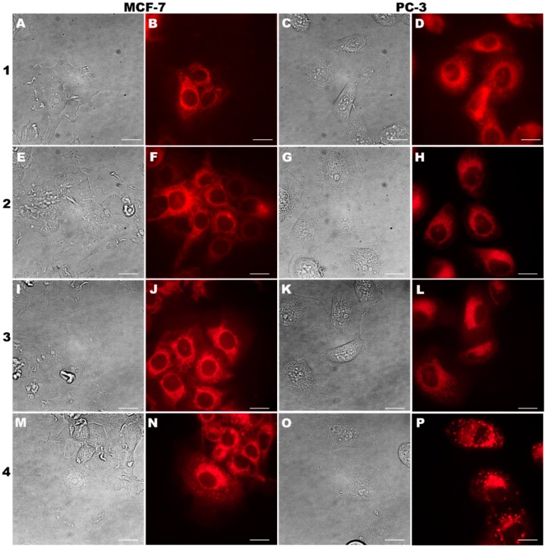
Figure 2.
Fluorescence microscopy images of intracellular localization of purpurin 18 (compound 1) and its derivatives (compounds 2–4) at 0.5 µM concentration in human cancer cell lines of MCF-7 (breast carcinoma) and PC-3 (prostate carcinoma) after 24 h incubation. In the first and third columns, there are bright field images; the second and fourth columns show compound localization. The scale bars represent 20 µm.

