Abstract
Background:
We describe successful surgical treatment of superior oblique myokymia, which had recurred after superior oblique tenectomy.
Methods:
Single case report.
Results:
The distal stump of the superior oblique tendon was extirpated by stripping it from the globe. The ipsilateral superior rectus muscle also was recessed, to correct a hypertropia that had resulted from the original superior oblique tenectomy.
Conclusions:
Complete removal of the distal superior oblique muscle tendon provided definitive relief of superior oblique myokymia. Superior rectus muscle recession, combined with previous inferior oblique myectomy, compensated effectively for loss of superior oblique function.
Surgical treatment is a last resort for medically intractable superior oblique myokymia (1). The usual approach is a superior oblique tenotomy or tenectomy, combined with anticipatory inferior oblique recession or myectomy (2). Superior oblique myectomy with trochlear resection from an anterior orbitotomy approach also has been described(3), as well as nasal displacement (inverse Harada–Ito) of the anterior portion of the tendon (4).
Occasionally, superior oblique myokymia can return after surgery. We treated a patient with recurrent superior oblique myokymia by excising the stump of the superior oblique tendon. The ipsilateral superior rectus muscle also was recessed to correct a residual hypertropia.
CASE REPORT
A 52-year-old woman was referred to the UCSF Neuro-Ophthalmology Clinic with a 4-year history of right superior oblique myokymia, occurring so frequently that it interfered with reading, driving, and work. No inciting factors could be identified. Systemic treatment with propranolol, carbamazepine, and gabapentin was unsuccessful. Visual acuity was 20/20 bilaterally. Eye movements were full and alignment was orthotropic in all gaze fields. The right eye exhibited 2–3-Hz fast intorting movements alternating with slow relaxing extorting phases, averaging 2° in amplitude, with occasional jerks up to 5° (See Supplemental Digital Content, Video 1, http://links.lww.com/WNO/A356). Contrast-enhanced brain MRI showed no blood vessel impinging on the right fourth nerve.
The patient underwent a right superior oblique tenectomy, approaching nasal to the superior rectus muscle. A 5-mm segment of the superior oblique tendon was isolated between 2 small muscle clamps. Hot cautery was used to excise the trapped piece. It appeared as a glistening white cord of connective tissue. After release of the muscle clamps, the ends of the superior oblique tendon retracted into the orbit. Next, an ipsilateral inferior oblique myectomy was performed. Two days after surgery, there were 10 prism-diopter of right hypertropia in primary gaze. The right hypertropia measured 4 prism-diopters in upgaze and 16 prism-diopters in downgaze. The presence of a large right hypertropia, despite inferior oblique muscle weakening, confirmed that the superior oblique muscle had undergone successful tenectomy.
Three weeks later, the patient reported recurrence of superior oblique myokymia. Slit-lamp examination revealed tiny but unmistakable oscillations, approximately 0.2° in amplitude. After a 3-month observation period, the vertical misalignment improved, with fusion in upgaze, 6 prism-diopters of right hypertropia in primary gaze, and 10 prism-diopters of right hypertropia in downgaze. Ductions were full, except for slight reduction of movement in the field of action of the right superior oblique muscle. Superior oblique myokymia was still present, unchanged in frequency although reduced in amplitude.
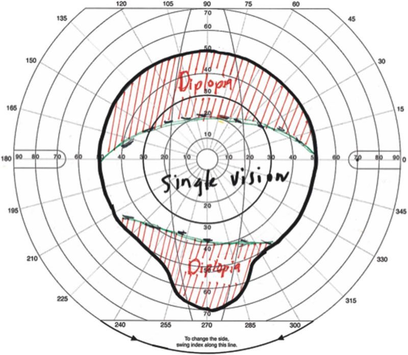
Because the patient found the ocular tremor intolerable, a second operation was performed. The superior rectus muscle was disinserted from the globe. This exposed the underlying distal tendon stump of the superior oblique muscle, fanning out to insert in the globe. It appeared intact, despite the previous tenectomy. The superior oblique tendon stump was excised completely by stripping it off the sclera and dissecting it back to the margin of the tenectomy. Next, the superior rectus muscle was recessed 3 mm, using an adjustable suture technique, to correct the right hypertropia. Postoperatively, alignment was orthotropic in primary gaze and superior oblique myokymia was abolished. Three months later, the patient had a vertical fusional range of over 50° (Fig. 1). Her oscillopsia had resolved and there was weakness of elevation, depression, and movement in the field of the right superior oblique muscle (See Supplemental Digital Content, Video 2, http://links.lww.com/WNO/A357). Nearly 2 years later, the patient remained free of superior oblique myokymia.
FIG. 1.
Field of single binocular vision plotted at the Goldmann perimeter after superior oblique tenotomy, inferior oblique myectomy, and superior rectus recession. Single vision was present from 20° upgaze to 35° downgaze.
DISCUSSION
Gittinger was the first to report that superior oblique tenotomy may provide only temporary relief of superior oblique myokymia (5). Others have documented that superior oblique myokymia may persist after surgery (2,6). Contractions of the superior oblique muscle presumably can be transmitted to the globe by either tendon remnants, fascial, or sheath connections, which heal after surgery. Agarwal and Kushner (2) advocate a tenectomy, rather than a tenotomy, to avoid recurrence of oscillopsia after surgery. In our patient, a superior oblique tenectomy was performed. Nonetheless, superior oblique myokymia recurred, although the amplitude was reduced by 90%. Parks (7) observed that “Tenotomy or tenectomy of the cord portion of the tendon does not completely eliminate the function of the superior oblique muscle.” Ruttum and Harris (3) confirmed Park’s observation, by reporting several patients with recurrent superior oblique myokymia after excision of a 5–7-mm segment of the tendon. Even after tenectomy, therefore, it seems that superior oblique contractions can sometimes be transmitted to the globe. Ruttum and Harris recommend removal of the trochlea and an 8–10-mm segment of the superior oblique muscle through an anterior orbitotomy to prevent recurrence of myokymia. Our experience indicates that complete extirpation of the distal superior oblique tendon stump from the dorsal globe also may eliminate residual oscillations. Why this procedure was effective remains unclear. It may have lysed residual connections to the globe that occurred from postsurgical adhesions or partial healing of the superior oblique tendon sheath.
Diplopia is the most common complication of eye muscle surgery for superior oblique myokymia, even with anticipatory inferior oblique weakening. In the largest surgical series, no patient developed a hypertropia in primary gaze after superior oblique tenectomy and ipsilateral inferior oblique myectomy (2). However, a hypertropia limited to downgaze occurred in 5 of 12 patients. It is surprising that diplopia in primary gaze is so rare, when one considers that a full-blown superior oblique palsy often requires surgery on 2 muscles for adequate correction (8,9). It is possible that simultaneous inferior oblique weakening is more effective than delayed inferior oblique weakening at treating vertical misalignment caused by impairment of superior oblique function.
When 2-muscle surgery is needed to treat a superior oblique muscle palsy, the usual approach is to recess the contralateral inferior rectus muscle. This is logical, because the hypertropia increases on downgaze (8,9). Weakening the inferior rectus muscle also has the benefit of reducing excyclotorsion. However, it requires operating on the uninvolved eye. Furthermore, inferior rectus recession entails some risk of lower lid retraction and late muscle slippage.
We elected to recess the ipsilateral superior rectus muscle to address the hypertropia induced by superior oblique tenotomy. The superior rectus had been disinserted to expose the remnants of the superior oblique tendon. Once it was off the globe, the simplest approach was to recess it. Although recession of the superior rectus muscle has not been reported previously in the context of superior oblique myokymia, it has been shown to be an effective treatment for the hypertropia that often remains after an initial inferior oblique muscle weakening in patients with severe superior oblique palsy (10).
Postoperative diplopia is a risk that some patients are willing to accept for relief of superior oblique myokymia. Having accepted this potential tradeoff, it may not occur to the surgeon or to the patient that superior oblique tenectomy can fail. When superior oblique myokymia does persist or recur, the solution is unclear. In our patient, removal of the distal superior oblique tendon at its insertion into the globe was effective at preventing transmission of superior oblique contractions to the eye. In this setting, we propose recession of the ipsilateral superior rectus muscle with an adjustable suture, rather than recession of the contralateral inferior rectus muscle, to treat any residual vertical misalignment.
Supplementary Material
Acknowledgments
Supported by grants EY10217 (J.C.H.), EY02162 (Beckman Vision Center) from the National Eye Institute, and by an unrestricted grant and a Physician Scientist Award from Research to Prevent Blindness. Jessica Wong assisted with video editing.
Footnotes
The authors report no conflicts of interest.
Supplemental digital content is available for this article. Direct URL citations appear in the printed text and are provided in the HTML and PDF versions of this article on the journal’s Web site (www.jneuro-ophthalmology.com).
REFERENCES
- 1.Zhang M, Gilbert A, Hunter D. Superior oblique myokymia. Surv Ophthalmol. 2018;63:507–517. [DOI] [PubMed] [Google Scholar]
- 2.Agarwal S, Kushner BJ. Results of extraocular muscle surgery for superior oblique myokymia. J AAPOS. 2009;13:472–476. [DOI] [PubMed] [Google Scholar]
- 3.Ruttum MS, Harris GJ. Superior oblique myectomy and trochlear resection for superior oblique myokymia. Am J Ophthalmol. 2009;148:563–565. [DOI] [PubMed] [Google Scholar]
- 4.Kosmorsky GS, Ellis BD, Fogt N, Leigh RJ. The treatment of superior oblique myokymia utilizing the Harada-Ito procedure. J Neuroophthalmol. 1995;15:142–146. [PubMed] [Google Scholar]
- 5.Keltner JL. The monocular shimmers—your patient isn’t deluded! Surv Ophthalmol. 1983;27:313–316. [DOI] [PubMed] [Google Scholar]
- 6.Palmer EA, Shults WT. Superior oblique myokymia: preliminary results of surgical treatment. J Pediatr Ophthalmol Strabismus. 1984;21:96–101. [DOI] [PubMed] [Google Scholar]
- 7.Parks MM. Ocular Motility and Strabismus. Hagerstown, MD: Harper and Row, 1975. [Google Scholar]
- 8.Nash DL, Hatt SR, Leske DA, May L, Bothun ED, Mohney BG, Brodsky MC, Holmes JM. One- versus two-muscle surgery for presumed unilateral fourth nerve palsy associated with moderate angle hyperdeviations. Am J Ophthalmol. 2017;182:1–7. [DOI] [PMC free article] [PubMed] [Google Scholar]
- 9.Nejad M, Thacker N, Velez FG, Rosenbaum AL, Pineles SL. Surgical results of patients with unilateral superior oblique palsy presenting with large hypertropias. J Pediatr Ophthalmol Strabismus. 2013;50:44–52. [DOI] [PMC free article] [PubMed] [Google Scholar]
- 10.Ahn SJ, Choi J, Kim SJ, Yu YS. Superior oblique muscle recession for residual head tilt after inferior oblique muscle weakening in superior oblique palsy. Korean J Ophthalmol. 2012;26:285–289. [DOI] [PMC free article] [PubMed] [Google Scholar]
Associated Data
This section collects any data citations, data availability statements, or supplementary materials included in this article.


