ABSTRACT
Neuroendocrine cell tumors of the esophagus are rare forms of cancer. Incidence of squamous cell cancer of the esophagus is low in the United States. Combined tumors with components of both neuroendocrine and squamous cell cancer which are very rarely seen have not been reported in the United States. We present a unique case of a composite tumor of the esophagus with squamous cell carcinoma and neuroendocrine carcinoma.
INTRODUCTION
Neuroendocrine cell tumors of the esophagus occur rarely with a reported incidence of less than 2%.1 Squamous cell cancer of the esophagus is uncommon in the United States.2 Combined tumors with components of both neuroendocrine and squamous cell cancer are exceedingly rare and have not been reported in the United States. We present a unique case of a composite tumor of the esophagus with squamous cell carcinoma and neuroendocrine carcinoma.
CASE REPORT
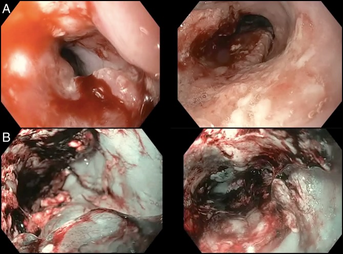
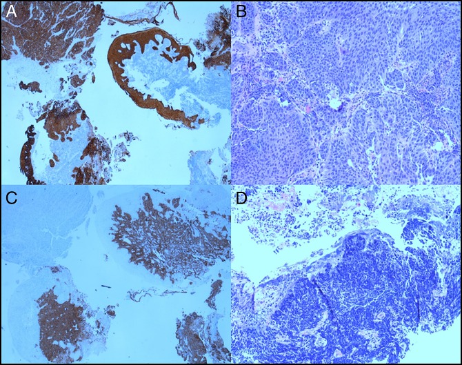
A 67-year-old man with achalasia treated with surgical myotomy 10 years ago presented with complaints of progressive dysphagia to solids and odynophagia. The initial upper endoscopy was notable for a partially obstructing 11 cm mass extending from 28 to 39 cm from the incisors (Figure 1). The esophageal-gastric junction was noted 1 cm distal to the inferior margin of the mass and was uninvolved (Video 1). Lugol iodine was sprayed to provide negative contrast, and the lack of uptake was seen. Biopsies from the mass demonstrated the presence of contiguous areas with squamous cell carcinoma and large cell neuroendocrine carcinoma. Hematoxylin and eosin-stained sections demonstrated atypical cells with squamous differentiation and increased mitosis along with a contiguous segment of atypical cells with hyperchromatic nuclei and increased nuclear to cytoplasmic ratios. Immunostaining for p63 and cytokeratin 5/6 markers confirmed the diagnosis of squamous cell carcinoma. Immunostaining with synaptophysin and CD56 confirmed neuroendocrine differentiation (Figure 2). Diagnostic endoscopic ultrasound was performed and staged the mass as T3 N1 MX. The patient underwent dilatation and palliative stenting and then decided to pursue comfort care measures.
Figure 1.
Endoscopic appearance of the esophageal tumor. (A) Under high-definition white light endoscopy and (B) narrow-band imaging.
Figure 2.
Squamous cell carcinoma of the esophagus demonstrating (A) atypical cells with positive staining for cytokeratin 5/6 immunostain; (B) squamous differentiation with increased mitosis (hematoxylin and eosin stain, 10×); (C) positive staining for sign apt to 57 within the same tumor confirms neuroendocrine differentiation; and (D) the neuroendocrine tumor demonstrates atypical cells with hyperchromatic nuclei and increased nuclear/cytoplasmic (hematoxylin and eosin stain, 10×).
Video 1.
Esophageal-gastric junction noted 1 cm distal to the inferior margin of the mass and was uninvolved. Watch the video at http://links.lww.com/ACGCR/A16.
DISCUSSION
Esophageal tumors with 2 different histological subtypes are very rare.3,4 Tumors with 2 different histological morphologies are classified as either composite or collision tumors. A collision tumor is composed of 2 independent neoplasms which coexist and have different behavioral, genetic, and histological features that are clearly demarcated.5 The different neoplasms do not have tissue admixture. Composite tumors also have 2 morphologically and immunohistochemically distinct neoplasms that coexist in the same organ. However, the neoplasms do demonstrate cellular intermingling and a common mutation that results in 2 different histologies. In our patient, cellular intermingling was present, and we consider this to be a composite tumor. To our knowledge, this is the first case of combined squamous cell cancer and neuroendocrine cancer of the esophagus to be diagnosed in the United States. Worldwide, only 3 other cases have been published in the literature.
Esophageal squamous cell carcinoma is the most common histologic subtype of esophageal cancer worldwide and is frequently diagnosed in underdeveloped countries. However, it is not frequently diagnosed in the United States and other industrialized nations. Risk factors for esophageal squamous cell cancer include smoking, alcohol consumption, caustic ingestion, achalasia, Chagas disease, older age, male sex, black race, and tylosis, among others. Achalasia, a risk factor present in our patient, is a relatively rare condition with an annual incidence rate of 0.5–1.2 per 100,000 individuals.6 A recent meta-analysis by Tustumi et al who looked at the incidence of esophageal cancer in patients with achalasia showed an absolute risk increase of 308.1.7 This study showed the prevalence of squamous cell carcinoma in 26 cases of 1,000 patients with achalasia (95% confidence interval, 18–39).
Neuroendocrine tumors of the esophagus are very rare but aggressive tumors with a poor prognosis when left untreated.8 The prevalence of esophageal neuroendocrine tumors is approximately 0.04%–4.6%.9 Dysphagia and weight loss are the common clinical features. Retrosternal and epigastric pains, odynophagia, dysphonia, dyspnea, and digestive bleeding (hematemesis and melena) are less frequent symptoms.10 Sometimes the initial symptoms are related to metastatic sites because these tumors are very aggressive. Paraneoplastic symptoms can occur rarely as well.11 Lymph node and distant metastasis to other organs is common at presentation.12
We report the first case of a patient with a composite tumor comprising squamous cell cancer and neuroendocrine cancer in the esophagus. Our patient had a history of achalasia which increased the risk of squamous cell cancer. However, the presence of both squamous cell and neuroendocrine components is unique.
DISCLOSURES
Author contributions: CS Dasari wrote the manuscript, U. Ozlem provided the pathology images, and DR Kohli revised the manuscript and is the article guarantor.
Financial disclosure: None to report.
Informed consent was obtained for this case report.
REFERENCES
- 1.Modlin IM, Sandor A. An analysis of 8305 cases of carcinoid tumors. Cancer. 1997;79(4):813–29. [DOI] [PubMed] [Google Scholar]
- 2.Kim A, Ashman P, Ward-Peterson M, Lozano JM, Barengo NC. Racial disparities in cancer-related survival in patients with squamous cell carcinoma of the esophagus in the US between 1973 and 2013. PLoS One. 2017;12(8):e0183782. [DOI] [PMC free article] [PubMed] [Google Scholar]
- 3.Fujihara S, Kobayashi M, Nishi M, et al. Composite neuroendocrine carcinoma and squamous cell carcinoma with regional lymph node metastasis: A case report. J Med Case Rep. 2018;12(1):227. [DOI] [PMC free article] [PubMed] [Google Scholar]
- 4.Sung CT, Shetty A, Menias CO, et al. Collision and composite tumors; radiologic and pathologic correlation. Abdom Radiol. 2017;42(12):2909–26. [DOI] [PubMed] [Google Scholar]
- 5.Yazici O, Aksoy S, Ozhamam EU, Zengin N. Squamous cell and neuroendocrine carcinoma of esophagus: Collision versus composite tumor: A case report and review of literature. Indian J Cancer. 2015;52(4):603–4. [DOI] [PubMed] [Google Scholar]
- 6.Torres-Aguilera M, Remes Troche JM. Achalasia and esophageal cancer: Risks and links. Clin Exp Gastroenterol. 2018;11:309–16. [DOI] [PMC free article] [PubMed] [Google Scholar]
- 7.Tustumi F, Bernardo WM, da Rocha JRM, et al. Esophageal achalasia: A risk factor for carcinoma. A systematic review and meta-analysis. Dis Esophagus. 2017;30(10):1–8. [DOI] [PubMed] [Google Scholar]
- 8.Estrozi B, Bacchi CE. Neuroendocrine tumors involving the gastroenteropancreatic tract: A clinicopathological evaluation of 773 cases. Clinics. 2011;66(10):1671–5. [DOI] [PMC free article] [PubMed] [Google Scholar]
- 9.Kuntegowdanahalli LC, Kanakasetty GB, Thanky AH, et al. Prognostic and predictive implications of Sokal, Euro and EUTOS scores in chronic myeloid leukaemia in the imatinib era-experience from a tertiary oncology centre in Southern India. Ecancermedicalscience. 2016;10:679. [DOI] [PMC free article] [PubMed] [Google Scholar]
- 10.Kim SO, Yun SJ, Lee EH. The water extract of adlay seed (Coix lachrymajobi var. mayuen) exhibits anti-obesity effects through neuroendocrine modulation. Am J Chin Med. 2007;35(2):297–308. [DOI] [PubMed] [Google Scholar]
- 11.Saif MW, Vethody C. Poorly differentiated neuroendocrine tumor of the esophagus with hypertrophic osteoarthropathy and brain metastasis: A success story. Cureus. 2016;8(6):e646. [DOI] [PMC free article] [PubMed] [Google Scholar]
- 12.Egashira A, Morita M, Kumagai R, et al. Neuroendocrine carcinoma of the esophagus: Clinicopathological and immunohistochemical features of 14 cases. PLoS One. 2017;12(3):e0173501. [DOI] [PMC free article] [PubMed] [Google Scholar]


