

Abstract
Context:
Micronuclei, tannery workers, chromium, papanicolaou (PAP) stain.
Aims:
The presence of micronuclei are biomarkers broadly used; the detection of micronuclei offers a great opportunity to monitor individuals or populations exposed to mutagenic, genotoxic or teratogenic events, mainly the evaluation of micronucleogenic cells presence in epithelial tissues.
Settings and Design:
Tannery workers with and without tobacco usage was considered for microneuceli assessment using PAP stain, 50 patients without usage of tobacco was taken and 50 patients with usage of tobacco was considered.
Subjects and Methods:
Cytological smears were produced and stained with PAP stain for the evaluation of micronuclei.
Statistical Analysis Used:
Students t-test, analysis of variance.
Results:
Our findings concluded that chromium exposure causes instability of the genetic material in the tannery workers and can be taken as an indication that these individuals have increased cancer risks. The present study also concluded that PAP could be used as a specific marker for micronuclei assessment.
Conclusions:
In our case study, we have seen increasing number of micronuclei frequencies in tobacco user's tannery workers than nonuser's tannery workers, which we can use as a predictor marker of premalignant and malignant lesion.
Keywords: Chromium, micronuclei, papanicolaou stain, tannery workers
INTRODUCTION
The tanning industry is known to be very polluting, especially through effluents high in organic and inorganic dissolved and suspended solids content accompanied by propensities for high oxygen demand and containing potentially toxic metal salt residues. A significant part of the chemical used in the leather processing is not actually absorbed in the process but is discharged into the environment.[1]
Leather tanning is the process of converting rawhides of skins into leather. Tanning is essentially the reaction of collagen fibers in the hide with tannins, chromium, alum or other chemical agents. Approximately 90% of all leather is produced by chrome tanning. A person spends, on average, one-third of his life at his workplace; and therefore, the environment in which he works can be a major factor in determining health.[2]
The use of tobacco in developing countries like India has been on a constant rise, tobacco is used in smoke, and smokeless forms and both these forms have a direct impact on the health of the individual, involving the lungs, larynx, pharynx and the oral cavity. The lesions developing in the oral cavity include potentially malignant disorders, squamous cell carcinomas, dental caries, periodontal diseases, tooth loss, pigmentations and a wide range of several other lesions.[3]
Oral cavity reflects the various events occurring in the body, and this is reflected by cytomorphological and nucleomorphological variations in the exfoliated cells. Cytological specimens are recently analyzed for nuclear DNA content, immunohistochemical tumor cell marker identification and molecular analysis.[4] Oral mucosa could be used for early detection of a premalignant or cancerous oral lesion promises to improve the survival and the morbidity of patients.[5]
Micronucleus (MN) is a recently upgraded topic, especially in the field of oral cancer. MN takes its origin from chromosome fragments or whole chromosomes, which lag behind at anaphase during nuclear division. Various studies have shown the correlation of frequency of micronuclei and severity of this genotoxic damage.[6]
The present study with an aim to compare the frequency of micronuclei among tannery industrial workers with and without tobacco habits and to evaluate the role of micronuclei assay in cytological smears of tannery workers for the assessment of genotoxic damage caused by the chromium and to assert if the changes of these epithelial cells at the molecular level is accelerated by tobacco products or not. To even study and compare the different factors such as size, texture, focal plane, shape, color and separation by papanicolaou (PAP) stain which is easy to use, economical, less time-consuming stain to stain the MN. All these tools can be used for enhancing better life susceptibility of tannery workers.
SUBJECTS AND METHODS
The present study was conducted in the Department of Oral Pathology and Microbiology, Career Post Graduate Institute of Dental Sciences and Hospital, Lucknow, Uttar Pradesh, India. The study subjects were the volunteers who work in tannery industries, especially in Kanpur. Group I: 25 healthy individuals without the habit of tobacco (control group), Group II: 50 tannery industrial workers without the habit of tobacco and Group III: 50 tannery industrial workers with the habit of tobacco.
Inclusion criteria that were considered for the study are individual between the age group of 25–50 years unbiased of gender, having work experience of 5 years, individual with the habit of tobacco chewing without any premalignant disorders. In exclusion criteria individual with a history of systemic disorder, radiation exposure and mental illness along with cases of premalignant and malignant disorders were excluded.
Methodology
Buccal cells (BCs) were collected from all the consented volunteers of the study at the end of the work shift according to the criteria established. Prior to BC collection, the volunteer has rinsed the mouth; cytobrush was used for collection of BCs, BCs were transferred to phosphate-buffered saline (PBS). These BCs were transferred into blood sample collection tubes with PBS at pH 7.0 and centrifuged for 10 min at 2000 rpm, the same cycle was repeated once more. After discarding the supernatant pellet was smeared on a glass slide and fixed in cold methanol: acetic acid (3:1) for 15 min. Slides were air-dried for 10 min. Slide from each sample was prepared.
All the prepared microslides from each study group were subjected for staining with PAP stain for the micronuclei evaluation. After preparation of the slides, they were coded and given to three different observers in a random order, and they evaluated all the slides based on the analysis criteria. After assessing the various factors, the micronuclei were counted under ×100. Thus, obtained data were tabulated and analyzed statistically to compare within the study groups.
The following criteria for MN assay analysis were used in oral epithelial cells, according to Sellappa et al.[2] Micronucleus might be of one-third of the main nucleus in size; must be on the same focal plane; must have the same color and texture (smooth or rough) as the main nucleus; must have an oval or round in shape; and must be clearly separated from the main nucleus.
RESULTS
The present study showed that PAP is a good stain for the MN assay screening of the exfoliated buccal mucosa cells. Further, severity of the buccal mucosal changes which were associated with tobacco use was found to be more than that in nonsmokers, thus indicating the synergistic effect of tobacco along with chromium damage [Figures 1–3]. The mean MN frequency in oral exfoliated cells was significantly increased in Group III (tannery workers with habits of tobacco) as compared to the other groups. In our case study, we have seen increasing number of micronuclei frequencies in tobacco user's tannery workers than nonuser's tannery workers.
Figure 1.
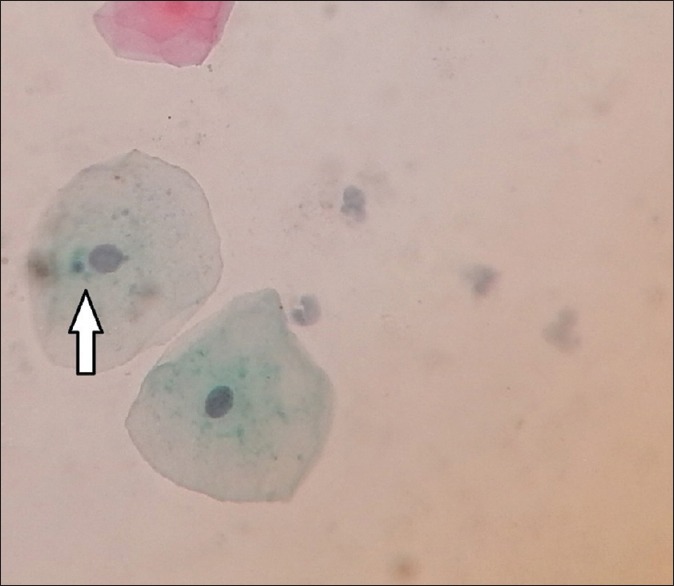
Pap smear showing microneuceli in healthy individual
Figure 3.
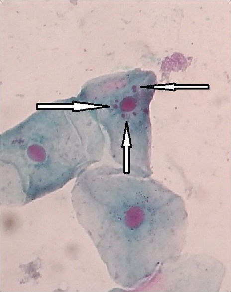
Pap smear showing microneuceli in tannery worker with tobacco habit
Figure 2.
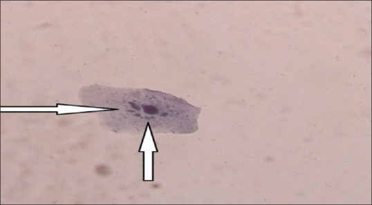
Pap smear showing micronuclei in tannery worker without tobacco habbit
To summarize the results, micronuclei can be seen in normal mucosal cells. The frequency of micronuclei in oral mucosal cells of tannery workers was four-fold to five-fold higher as compared with the control group.
Group I
Each smear was observed by three different observers to record the specificity of the stains in staining micronuclei.
Table 1 shows intergroup comparison of smear examination by PAP stain in all samples was found to be P < 0.001 very highly significant (VHS). All the criteria seem to be placed at the same level.
Table 1.
Intergroup comparison of smear examination by PAP stain (all samples)
| Group I (n=75), n (%) | Group II (n=150), n (%) | Group III (n=150), n (%) | |
|---|---|---|---|
| Size | |||
| Less than one-third of nucleus* | 51 (68.00) | 0 (0.00) | 0 (0.00) |
| One-third of nucleus | 24 (32.00) | 150 (100.00) | 150 (100.00) |
| χ2; P | 236.111; <0.001 | ||
| Texture | |||
| None | 51 (68.00) | 0 (0.00) | 0 (0.00) |
| Rough | 7 (9.33) | 18 (12.00) | 25 (16.67) |
| Smooth | 17 (22.67) | 132 (88.00) | 125 (83.33) |
| χ2; P | 239.166; <0.001 | ||
| Focal plane* | |||
| NA* | 57 (76.00) | 22 (14.67) | 18 (12.00) |
| Same | 18 (24.00) | 128 (85.33) | 132 (88.00) |
| χ2; P | 123.155; <0.001 | ||
| Shape | |||
| NA* | 51 (68.00) | 0 (0.00) | 0 (0.00) |
| Oval | 8 (10.67) | 39 (26.00) | 58 (38.67) |
| Round | 16 (21.33) | 111 (74.00) | 92 (61.33) |
| χ2; P | 242.474; <0.001 | ||
| Color | |||
| NA* | 58 (77.33) | 21 (14.00) | 20 (13.33) |
| Same | 17 (22.67) | 129 (86.00) | 130 (86.67) |
| χ2; P | 125.185; <0.001 | ||
| Separation | |||
| NA* | 51 (68.00) | 0 (0.00) | 0 (0.00) |
| Separated | 24 (32.00) | 150 (100.00) | 150 (100.00) |
| χ2; P | 236.111; <0.001 |
NA: Not available, PAP: Papanicolaou
Table 2 shows intergroup comparison of number of micronuclei in PAP stain was found to be P < 0.001 (VHS). Intergroup comparison of number micronuclei in PAP stain was high in Group III.
Table 2.
Intergroup comparison of number of micronuclei (by PAP stain)
| Group | Number of specimens | Minimum | Maximum | Mean | SD |
|---|---|---|---|---|---|
| Group I | 24 | 1 | 2 | 1.08 | 0.28 |
| Group II | 150 | 1 | 6 | 3.26 | 1.33 |
| Group III | 150 | 1 | 16 | 4.19 | 2.94 |
| Total | 324 | 1 | 16 | 3.53 | 2.34 |
F=22.627, P<0.001. SD: Standard deviation, PAP: Papanicolaou
Table 3 shows the difference in number of micronuclei (PAP stain) in between all groups were found to be P < 0.001 (VHS) but the difference was higher in-between Group I and Group III.
Table 3.
Between-group difference in number of micronuclei (by PAP stain) (Tukey highly significant difference test)
| Mean difference | SE | Significant | |
|---|---|---|---|
| Group I versus Group II | −2.18 | 0.484 | <0.001 |
| Group I versus Group III | −3.10 | 0.484 | <0.001 |
| Group II versus Group III | −0.93 | 0.254 | 0.001 |
SE: Standard error, PAP: Papanicolaou
Statistical analysis
Mean and standard deviation (SD) was calculated for biomarker. The significance of the differences between control and worker end-point means was analyzed using Student's t-test, whereas simple and multiple linear regression analyses were performed to assess the association between end-points and the independent variables. All calculations were performed using Windows statistical package, version 11.5 (SPSS, IL, USA). Mean values and SDs were computed for the scores, and the statistical significance (P < 0.05) of effects (exposure and tobacco) was determined using analysis of variance.
DISCUSSION
A tannery worker spends, on average, one-third of his life at his workplace; and therefore, the environment in which he works can be a major factor in determining health. Chromium has been recognized as one of the most effective tanning agents and has been widely employed in the leather industry since its discovery more than 100 years ago. Since then, some 85% of the leather produced worldwide is tanned with chromium salts, either alone or in combination with other tanning agents.[2]
The usefulness of an MN test to detect and quantitate the genotoxic action of carcinogens and mutagens has been well established in vitro and in vivo studies. The sensitivity of the MN test is comparable to that of scoring chromatid breaks and exchanges. A reasonable relationship between the carcinogenicity of chemicals and their capacity to induce micronuclei, as well as the ease of scoring, stimulated the application of the MN test to exfoliate human cells.[7]
The MN assay can be used for exfoliated cells, which offers the advantage of conducting a genotoxicity test on material from an intact organism with its multitude of defense systems.[7]
The present study comprised of a control group (Group I) 25 sample and two study groups (Group II), tannery industrial workers without the habit of tobacco and Group III, Tannery industrial workers with the habit of tobacco) 50 samples each.
In our study [Table 1], intergroup comparison of smear examination by PAP stain in all samples was found to be P < 0.001 (VHS). All the criteria seem to be placed at the same level. All observers observe that more than one-third of the size of nucleus was seen in 24 normal individuals. While 51 normal individuals size showed less than one-third of the main nucleus. In volunteers of Group II and Group III showed MN size more than one-third of the main nucleus in all volunteers. This size change could be attributed to change in the content of the nucleus, as a result of genetic disturbance caused by the genotoxic agent such as chromium. On analysis of the texture of the MN, the rough texture was seen in 9.3% of the cases in normal individuals, 12% of the cases in Group II volunteers and 25% of the Group III volunteers. These changes in the texture of the nucleus were assessed by Colomb et al. They asserted that changes in the texture of the nucleus are caused by changes in the chromatin content in the nucleus. While these chromatin changes are attributed to factors such as drugs and toxins which affect the cell cycle of a cell.[8]
Changes in focal plane and shape of the MN were observed more in volunteers of Group II and Group III as compared to normal individuals. The change in the shape of MN is caused by the change in the content of the nucleus. This change in the shape of MN causes distortion of the image formed by the light microscope. This three dimensional changes in the shape of the MN causes the change in the focal plane of the MN. The same explanation was given in text book's molecular biology of the cell, 4th edition.[9]
The P < 0.001 value for color was highly significant, indicating that the PAP stain color of micronuclei is depending on the content of nucleus. As we see micronuclei in Group II and Group III volunteers, the micronuclei that we considered more than one-third of the main nucleus. Hence, the color variation was observed. The highly significant P < 0.001 value seen in separation in PAP stain because we have taken the major criteria for assessment of chromium genotoxic activity by assessment of micronuclei formation.
In the present study [Table 2], intergroup comparison of number of micronuclei in PAP stain were found to be P < 0.001 (VHS). We have done intergroup comparison of micronuclei using PAP stain. The maximum number of micronuclei was seen in tannery workers with habits of tobacco (Group III). While least number of micronuclei was seen in normal individuals (Group I). In earlier study which was done by Kashyap and Reddy, they have seen that genomic damage is produced by environmental exposure to genotoxin, medical procedure, micronutrient deficiency, lifestyle factors, and genetic factors. Cell death as a result of genome damage can be accessed using microneuclei in contex of cytotoxic and cytostatic effect. Thus, it is well-proven fact that any insult to nuclear content by chromium might cause increased number of micronuclei in tannery workers with the habits tobacco. As shown in Table 3, we have observed a statistically significant P < 0.001 value in comparison between Group I and Group II and Group I and Group III. The reason of this could be the Group III volunteers have experienced double genetic damage one from chromium compound of tannery and second from tobacco.[10]
In the present study, it is clearly demonstrated that MN assay in oral exfoliated cells can be used as a simple reliable marker to assess the genotoxicity and for the early diagnosis of premalignant and malignant lesions. MN assay is, thus, an easy tool for the early detection of cancer.
Financial support and sponsorship
Nil.
Conflicts of interest
There are no conflicts of interest.
REFERENCES
- 1.Gupta S, Gupta R, Tamra R. Challenges Faced By Leather Industry in Kanpur. A Project Report from IIT Kanpur. 2007 [Google Scholar]
- 2.Sellepa S, Parthyuman S, Joseph S, Keyan K. Microneucleus test in exfoliated buccal cells from chromium exposed tannery workers. Int J Biosci Biochem Bioinform. 2011;1:58–62. [Google Scholar]
- 3.Pillai HS, Jangannathan N. Tobacco a potential threat to oral cavity. Int J Pharma Sci. 2014;6:38–40. [Google Scholar]
- 4.Kumarseran GD, Janghanathan N. Exfoliative Cytology- a predictive diagnostic tool. Int J Pharm Pharm Sci. 2014;6:1–3. [Google Scholar]
- 5.Kaur M, Saxena S, Samantha YP, Chawla G, Yadav G. Usefulness of oral exfoliative cytology in dental practice. J Oral Health Community Dent. 2013;7:161–5. [Google Scholar]
- 6.Jadhav K, Gupta N, Ahmed MB. Micronuclei: An essential biomarker in oral exfoliated cells for grading of oral squamous cell carcinoma. J Cytol. 2011;28:7–12. doi: 10.4103/0970-9371.76941. [DOI] [PMC free article] [PubMed] [Google Scholar]
- 7.Palaskar S, Jindal C. Evaluation of micronuclei using Papanicolaou and may Grunwald Giemsa stain in individuals with different tobacco habits – A comparative study. J Clin Diagn Res. 2010;4:3607–13. [Google Scholar]
- 8.Colomb E, Dussert C, Martin PM. Nuclear texture parameters as discriminant factors in cell cycle and drug sensitivity studies. Cytometry. 1991;12:15–25. doi: 10.1002/cyto.990120104. [DOI] [PubMed] [Google Scholar]
- 9.4th ed. Washington: Garland publishing; 2005. Albert's, Jhonson, Visualizing cells, molecular biology of cell; pp. 1410–6. [Google Scholar]
- 10.Kashyap B, Reddy PS. Micronuclei assay of exfoliated oral buccal cells: Means to assess the nuclear abnormalities in different diseases. J Cancer Res Ther. 2012;8:184–91. doi: 10.4103/0973-1482.98968. [DOI] [PubMed] [Google Scholar]