

Abstract
Many options have been developed to treat irreparable massive cuff tears. Superior capsular reconstruction has been reported as one of the treatment options for relatively young patients with irreparable massive cuff tear. However, this original technique has a disadvantage of requiring a tensor facia lata autograft. It requires another incision at the lateral thigh and can be a cause of thigh pain. This article describes our modified technique for arthroscopic superior capsule reconstruction using the biceps autograft to preserve the long head of the biceps tendon anchors to the glenoid labrum (the snake technique).
Introduction
The treatment of massive irreparable rotator cuff tears is a challenge to shoulder surgeons.1, 2, 3 There are many treatment options, such as conservative treatment,4 arthroscopic debridement and biceps tenotomy,5, 6 rotator cuff partial repair,6, 7, 8 patch augmentation,9, 10, 11, 12 reverse total shoulder arthroplasty,13, 14, 15 and superior capsular reconstruction.16, 17, 18, 19, 20 Superior capsular reconstruction is a good treatment option for relatively young patients with irreparable massive cuff tear because the superior capsule is the superior restraint to proximal migration of the humeral head.21 However, the superior capsular reconstruction technique requires the additional surgical procedure of tensor fascia lata autograft harvesting from the lateral thigh or using a dermal allograft.17, 19 The technique described here is an arthroscopic reconstruction of the superior capsule with the biceps tendon autograft preserving the long head of the biceps tendon (LHBT) anchors to the labrum. We called it the snake technique because the reconstructed superior capsule resembles a snake (Video 1).
Surgical Technique
Preoperative Workup
The indications for the snake technique are irreparable massive rotator cuff tears (supraspinatus tear and/or infraspinatus tear and/or subscapularis tear), normal attachment of the LHBT to the glenoid labrum or <20% partial tear of the LHBT, good deltoid muscle, and minimal/no glenohumeral arthritis (Fig 1, Table 1). It is important to check the quality of the LHBT anchors to the labrum with the use of preoperative magnetic resonance imaging or magnetic resonance arthrography (Fig 2).
Fig 1.

(A) The snake technique uses both intra-articular and extra-articular portions of the long head of the biceps tendon, preserving biceps tendon anchors to the glenoid labrum. (B) Diagram of the snake technique.
Table 1.
Indications for the Snake Technique
| Irreparable supraspinatus and/or infraspinatus tears |
| Severe shoulder pain with failed conservative management |
| Good quality of the long head of the biceps tendon anchors to the glenoid labrum (normal or <20% partial tear) Minimal to no glenohumeral arthritis |
Fig 2.
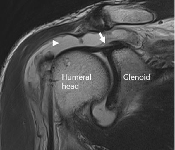
Preoperative T2-weighted oblique-coronal magnetic resonance (MR) images from a 61-year-old male. It shows massive retracted cuff tear (arrowhead) and intact attachment of the long head of the biceps tendon to the glenoid labrum (arrow).
Patient Position and Diagnostic Arthroscopy
The patient was positioned in the beach-chair table with arms externally rotated and 30° abducted and fixed with the use of a padded arm sleeve (STAR sleeve; Arthrex, Naples, FL) while under general anesthesia. Suprascapular nerve block was added to help the immediate postoperative pain control and initial rehabilitation process.
Posterior viewing portal was made approximately 2 cm medial and 2 cm inferior to the posterolateral corner of the acromion. A standard 30° arthroscope (Arthrex) was introduced, and an anterior portal was made. Diagnostic arthroscopy was performed to examine the glenohumeral joint, the articular side of the supraspinatus tendon, the subscapularis tendon, and the integrity of the LHBT. If the quality of LHBT anchor to the glenoid labrum was not good enough to perform the snake superior capsular reconstruction or the LHBT was partially teared >20%, we chose a different treatment option.
The arthroscope was moved into the subacromial space through the posterior portal to examine the subacromial space. A lateral portal was made approximately 4 cm lateral to the anterolateral corner of the acromion. Bursectomy was performed with a shaver (Arthrex) through the lateral portal and then through the posterior portal. An acromioplasty and a coracoacromial ligament release were performed with the shaver (Arthrocare; Smith & Nephew, Andover, MA) and a burr (Arthrex) if needed. The arthroscope was moved through the lateral portal, a bursectomy was performed, and a portal of Wilmington (viewing portal) was created.
Open Subpectoralis Biceps Tenodesis and Biceps Autograft Preparation
After confirmation that the quality of the LHBT was sufficient, an approximately 3-cm longitudinal skin incision was made at the inferior border of the pectoralis major tendon and anteromedial aspect of the humerus. The subcutaneous tissues were dissected with use of an electrical cautery device (Megadyne; Ethicon, Somerville, NJ). Dissection was directed to the humerus medially so as not to touch the neurovascular structure. We identified the LHBT through palpation of the bicipital groove. The snake technique requires approximately 14 cm of the LHBT. We decided the biceps autograft (BAG) length based on the tenotomy level. Running-locking sutures (Ethibond Excel; Ethicon) were placed above and under the biceps tenotomy level, and then biceps tenotomy was performed. Subpectoralis biceps tenodesis was performed with the 5.5-mm Corkscrew FT anchor (Arthrex). We performed finger sweeps through the bicipital groove from the subpectoralis tenotomy area to the subacromial space. While pulling the BAG in the distal direction, we peeled the surrounding soft tissue including the mesotendon attached to the BAG with the use of our fingers and a curved Metzenbaum scissor.
The arthroscope was placed at a viewing portal, and the wire-passer was inserted from the anterior portal along the biceps groove into the subpectoralis biceps tenodesis site. Next, the BAG was retrieved and moved from the subpectoralis lesion to the subacromial space by using the wire-passer or Ethibond suture (Fig 3). The diameter and length of the harvested BAG were measured with the use of a ruler. The fixation position of the BAG on the greater tuberosity of the humerus was determined by considering the rotator cuff defect size and the diameter and length of the BAG.
Fig 3.

Harvesting of biceps autograft (BAG) and open subpectoralis tenodesis. (A) Running-locking sutures above and under biceps tendon. (B) Subpectoralis biceps tenodesis (arrow) performed with the 5.5-mm Corkscrew FiberTape anchor (Arthrex). (C) BAG was pulled through the shoulder joint.
An additional posterior portal (posteromedial [PM] portal) was created approximately 2 cm medial to the posterior portal, and an additional anterior portal (anteromedial [AM] portal) was created approximately 2 cm medial to the anterior portal (Fig 4). The posterosuperior glenoid and greater tuberosity of the humerus bone beds were decorticated for BAG attachment by using a shaver and burr. The anterosuperior labrum where the LHBT anchors to the glenoid labrum was left intact. We used a laser-marked probe (Arthrex) to measure the anterior-to-posterior and medial-to-lateral cuff tear sizes, diameter of the LHBT, and length of the intra-articular portion of LHBT through the anterior portal and the viewing portal (Fig 5).
Fig 4.

Portals used in the snake technique.
Fig 5.

(A) Posterosuperior glenoid was decorticated with a burr. The long head of the biceps tendon (LBHT) condition must be checked before superior capsular reconstruction. (B) Laser-marked probe was used to measure anterior-to-posterior and medial-to-lateral cuff tear size, diameter of the LHBT, and length of intra-articular portion of LHBT.
Next, 8.25-mm × 7-cm cannulas (Arthrex) were inserted in the AM, PM, and lateral portals to facilitate instrumentation. A Neviaser portal was created, and a 5.5-mm SwiveLock anchor with attached swedged FiberTape (Arthrex) was placed percutaneously through a Neviaser portal on the PM lesion of the glenoid. If the size of the glenoid bone was not large enough to insert a 5.5-mm SwiveLock anchor with attached swedged FiberTape, we used an all-suture anchor (2.8-mm Q-FIX; Smith & Nephew) (Fig 6). Through anchor portals, we placed 2 SwiveLock 5.5-mm anchors with attached swedged FiberTape into the lateral margin of the footprint of the greater tuberosity of the humerus both anteriorly and posteriorly.
Fig 6.

(A) Glenoid preparation and a 5.5-mm SwiveLock anchor with attached swedged FiberTape. (B) If glenoid was too small to insert a 5.5-mm SwiveLock anchor, we used an all-suture anchor.
First Bundle (Fixation on Anterior Portion of the Greater Tuberosity of the Humerus)
The distal part of the harvested BAG was pulled to the subacromial space. A 5.5-mm SwiveLock anchor with attached swedged FiberTape was inserted by using an anchor portal. The first bundle of the BAG was affixed at the greater tuberosity of humerus by using inserted FiberTape and a knotless anchor (5.5-mm SwiveLock; Arthrex) while pulling the BAG in the lateral direction (Fig 7A). If the fixation was insufficient, we used additional sutures with FiberWire loaded together with a 5.5-mm SwiveLock anchor and attached swedged FiberTape (Fig 7B). After fixation, we checked that the tension of the first bundle was correct and that the attachment of the LBHT to the glenoid labrum was not detached from the glenoid.
Fig 7.

First bundle. (A) FiberTape and knotless fixation. (B) Additional suture.
Second Bundle (Fixation on the Glenoid)
A 5.5-mm SwiveLock anchor with attached swedged FiberTape was inserted into the posterior glenoid through the Neviaser portal. We inserted it into the medial side of the glenoid as far as possible. The distal part of the BAG was pulled through a PM portal. While we were maintaining the graft tension through a PM portal, the BAG was fixed using FiberTape and a knotless anchor (Swivelock; Arthrex) at the articular surface margin of the glenoid through the posterior portal. If the glenoid was too small or too weak to insert 5.5-mm SwiveLock anchor with attached swedged FiberTape, we used an all-suture anchor (Fig 8).
Fig 8.

Second bundle. (A) FiberTape and knotless fixation. (B) All-suture anchor fixation. (C) If it was sufficient to restore a superior capsule with 2 bundles, we made 2 bundles only.
Third Bundle (Fixation on Posterior Portion of the Greater Tuberosity of the Humerus)
If it is not sufficient to restore a superior capsule with 2 bundles, we made a third bundle. A 5-mm SwiveLock anchor with attached swedged FiberTape was inserted into the greater tuberosity of the humerus posteriorly considering the second bundle position. The remainder of the BAG was pulled through the lateral portal, and we applied the appropriate tension during fixation of the BAG using FiberTape and a knotless anchor. If the BAG was not long enough for knotless fixation, a single stitch was performed (Fig 9A). After performing superior capsular reconstruction, remnant rotator cuff partial repair was performed to the posterior reconstructed superior capsule (Fig 9B). The stability of the reconstructed superior capsule was checked using a probe (Table 2).
Fig 9.

(A) Third bundle fixation. If the long head of the biceps tendon was not sufficient for FiberTape and knotless fixation, single-row repair was performed. (B) Partial repair was performed to posterior reconstructed superior capsule.
Table 2.
Surgical Key Steps With Pearls and Pitfalls
| Key Steps | Pearls | Pitfalls |
|---|---|---|
| Diagnostic arthroscopy | Check the attachment of the long head of the biceps tendon to the glenoid labrum. | If >20% of the LBHT partial tear or <5 mm of the LBHT thickness, this technique cannot be performed with BAG. |
| Acromioplasty and coracoacromial ligament release, bursectomy | Bursectomy is performed enough to view entire cuff tear and glenoid. | |
| Humeral and glenoid bone bed preparation and anchors insertion | A spinal needle should be used to localize the portals. Bone bed should be debrided sufficiently. |
If glenoid anchors are inserted too laterally, a fracture in the glenoid can occur. |
| Biceps tenodesis and Biceps autograft preparation | For graft passage, soft tissues around the biceps tendon should be completely released for easy BAG harvesting and passing into the shoulder joint. | If incision is made too medially, the musculocutaneous nerve can be injured. When performing biceps tenodesis, be careful not to cause a humerus shaft fracture when fixing the suture anchor. |
| First bundle | The biceps should be fixed with the arm in neutral rotation at 30° of abduction. Shorten the operation time and maximize healing surface area with knotless fixation using FiberTape. |
Be careful with the anchor malposition or pull out. |
| Second bundle | Pull the BAG through a posteromedial portal and fix it with proper tension. | If the size of the glenoid bone is not large enough to insert a SwiveLock anchor, use an all-suture anchor. |
| Third bundle (if needed) | Pull the BAG through the lateral portal and fix it with tension. | If the length of the biceps autograft is not long enough for FiberTape knotless fixation, perform single-row suture. |
| Partial repair (if needed) and final inspection, portal closing | Do not apply too much tension when performing partial repair. If rotator cuff is not sufficiently mobile, try a partial repair at the greater tuberosity just posterior the third bundle. |
Do not attempt to repair one bundle with another bundle. |
BAG, biceps autograft; LBHT, superior capsular reconstruction.
Postoperative Care
Postoperatively, the patient was treated by our hospital's massive cuff repair protocol. After surgery, the patient was applied with shoulder abduction brace immediately. The patient was restricted with shoulder joint motion and because we performed open sub-pectoralis biceps tenodesis in all patients, even passive motion of the elbow was prohibited during the entire brace application period. Passive motion was started 6 to 8 weeks postoperatively after removal of the brace. Active range of motion typically started after 12 weeks, and external rotation strengthening exercises started after 16 weeks.
Discussion
The superior capsule is a static stabilizer of the glenohumeral joint.20, 22 The absence of a superior capsule causes superior migration of the humeral head and accelerates cuff tear arthropathy.20 Mihata et al.19 reported use of a superior capsular reconstruction technique with tensor fascia lata autograft to restore the normal restraint to superior translation that leads to deficient rotator cuff. This technique is especially good for relatively young irreparable massive cuff tear patients in that it has few complications and it is possible to switch to a salvage procedure such as reverse total shoulder arthroplasty when retear or reoperation is needed. However, this technique has the disadvantage of requiring tensor facia lata autograft harvesting and donor site morbidity such as lateral thigh pain.16, 17, 18, 19, 20, 21, 22
Many surgeons have tried to use the LHBT to treat patients with irreparable massive cuff tear. Sano et al.23 reported the clinical outcomes using the LBHT as a patch graft. However, this kind of interposition technique differs from superior capsular reconstruction (SCR). Recently, Kim et al.24 reported in situ SCR via LBHT rerouting. By rerouting the LBHT, the superior migration of the humeral head is restricted through the application of a downward force on the humeral head, and the LBHT itself forms part of the superior capsule, but it is difficult to reconstruct the complete superior capsule. In both studies, only the intra-articular part of the LBHT had used. However, we used not only intra-articular side but also the extra-articular side of the LHBT to obtain sufficient graft.
We demonstrated a new SCR technique in this report. We harvested an average 14-cm length (range 12 to 17 cm) of the BAG, which is long and thick (average 6.5 mm, range 5 to 10 mm) enough to make 2 or 3 bundles with our technique. LHBT can be taken easily during shoulder surgery, and it is unnecessary to prepare other parts of the body such as the hip and knee for autograft harvesting. This technique does not require additional incisions other than shoulder. Unlike dermal allografts, this is a BAG, so the risk of complications due to allografts such as postoperative infection is relatively low. One of the greatest advantages of the snake technique is that the size of reconstructed capsule can be modified by adjusting the number of bundles in the middle of the operation, thus reducing the operation time required for rotator cuff tear size measurements. In most cases, SCR was possible with 2 bundles, although occasionally 3 bundles were required.
Another advantage of the snake technique is that the BAG passage is easy. Mihata et al.19 recommended making autografts with a thickness of ≥8 mm, which is technically difficult to pass through the lateral portal. The total bundle thickness using BAG varies from 5 to 10 mm, but the graft passage is relatively easy because it is fixed by dividing each bundle. In addition, preservation of the LBHT anchors to the labrum will result in a better proprioception and more favorable healing of the reconstructed superior capsule because the blood supply is maintained. Compared with preoperative radiographs, postoperative radiographs showed restored center of rotation of humeral head (Fig 10).
Fig 10.

(A) Preoperative and postoperative shoulder Rockwood view radiographs from a 61-year-old male. (A) Preoperative radiograph shows superior humeral head migration (Hamada classification grade II). (B) Postoperative radiograph shows inferior humeral head migration (5 to 13 mm) compared with preoperative radiograph.
A limitation is that this technique requires a good-quality LBHT. This technique cannot be indicated in patients with >20% partial tear of biceps tendon itself or severe biceps tendinitis. We also excluded patients who had biceps tendon <5 mm thick. Thus, it is important to check the quality of anchors of LHBT with preoperative magnetic resonance imaging and physical examination.
We fixed each bundle with a knotless technique using FiberTape, which enabled strong fixation by pressing a wide cross section during a relatively short operation time. The snake SCR technique with BAG can be one of the useful surgical treatment options for irreparable massive rotator cuff tears.
Footnotes
The authors report that they have no conflicts of interest in the authorship and publication of this article. Full ICMJE author disclosure forms are available for this article online, as supplementary material.
Supplementary Data
This video demonstrates the modified technique for superior capsular reconstruction using the biceps autograft (BAG). Diagnostic arthroscopy is performed through the posterior portal. It is important to check the quality of the long head of the biceps tendon (LHBT) anchors to the labrum. Now the arthroscope is moved to the subacromial space, bursectomy and acromioplasty can be performed if needed. Fraying tissues are debrided for clear visualization of the shoulder joint. In this case, the patient has a massive rotator cuff tear and the teared cuff could not be completely reduced to the footprint. An about 3-cm skin incision is made at the inferior border of the pectoralis major tendon, and running-locking sutures are placed above and under the biceps tenotomy level. Biceps tenodesis is performed using 5.5-mm Corkscrew anchor. Surrounding soft tissue is peeled, including the mesotendon attached to the BAG, with ArthroCare and fingers. The wire-passer is inserted into the anterior portal to the tenodesis site. The BAG is harvested from the subpectoralis lesion to the subacromial space. Diameter and length of the harvested BAG are measured using the probe and the ruler. Bone beds of the posterosuperior glenoid and the greater tuberosity of the humerus are decorticated for the BAG attachment. The 5.5-mm SwiveLock anchors loaded with FiberTape are inserted into the posterosuperior glenoid, anterior, and posterior greater tuberosity. The first bundle of the snake is made at the anterior portion of the greater tuberosity of the humerus using the FiberTape and another 5.5-mm SwiveLock anchor. After fixation, tension of the first bundle is checked using the probe. Next, the second bundle of the snake is made at the posterosuperior portion of the glenoid using the FiberTape and another 5.5-mm SwiveLock anchor. If the glenoid is too small or too weak to insert 5.5-mm SwiveLock anchors, all-suture anchors can be used. If it is not sufficientto restore a superior capsule with 2 bundles, a third bundle is made at the posterior portion of the greater tuberosity in the same manner. Additional repair at the end of the third bundle was done in this case. Partial repair is performed with not too much tension to the rotator cuff. The final reconstructed capsule is visualized via the viewing portal.
References
- 1.Schmidt C.C., Jarrett C.D., Brown B.T. Management of rotator cuff tears. J Hand Surg Am. 2015;40:399–408. doi: 10.1016/j.jhsa.2014.06.122. [DOI] [PubMed] [Google Scholar]
- 2.Thorsness R., Romeo A. Massive rotator cuff tears: Trends in surgical management. Orthopedics. 2016;39:145–151. doi: 10.3928/01477447-20160503-07. [DOI] [PubMed] [Google Scholar]
- 3.Oh J.H., Park M.S., Rhee S.M. Treatment strategy for irreparable rotator cuff tears. Clin Orthop Surg. 2018;10:119–134. doi: 10.4055/cios.2018.10.2.119. [DOI] [PMC free article] [PubMed] [Google Scholar]
- 4.Javed M., Robertson A., Evans R. Current concepts in the management of irreparable rotator cuff tears. Br J Hosp Med (Lond) 2017;78:27–30. doi: 10.12968/hmed.2017.78.1.27. [DOI] [PubMed] [Google Scholar]
- 5.Hawi N., Schmiddem U., Omar M. Arthroscopic debridement for irreparable rotator cuff tears. Open Orthop J. 2016;10:324–329. doi: 10.2174/1874325001610010324. [DOI] [PMC free article] [PubMed] [Google Scholar]
- 6.Franceschi F., Papalia R., Vasta S., Leonardi F., Maffulli N., Denaro V. Surgical management of irreparable rotator cuff tears. Knee Surg Sports Traumatol Arthrosc. 2015;23:494–501. doi: 10.1007/s00167-012-2317-7. [DOI] [PubMed] [Google Scholar]
- 7.Castricini R., Galasso O., Riccelli D.A. Arthroscopic partial repair of irreparable, massive rotator cuff tears. Arthrosc Tech. 2017;6:e143–e147. doi: 10.1016/j.eats.2016.09.020. [DOI] [PMC free article] [PubMed] [Google Scholar]
- 8.Pandey R., Tafazal S., Shyamsundar S., Modi A., Singh H.P. Outcome of partial repair of massive rotator cuff tears with and without human tissue allograft bridging repair. Shoulder Elbow. 2017;9:23–30. doi: 10.1177/1758573216665114. [DOI] [PMC free article] [PubMed] [Google Scholar]
- 9.Ciampi P., Scotti C., Nonis A. The benefit of synthetic versus biological patch augmentation in the repair of posterosuperior massive rotator cuff tears: a 3-year follow-up study. Am J Sports Med. 2014;42:1169–1175. doi: 10.1177/0363546514525592. [DOI] [PubMed] [Google Scholar]
- 10.Petri M., Warth R.J., Horan M.P., Greenspoon J.A., Millett P.J. Outcomes after open revision repair of massive rotator cuff tears with biologic patch augmentation. Arthroscopy. 2016;32:1752–1760. doi: 10.1016/j.arthro.2016.01.037. [DOI] [PubMed] [Google Scholar]
- 11.Yoon J.P., Chung S.W., Kim J.Y. Outcomes of combined bone marrow stimulation and patch augmentation for massive rotator cuff tears. Am J Sports Med. 2016;44:963–971. doi: 10.1177/0363546515625044. [DOI] [PubMed] [Google Scholar]
- 12.Petri M., Greenspoon J.A., Moulton S.G., Millett P.J. Patch-augmented rotator cuff repair and superior capsule reconstruction. Open Orthop J. 2016;10:315–323. doi: 10.2174/1874325001610010315. [DOI] [PMC free article] [PubMed] [Google Scholar]
- 13.Tokish J.M., Alexander T.C., Kissenberth M.J., Hawkins R.J. Pseudoparalysis: A systematic review of term definitions, treatment approaches, and outcomes of management techniques. J Shoulder Elbow Surg. 2017;26:e177–e187. doi: 10.1016/j.jse.2017.02.024. [DOI] [PubMed] [Google Scholar]
- 14.Hartzler R.U., Steen B.M., Hussey M.M. Reverse shoulder arthroplasty for massive rotator cuff tear: Risk factors for poor functional improvement. J Shoulder Elbow Surg. 2015;24:1698–1706. doi: 10.1016/j.jse.2015.04.015. [DOI] [PubMed] [Google Scholar]
- 15.Al-Hadithy N., Domos P., Sewell M.D., Pandit R. Reverse shoulder arthroplasty in 41 patients with cuff tear arthropathy with a mean follow-up period of 5 years. J Shoulder Elbow Surg. 2014;23:1662–1668. doi: 10.1016/j.jse.2014.03.001. [DOI] [PubMed] [Google Scholar]
- 16.Burkhart S.S., Denard P.J., Adams C.R., Brady P.C., Hartzler R.U. Arthroscopic superior capsular reconstruction for massive irreparable rotator cuff repair. Arthrosc Tech. 2016;5:e1407–e1418. doi: 10.1016/j.eats.2016.08.024. [DOI] [PMC free article] [PubMed] [Google Scholar]
- 17.Hirahara A.M., Adams C.R. Arthroscopic superior capsular reconstruction for treatment of massive irreparable rotator cuff tears. Arthrosc Tech. 2015;4:e637–e641. doi: 10.1016/j.eats.2015.07.006. [DOI] [PMC free article] [PubMed] [Google Scholar]
- 18.Petri M., Greenspoon J.A., Millett P.J. Arthroscopic superior capsule reconstruction for irreparable rotator cuff tears. Arthrosc Tech. 2015;4:e751–e755. doi: 10.1016/j.eats.2015.07.018. [DOI] [PMC free article] [PubMed] [Google Scholar]
- 19.Mihata T., Lee T.Q., Watanabe C. Clinical results of arthroscopic superior capsule reconstruction for irreparable rotator cuff tears. Arthroscopy. 2013;29:459–470. doi: 10.1016/j.arthro.2012.10.022. [DOI] [PubMed] [Google Scholar]
- 20.Ishihara Y., Mihata T., Tamboli M. Role of the superior shoulder capsule in passive stability of the glenohumeral joint. J Shoulder Elbow Surg. 2014;23:642–648. doi: 10.1016/j.jse.2013.09.025. [DOI] [PubMed] [Google Scholar]
- 21.Mihata T., McGarry M.H., Pirolo J.M., Kinoshita M., Lee T.Q. Superior capsule reconstruction to restore superior stability in irreparable rotator cuff tears: A biomechanical cadaveric study. Am J Sports Med. 2012;40:2248–2255. doi: 10.1177/0363546512456195. [DOI] [PubMed] [Google Scholar]
- 22.Mihata T., Bui C.N.H., Akeda M. A biomechanical cadaveric study comparing superior capsule reconstruction using fascia lata allograft with human dermal allograft for irreparable rotator cuff tear. J Shoulder Elbow Surg. 2017;26:2158–2166. doi: 10.1016/j.jse.2017.07.019. [DOI] [PubMed] [Google Scholar]
- 23.Sano H., Mineta M., Kita A., Itoi E. Tendon patch grafting using the long head of the biceps for irreparable massive rotator cuff tears. J Orthop Sci. 2010;15:310–316. doi: 10.1007/s00776-010-1453-5. [DOI] [PubMed] [Google Scholar]
- 24.Kim Y.S., Lee H.J., Park I., Sung G.Y., Kim D.J., Kim J.H. Arthroscopic in situ superior capsular reconstruction using the long head of the biceps tendon. Arthrosc Tech. 2018;7:e97–e103. doi: 10.1016/j.eats.2017.08.058. [DOI] [PMC free article] [PubMed] [Google Scholar]
Associated Data
This section collects any data citations, data availability statements, or supplementary materials included in this article.
Supplementary Materials
This video demonstrates the modified technique for superior capsular reconstruction using the biceps autograft (BAG). Diagnostic arthroscopy is performed through the posterior portal. It is important to check the quality of the long head of the biceps tendon (LHBT) anchors to the labrum. Now the arthroscope is moved to the subacromial space, bursectomy and acromioplasty can be performed if needed. Fraying tissues are debrided for clear visualization of the shoulder joint. In this case, the patient has a massive rotator cuff tear and the teared cuff could not be completely reduced to the footprint. An about 3-cm skin incision is made at the inferior border of the pectoralis major tendon, and running-locking sutures are placed above and under the biceps tenotomy level. Biceps tenodesis is performed using 5.5-mm Corkscrew anchor. Surrounding soft tissue is peeled, including the mesotendon attached to the BAG, with ArthroCare and fingers. The wire-passer is inserted into the anterior portal to the tenodesis site. The BAG is harvested from the subpectoralis lesion to the subacromial space. Diameter and length of the harvested BAG are measured using the probe and the ruler. Bone beds of the posterosuperior glenoid and the greater tuberosity of the humerus are decorticated for the BAG attachment. The 5.5-mm SwiveLock anchors loaded with FiberTape are inserted into the posterosuperior glenoid, anterior, and posterior greater tuberosity. The first bundle of the snake is made at the anterior portion of the greater tuberosity of the humerus using the FiberTape and another 5.5-mm SwiveLock anchor. After fixation, tension of the first bundle is checked using the probe. Next, the second bundle of the snake is made at the posterosuperior portion of the glenoid using the FiberTape and another 5.5-mm SwiveLock anchor. If the glenoid is too small or too weak to insert 5.5-mm SwiveLock anchors, all-suture anchors can be used. If it is not sufficientto restore a superior capsule with 2 bundles, a third bundle is made at the posterior portion of the greater tuberosity in the same manner. Additional repair at the end of the third bundle was done in this case. Partial repair is performed with not too much tension to the rotator cuff. The final reconstructed capsule is visualized via the viewing portal.