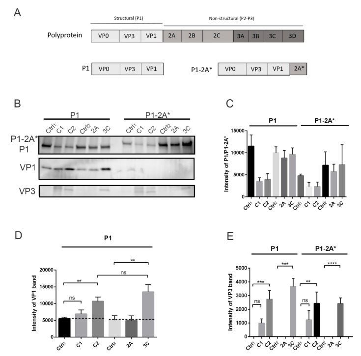
Figure 3.
Processing of P1 and P1-2A* constructs by calpains and viral proteases in vitro: P1 and P1-2A* constructs were incubated with purified calpain 1 (C1) or 2 (C2) at +25 °C for 2 h or with viral proteases 2A or 3C at 22 °C for 18 h. Negative controls where no proteases were added were also included: ctrl1 and ctrl2 for calpain and viral protease reactions, respectively. (A) Schematic image of P1 and P1-2A* constructs. (B) Representative image of P1, P1-2A*, VP1, and VP3 bands revealed by immunolabeling western blots with antibodies against VP1 and VP3. (C–E) Quantification of P1, P1-2A*, VP1, and VP3 signal from Figure 3B. Data are presented as means ± SEM from three separate experiments. Significance was determined with one-way ANOVA with Bonferroni’s multiple comparisons test (** p < 0.01; *** p < 0.001; **** p < 0.0001; ns, non-significant).

