Abstract
Tuberculosis is the leading cause of death due to infectious disease worldwide. There is an urgent need for more effective compounds against this pathogen to control the disease. Investigation of the anti-mycobacterial activity of a deep-water sponge of the genus Plakina revealed the presence of a new steroidal alkaloid of the plakinamine class, which we have given the common name plakinamine P. Its structure is most similar to plakinamine L, which also has an acyclic side chain. Careful dissection of the nuclear magnetic resonance data, collected in multiple solvents, suggests that the dimethyl amino group at the 3 position is in an equatorial rather than axial position unlike previously reported plakinamines. Plakinamine P was bactericidal against M. tuberculosis, and exhibited moderate activity against other mycobacterial pathogens, such as M. abscessus and M. avium. Furthermore, it had low toxicity against J774 macrophages, yielding a selectivity index (SI, or IC50/MIC) of 8.4. In conclusion, this work provides a promising scaffold to the tuberculosis drug discovery pipeline. Future work to determine the molecular target of this compound may reveal a pathway essential for M. tuberculosis survival during infection.
Keywords: marine compounds, tuberculosis, drug discovery
1. Introduction
Mycobacterium tuberculosis (Mtb) is the causative agent of the primarily pulmonary disease tuberculosis (TB). Approximately 10 million new cases of TB are diagnosed every year worldwide, and 5% of these are caused by multidrug-resistant strains of Mtb, rendering the disease extremely difficult to control [1]. TB treatment entails a combination of four antibiotics taken for at least six months in order to achieve a relapse-free cure [2]. Considering this, there is an urgent need for novel compounds that are more effective against this pathogen, capable of shortening treatment time, and killing drug-resistant strains of Mtb.
Recently, a large-scale screen of a marine natural products (MNP) library containing 4400 pre-fractionated peak fraction samples yielded highly potent inhibitors of Mtb [3]. A sample from the marine invertebrate Plakina sp. was highly active in the primary screen and was chosen for further deconvolution in this study. A novel plakinamine was discovered with potent bactericidal activity against Mtb. We have elucidated the structure of this novel compound and further characterized its activity against Mtb as well as other important mycobacterial pathogens, including M. abscessus, and two species belonging to the M. avium complex. The plakinamine compound was selectively active against Mtb, though moderate activity was observed against the other mycobacteria tested. Investigating the mechanism of action of this compound may reveal novel druggable Mtb targets. The structural similarity of plakinamine P and cholesterol, coupled with the essential nature of the cholesterol catabolism pathway for Mtb survival during infection, poses an interesting pool of putative targets for the novel structure described in this study [4].
2. Results
2.1. Chemical Analysis
A specimen of sponge identified as a new species of Plakina was collected using the Johnson Sea Link I manned submersible at a depth of 93 m off Crooked Island, the Bahamas, and stored at −20 °C until workup. The frozen sponge was extracted using a Dionex Accelerated Solvent Extractor and a series of solvents with increasing polarity. The CH3OH:H2O (3:1 v/v) extract was further purified using reverse-phase flash chromatography on a Teledyne Isco CombiFlash Rf 4x, followed by semi-preparative HPLC leading to the isolation of plakinamine P (1) as a tan oil [α]D20 = + 16.9 (c 0.11 in MeOH).
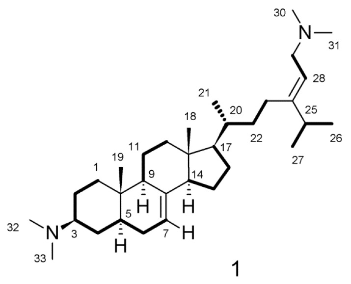
Direct analysis in real time–high resolution mass spectrometry (DART-HRMS) analysis of 1 coupled with interpretation of the 13C nuclear magnetic resonance (NMR) spectrum suggested a molecular formula of C33H58N2 requiring six degrees of unsaturation. The 1H NMR spectra of 1 had substantial overlap and, therefore, data were collected in methanol-d4 and dimethyl-sulfoxide (DMSO)-d6 to allow for assignment of all atoms (Supplementary Figures S1–S24, Table S1 and S2). The 1H and 13C NMR spectra collected in methanol-d4 showed the presence of four sp2 hybridized carbons [δC 159.9, 140.7, 118.4 and 111.7] consistent with two double bonds. No additional unsaturation was apparent from the NMR data, suggesting that 1 has four rings. The 1H NMR spectrum showed the presence of an isopropyl group [δH 2.38 sep (J = 6.9 Hz), δH 1.10 3H d (J = 6.9 Hz), δH 1.09 d 3H (J = 6.9 Hz)]. The presence of two dimethylamino groups in 1 was suggested by the resonances observed as two broadened singlets (δH 2.84 and δH 2.85) integrating for 12 protons attached to carbon resonances observed at δC 42.7 and 40.5. A literature search suggested that similar resonances are found in the plakinamine class of natural products [5,6,7,8,9,10,11,12,13,14,15] and comparison of the NMR data to the published data suggested that 1 is most similar to plakinamines L and M [14,15]. Other characteristic resonances observed in the 1H NMR spectrum of 1 were the methyl resonances observed at δH 0.59 (H3-18, s) and δH 0.86 (H3-19, s), which are consistent with the angular methyl groups of a steroidal alkaloid. Analysis of the 2D-COSY and 2D-edited gHSQC NMR spectra allowed for the assignment of five spin systems shown in Figure 1 as thickened bonds: C-1 through C-7; C-9 → C-11 → C-12; C-14 though C-17 → C-20 through C-23; C-25 → C-26 and C-27; and C-28 → C-29. The fragments were connected through the analysis of the 2D gHMBC spectrum (Figures S9–S12).
Figure 1.
Thick lines indicate spins systems defined through interpretation of the 2D-COSY and edited gHSQC NMR spectra.
Correlations observed in the HMBC spectrum from H-6a to C-8 assign the position of the olefinic carbon C-8. Correlations observed between H-7 and C-9; between H3-19 and C-1, C-5, C-9, and C-10, and between H-1b and C-19 defined the presence of fused rings A and B and the position of the C-19 methyl group. Correlations in the HMBC spectrum from H-7 to C-14; H-11ab to C-8; H-12a to C-13 and C-14; H-14 to C-8 and C-13, and from H3-18 to C-12, C-13, C-14, and C-17 established rings C and D as well as the position of C-18. The structure of the side chain attached at C-17 was established based on correlations in the HMBC spectrum from H-23ab to C-24, C-25, and C-28; from H-25 to C-23, C-24, and C-28; and from H-28 to C-23, C-24, and C-25. The placement of the two dimethylamino groups was determined based on HMBC correlations from H3-30/31 to C-29 in the acyclic side chain and from H3-32/33 to C-3 of ring A. Detailed results from NMR analysis are shown in Table 1.
Table 1.
1H and 13C NMR data for plakinamine P (1) (CD3OD, 600 MHz).
| Position. | δC, Type | δH (J in Hz) | COSY | HMBC a |
|---|---|---|---|---|
| 1a | 38.2, CH2 | 2.01 (ddd, 13.8, 4.1, 4.1) | 1b, 2ab | 3, 5, 10 |
| 1b | 1.21 (ddd, 13.8, 13.8, 3.4) | 1a, 2ab | 19 | |
| 2a | 23.7, CH2 | 1.95 (m) | 1ab, 2b, 3 | 1, 3, 4, 10 |
| 2b | 1.62 (m) | 1ab, 2a, 3 | 1, 3, 10 | |
| 3 | 66.8, CH | 3.21 (m) | 2ab, 4ab | |
| 4a | 29.7, CH2 | 1.84 (m) | 3, 4b, 5 | 2, 3, 10 |
| 4b | 1.51 (m) | 3, 4a, 5 | 3, 5 | |
| 5 | 41.9, CH | 1.51 (m) | 4ab, 6ab | 3 |
| 6a | 30.6, CH2 | 1.86 (m) | 5, 7 | 7, 8 |
| 6b | 1.30 (m) | 5, 7 | 4, 5 | |
| 7 | 118.4, CH | 5.20 (br d, 2.8) | 6ab | 5, 6, 9, 14 |
| 8 | 140.7, C | |||
| 9 | 50.5, CH | 1.73 (m) | 11ab | 8, 11 |
| 10 | 35.5, C | |||
| 11a | 22.7, CH2 | 1.63 (m) | 9, 11b, 12ab | 8, 9 |
| 11b | 1.52 (m) | 9, 11a, 12ab | 8, 9, 10, 13 | |
| 12a | 40.9, CH2 | 2.09 (m) | 11ab, 12b | 9, 11, 13, 14 |
| 12b | 1.28 (m) | 11ab, 12a | ||
| 13 | 44.7, C | |||
| 14 | 56.3, CH | 1.87 (m) | 15ab | 7, 8, 13 |
| 15a | 24.2, CH2 | 1.57 (m) | 14, 15b, 16ab | 16 |
| 15b | 1.46 (m) | 14, 15a, 16ab | ||
| 16a | 29.2, CH2 | 1.93 (m) | 15ab, 16b, 17 | 13, 15 |
| 16b | 1.33 (m) | 15ab, 16a, 17 | 15, 17, 20 | |
| 17 | 57.2, CH | 1.29 (m) | 16ab, 20 | 12, 13, 16, 18, 20, 21 |
| 18 | 12.4, CH3 | 0.59 (s) | 12, 13, 14, 17 | |
| 19 | 13.4, CH3 | 0.86 (s) | 1, 5, 9, 10 | |
| 20 | 38.2, CH | 1.45 (m) | 17, 21, 22ab | 22, 23 |
| 21 | 19.4, CH3 | 1.05 (d, 6.9) | 20 | 17, 20 |
| 22a | 36.8, CH2 | 1.46 (m) | 20, 22b, 23ab | |
| 22b | 1.17 (m) | 20, 22a, 23ab | 20, 21 | |
| 23a | 28.1, CH2 | 2.24 (ddd, 12.7, 12.7, 4.8) | 22ab, 23b | 22, 24, 25, 28 |
| 23b | 2.07 (m) | 22ab, 23a | 22, 24, 25, 28 | |
| 24 | 159.9, C | |||
| 25 | 36.1, CH | 2.38 (sep, 6.9) | 26, 27 | 23, 24, 26/27, 28 |
| 26 | 22.6, CH3 | 1.09 (d, 6.9) | 25 | 24, 25, 27 |
| 27 | 22.4, CH3 | 1.10 (d, 6.9) | 25 | 24, 25, 26 |
| 28 | 111.7, CH | 5.31 (t, 6.9) | 29ab | 23, 24, 25, 29 |
| 29 | 56.4, CH2 | 3.76 (dd, 7.6, 2.1) | 28, 29b | 24, 28, 30/31 |
| 30 | 42.7, CH3 | 2.84 (s) | 29, 30/31 | |
| 31 | 42.7, CH3 | 2.84 (s) | 29, 30/31 | |
| 32 | 40.5, CH3 | 2.85 (s) | 3, 32/33 | |
| 33 | 40.5, CH3 | 2.85 (s) | 3, 32/33 |
agHMBC correlations, optimized for 8 Hz, are from proton(s) stated to the carbons listed.
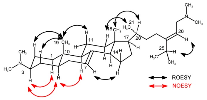
The relative configuration of 1 was defined based upon the ROESY and NOESY data (Figure 2 and Supplementary S13, S21-S23, Table S2) as well as comparison with previously described plakinamines [5,6,7,8,9,10,11,12,13,14,15]. NMR data were collected in two different solvents to fully resolve the A ring protons with DMSO- d6 yielding sufficient resolution. NOESY correlations observed between H-3 (δH 3.07), H-1b (δH 1.07), and H-5 (δH 1.37) were consistent with axial orientations for all three protons. This differs from other reported plakinamines in which the H-3 proton is equatorial. A trans-ring-fusion of the A and B rings indicating a chair conformation was established based on NOESY correlations between the axial protons H-5 (δH 1.37) and H-9 (δH 1.64) along with 1,3-diaxial ROESY correlations between H3-19 (δH 0.75), H-2b (δH 1.48), H-4b (δH 1.41), and H-11b (δH 1.42).
Figure 2.
Key ROESY and NOESY correlations for 1 observed in dimethyl-sulfoxide (DMSO)-d6.
A number of overlapping resonances in the 1H spectrum made the assignment of relative configuration at other centers difficult. Therefore, comparisons with previously reported plakinamine steroidal alkaloids were conducted. A transfused C/D ring, β-oriented side chain at C-17, and an α-methyl group at C-20 can be observed in all of the plakinamine steroidal alkaloids previously reported. By comparing the 13C NMR data for 1 (C-13, δC 44.7; C-14, δC 56.3; C-17, δC 57.2; C-20, δC 38.2) with the published data for structurally similar plakinamines [5,10,11,13,14], assignment of the relative configuration at these centers was possible as they fall within the ranges typically reported (C-13, δC 44.0 ± 0.8; C-14, δC 54.7 ± 1.6; C-17, δC 57.2 ± 1.6; C-20, δC 36.5 ± 2.0). The 2D-NOESY and ROESY data are consistent with the assigned structure. Plakinamines L and M are the only other compounds in the class that bear an acyclic side chain [13,14]. Plakinamine P differs due to a change in the position of the double bond from C-23/C-24 in plakinamines L and M to C-24/C-28 in 1. The Z geometry of the double bond was determined based on ROESY correlations observed from H-28 to H-25, H3-26, and H3-27.
2.2. Biological Activity
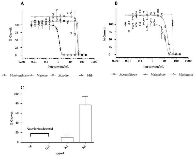
In a primary screen, the fraction from the marine organism of the genus Plakina sp. (HBOI.010.F07) inhibited actively growing M. tuberculosis CDC1551 by 97.4%, and 62% inhibition was detected against nonreplicating dormant bacteria. The data with nonreplicating bacteria were obtained using a multistress dormancy model which includes hypoxia, acidic pH, and starvation as the cues for Mtb to stop replicating [3]. Activity against dormant Mtb was absent when colony forming units (CFUs) were evaluated [16]. Plakinamine P was identified as the predominant compound in the mixture. Further purification yielded 1 mg of pure compound which did not retain activity against dormant Mtb, therefore the dormancy assay was no longer used for this compound. All the microbiological results reported were obtained with auto-luminescent mycobacteria expressing the luxABCDE operon as previously described [3]. Dose–response curves were conducted against Mtb as well as a panel of opportunistic non-tuberculous mycobacterial (NTM) pathogens, including both rapid-growing NTMs (M. massilliense, M. abscessus, M. fortuitum) and slow-growing NTMs (M. avium, M. intracellulare, M. simiae). All minimum inhibitory concentration (MIC) values are listed in Table 2 and dose–response curves are shown in Figure 3A,B. Plakinamine P was active against all mycobacterial species tested, with the lowest MIC of 1.8 µg/mL observed for Mtb and the highest of 57 µg/mL observed for M. intracellulare. The IC50 against J774 macrophages was 15.2 µg/mL, to produce a selectivity index (SI; IC50/MIC) of 8.5 against Mtb. The potent bactericidal activity of plakinamine P against Mtb was confirmed by plating samples for colony forming units (CFU) enumeration. These results demonstrated 90% bacterial killing by the compound at 3.1 µg/mL against Mtb. Furthermore, at the higher concentrations of 12.5 and 50 µg/mL, zero colonies were observed, indicating sterilization of the Mtb culture at those concentrations (Figure 3C).
Table 2.
Species used in this study and corresponding plakinamine P activity.
| Plasmid | Source | ID | MIC (µg/mL) |
|---|---|---|---|
| pMV306hsp+LuxG13 | Addgene plasmid #26161 [17] | N/A | |
| Fast-growing mycobacteria | |||
| M. massilliense | Mma | 57.6 | |
| M. fortuitum | Mfo | 32.45 | |
| M. abscessus | Mab | 22.16 | |
| Slow-growing mycobacteria | |||
| M. tuberculosis | [3] | Mtb | 1.84 |
| M. avium | Mav | 27.28 | |
| M. intracellulare | Min | 49.53 | |
| M. simiae | Msi | 48.25 |
N/A, non applicable. *Rifampicin MIC 0.01 µg/mL/Isoniazid MIC 0.04 µg/mL.
Figure 3.
Activity of plakinamine P against multiple mycobacterial pathogens. (A,B) Cultures were treated with 16-point, 2-fold serial dilutions of plakinamine P for 2 days (fast-growing mycobacteria – panel B) and 5 days (slow-growing mycobacteria – panel A) after which the luminescence was read. The Gompertz model was used to calculate MIC (99% killing). (C) Bactericidal activity of plakinamine P against Mtb was evaluated. Cultures treated with 50, 12.5, 3.1 and 0.8 μg/mL plakinamine P were plated on 7H10 OADC and incubated for 3 weeks before colony forming unit (CFU) enumeration. The data are presented as % growth relative to average CFU/mL of the 2% DMSO controls.
3. Discussion
The major global health threat posed by Mtb infections is intensified by the difficulty to treat TB. In this study, we have identified a novel scaffold bactericidal against Mtb with low toxicity towards mammalian cells. The structural characterization of this compound revealed it to be the novel steroidal alkaloid plakinamine P. Plakinamine P inhibited the growth of M. tuberculosis with an MIC of 1.8 µg/ml. It is most closely related to plakinamines L and M, which have been reported to inhibit the growth of M. tuberculosis strain H37Ra with MICs of 3.6 and 15.8 µg/mL, respectively [14]. Selectivity for mycobacteria versus mammalian cells and spectrum of activity against mycobacteria has not been reported for the other known plakinamines. In the current study, plakinamine P was most potent against Mtb, however, it was also moderately active against important opportunistic mycobacteria including M. abscessus and M. avium complex organisms. These are highly drug-tolerant pathogens known to cause chronic infections in immunocompromised individuals [18,19]. Nevertheless, the observed selectivity of plakinamine P for Mtb in comparison to other mycobacteria suggests it may target pathways uniquely essential for Mtb survival.
The delayed clearance of Mtb by current front-line TB drugs is often attributed to the distinctive physiological aspects of this intracellular pathogen during infection [20]. In light of this, many TB drug discovery studies are focusing efforts on finding scaffolds capable of inhibiting Mtb’s survival pathways during infection [21,22]. Plakinamine P described in this study is structurally similar to cholesterol, with a modified side chain. Previous work has shown the essentiality of cholesterol catabolism for the survival and persistence of Mtb during infection [4]. Inhibition of this pathway leads to death of intracellular Mtb [23]. Additionally, cholesterol analogs with undegradable side chains are capable of killing Mtb in culture [24]. Our data, combined with previously published work, suggest that plakinamine P may be causing mycobacterial death by inhibiting the cholesterol catabolism pathway. Additionally, inhibition may be due to toxic byproducts coming from the breakdown of plakinamine P via the cholesterol degradation pathway.
In conclusion, we have characterized an MNP-derived scaffold with potent antimycobacterial activity. The essentiality of the putative target of plakinamine P for Mtb survival and persistence in the host further highlights the potential of this compound in TB drug discovery. Future research will not only focus on confirming the molecular target of plakinamine P, but also demonstrating its activity against Mtb under in vivo-like conditions.
4. Materials and Methods
4.1. Chemical Ananlysis
Optical rotation was measured on a Rudolph Research Analytical AUTOPOL III automatic polarimeter. UV spectra were collected on a NanoDrop Spectrophotometer (Thermo Fisher Scientific, Inc., MA, USA). NMR data were collected on a JEOL ECA-600 spectrometer (JEOL USA, Peabody, MA, USA) operating at 600 MHz for 1H, and 150.9 for 13C. The edited gHSQC spectrum was optimized for 140 Hz and the gHMBC spectrum optimized for 8 Hz. Chemical shifts were referenced to solvent, e.g., CD3OD, δH observed at 3.31 ppm and δC observed at 49.1 ppm or DMSO- d6 δH observed at 2.50 ppm and δC observed at 39.5 ppm. High resolution mass spectrometry was performed on a JEOL AccuTOF-DART 4G using the DART source for ionization.
4.1.1. Biological Material
The steroidal alkaloid Plakinamine P, 1, was isolated from a sponge identified as Plakina n. sp. (S. 26, a picture of the sponge can be found in Figure S26A) (Phylum Porifera, Class Homoscleromorpha, Order Homosclerophorida, Family Plakinidae). The specimens (HBOI ID; 25-V-93-3-009, HBOI Museum Number 004.0001) were collected at a depth of 93 m using the Johnson Sea-Link I manned submersible in the Bahamas off the NW tip of Crooked Island near Pittstown (latitude 22 49.278’ N, longitude, 74 21.075’W). The external morphology is bulbous to globular (1–2 cm thick, 2–6 cm in diameter), with a single oscula per bulb. Oscules are less than 4 mm wide with a tubular and darkened membrane projection. The surface of the sponge is smooth and the consistency is gelatinous and compressible. The specimen is light brown externally and tan in color internally, both in life and preserved. Spicules are in very low abundance and consist of small ramified calthrops (tetralophose and trilophose), rare smooth non-lophose calthrops, and small diod microrhabs (7–10 µm in length and <1 µm in width). Calthrops are regular in size; less than 20 µm in total size, with rays 8–10 µm × 2–3 µm. Trilophose calthrops usually have deformed rays with variation in the pattern of ramification between rays. The lophose calthrops are typical of the genus Plakina. There are five Plakina species currently recognized for the Caribbean: Plakina elisa, Plakina nathaliae, Plakina tetralopha, Plakina jamaicensis, and Plakina arletensis [25]. The present Plakina specimen differs from those species in general morphology as it has a much thicker growth, bulbous shape, and unique oscula morphology. The spicule combination, the tendency to have deformed rays and diverse branching patterns within a spicule, and their low density in the body are unique characteristics of this sample that indicate that it may be an undescribed Plakina species. Further study of the histology and genetics of this specimen will allow its distinction from the other Caribbean species.
4.1.2. Extraction and Isolation
The sample was frozen immediately after collection and stored at −20 °C until extraction. The frozen sponge (92 g) was soaked overnight in CH3OH:H2O (1:1 v/v), filtered, and allowed to dry overnight. Extraction of the sponge was accomplished using a Dionex ASE 100® Accelerated Solvent Extractor (Dionex, Sunnyvale, CA, USA) using the following solvents: Extract 1: heptane, Extract 2: ethyl acetate:ethanol (1:1 v/v), Extract 3: ethanol, Extract 4: methanol:water (3:1 v/v) and washed with isopropyl alcohol. The methanol:water (3:1 v/v) extract and the isopropanol extract were combined and separated by medium pressure liquid chromatography using a Teledyne Isco Combiflash® Rf 4x equipped with PeakTrak software (Version 2.1.19, Teledyne Isco, Lincoln, NE, USA) as follows: 365 mg of the combined extract was absorbed onto approximately 2 g of C18 reversed-phase packing, dried, and then placed into a loading column. A Teledyne Isco 15.5 g C18 Redisep Rf Gold column operating at a flow rate of 30 mL/min and monitored at 225 and 280 nm and collected into 13 mm tubes was used for the separation. Solvent A was H2O:CH3CN (95:5), Solvent B was CH3CN, Solvent C was CH3OH, and Solvent D was CH2Cl2. The run lasted 15.9 mins and 37 column volumes. The column was first eluted with a mixture of A:B (94:6 v/v) for 2 column volumes. The column was then eluted over a linear gradient to 100% B over 21 column volumes and then held at 100% B for an additional 3 column volumes. The column was then washed with 100% Solvent C (CH3OH), followed by a rapid gradient of CH2Cl2 in CH3OH to wash the column. The anti-mycobacterial activity was observed in Fraction 7 eluting as a broad peak between column volumes 8 to 15 containing > 90% plakinamine P (0.040 g, 0.043% frozen weight). A portion of this material was further fractionated by semi-preparative HPLC at room temperature using a Vydac C18 protein and peptide column (10 × 250 mm, 10 µm particle size) and the following gradient Solvent A: H2O:CH3CN:TFA (95:5:0.1 v/v/v), and Solvent B: CH3CN:TFA (100:0.1 v/v) t = 0 min. A:B 4:1; t = 5 min. A:B 1:1, hold for 10 min; t = 20 min. 100% B, hold for 8 mins. Plakinamine P elutes at 12.5 mins.
Plakinamine P (1); tan oil; [α]20D = +16.9 (c 0.11 in MeOH); UV (MeOH) λmax (log ε) 195 nm (2.7); 1H and 13C NMR (Table 1, Figures S15 and S26); DART HRMS: C33H58N2 [m/z observed 483.4693 [M + H]+, calcd. 483.4678, Δ = -1.5 mmu], Figure S25.
4.2. Biological Activity
Four slow-growing and 3 fast-growing mycobacterial strains (Table 2) were cultured in Middlebrook 7H9 broth media, supplemented with 0.05% Tween 80, 10% OADC, and 50 µg/mL kanamycin when necessary for plasmid maintenance. The previously described Mtb-Lux strain constitutively expressing the luxCDABEG operon from the episomal plasmid pMV306hsp+LuxG13, was used [3]. pMV306hsp+LuxG13 was a gift from Brian Robertson & Siouxsie Wiles (Addgene plasmid # 26161; http://n2t.net/addgene:26161; RRID:Addgene_26161). All NTMs were transformed with the same plasmid to produce a stable luminescent signal. Log phase cultures of the mycobacterial strains expressing the lux operon were diluted to an OD600 of 0.01 and treated with serial dilutions of plakinamine P. This compound was resuspended in 100% dimethyl-sulfoxide (DMSO) at 10 mg/mL. Sixteen-point 2-fold serial dilutions of plakinamine P, starting at 200 µg/mL, were prepared in a final 2% DMSO concentration, 12 µg/mL rifampicin and 2% DMSO were used as controls. Treatments were performed in 384-well plates for 5 days at 37 °C (5% CO2, ambient O2, and humidity) for slow-growing mycobacteria and 2 days for fast-growing mycobacteria (Table 1), after which the luminescence was read using SynergyTM H4 plate reader (BioTeK). Samples of Mtb-Lux treated with 0.8, 3.1, 12.5 and 50 µg/mL plakinamine P were taken, diluted 10-fold in PBS-Tween80, and plated in 7H10 OADC. Plates were incubated for 3 weeks at 37 °C, after which colonies were enumerated.
J774A.1 (ATCC® TIB67™) macrophages were cultured in Dulbecco’s Modified Eagle Medium (DMEM, GIBCO), supplemented with 10% heat inactivated fetal calf serum (Atlanta Biologicals), 1 mM sodium pyruvate (Mediatech, Inc.), 2mM L-glutamine (Mediatech, Inc.), and 1% PenStrep (100 U/mL Penicillin, 100 µg/mL Streptomycin, GIBCO). Twenty-five thousand cells/well were seeded overnight in black 384-well plates, then treated with serial dilutions of plakinamine P as described above. Tryton X and 2% DMSO were used as controls in these experiments.
Data were normalized to highest and lowest output values in the dose response. MIC (99% killing) and IC50 were calculated using a Gompertz model, and a nonlinear regression—normalized response curve fit, respectively in GraphPad Prism 5 [26]. The selectivity index (SI) was calculated as IC50/MIC.
Supplementary Materials
The following are available online at https://www.mdpi.com/1660-3397/17/12/707/s1, Figure S1: 1H NMR (600 MHz, CD3OD) of Plakinamine P (1), Figure S2: Expansion of 1H NMR (600 MHz, CD3OD) of Plakinamine P (1), Figure S3: 13C NMR (150 MHz, CD3OD) of Plakinamine P (1), Figure S4: gDQF-COSY spectrum (600 MHz, CD3OD) of Plakinamine P (1), Figure S5: gCOSY spectrum expansion with key side chain correlations (600 MHz, CD3OD) of Plakinamine P (1), Figure S6: edited gHSQC spectrum (600 MHz, CD3OD) of Plakinamine P (1), Figure S7: Expansion 1 of edited gHSQC spectrum (600 MHz, CD3OD) Plakinamine P (1), Figure S8: Expansion 2 of edited gHSQC spectrum (600 MHz, CD3OD) of Plakinamine P (1), Figure S9: gHMBC spectrum (600 MHz, CD3OD) of Plakinamine P (1), Figure S10: gHMBC spectrum expansion of N,N dimethyl amino proton correlations (600 MHz, CD3OD) of Plakinamine P (1), Figure S11: gHMBC spectrum expansion with key side chain correlations (600 MHz, CD3OD) of Plakinamine P (1), Figure S12: Structure of plakinamine P (1) with key gHMBC correlations observed in CD3OD (600 MHz), Figure S13: gROESY spectrum (600 MHz, CD3OD) of Plakinamine P (1), Figure S14: Structure of plakinamine P (1) with chemical shifts in CD3OD (600 MHz), Figure S15: 1H NMR (600 MHz, DMSO-d6) of Plakinamine P (1), Figure S16: Expansion of 1H NMR (600 MHz, DMSO-d6) of Plakinamine P (1), Figure S17: 13C NMR (150 MHz, DMSO-d6) of Plakinamine P (1), Figure S18: gDQF-COSY spectrum (600 MHz, DMSO-d6) of Plakinamine P (1), Figure S19: edited gHSQC spectrum (600 MHz, DMSO-d6) of Plakinamine P (1), Figure S20: gHMBC spectrum (600 MHz, DMSO-d6) of Plakinamine P (1), Figure S21: NOESY spectrum (600 MHz, DMSO-d6) of Plakinamine P (1), Figure S22: Expansion of Plakinamine P (1) 2D-NOESY spectrum (600 MHz, DMSO-d6) showing key correlations for H-3. Figure S23: ROESY spectrum (600 MHz, DMSO-d6) of Plakinamine P (1), Figure S24: Structure of plakinamine P (1) with chemical shifts observed in DMSO-d6, Figure S25: High resolution DART mass spectrometry data of plakinamine P (1), Figure S26: Picture and taxonomic description of the Plakina sp. used in the study; Table S1: Table of 1H and 13C NMR Data for plakinamine P (1) (600 MHz, CD3OD), Table S2: Table of 1H and 13C NMR Data for plakinamine P (1) (600 MHz, DMSO-d6).
Author Contributions
Conceptualization, A.E.W. and K.H.R.; Data curation, C.R.F., R.G., M.C.D., S.A.P., A.E.W. and K.H.R.; Formal analysis, C.R.F., J.C.R., P.L.W., R.G., A.E.W. and K.H.R.; Funding acquisition, A.E.W. and K.H.R.; Investigation, C.R.F., J.C.R. and P.L.W.; Project administration, A.E.W. and K.H.R.; Resources, M.C.D. and S.A.P.; Supervision, A.E.W. and K.H.R.; Writing—original draft, C.R.F., A.E.W. and K.H.R.; Writing—review & editing, C.R.F., J.C.R., P.L.W., R.G., M.C.D., S.A.P. and K.H.R.
Funding
A.E.W. and K.H.R. received support from the National Institutes of Health (NIAID) grant number 1R21AI105977 and 4R33AI105977. K.H.R. also received support from University of Central Florida grant ORC2012. A.E.W received support for preparation of the enriched fraction library used in this study from the National Institutes of Health (NCCIH) grant number 1RC2AT005917.
Conflicts of Interest
The authors declare no conflict of interest.
References
- 1.World Health Organization . Global Tuberculosis Report. World Health Organization; Geneva, Switzerland: 2019. [Google Scholar]
- 2.D’Ambrosio L., Centis R., Sotgiu G., Pontali E., Spanevello A., Migliori G.B. New anti-tuberculosis drugs and regimens: 2015 update. ERJ Open Res. 2015;1 doi: 10.1183/23120541.00010-2015. [DOI] [PMC free article] [PubMed] [Google Scholar]
- 3.Rodrigues Felix C., Gupta R., Geden S., Roberts J., Winder P., Pomponi S.A., Diaz M.C., Reed J.K., Wright A.E., Rohde K.H. Selective Killing Of Dormant Mycobacterium tuberculosis By Marine Natural Products. Antimicrob. Agents Chemother. 2017 doi: 10.1128/AAC.00743-17. [DOI] [PMC free article] [PubMed] [Google Scholar]
- 4.Pandey A.K., Sassetti C.M. Mycobacterial persistence requires the utilization of host cholesterol. Proc. Natl. Acad. Sci. USA. 2008;105:4376–4380. doi: 10.1073/pnas.0711159105. [DOI] [PMC free article] [PubMed] [Google Scholar]
- 5.Rosser R.M., Faulkner D.J. Two steroidal alkaloids from a marine sponge, Plakina sp. J. Org. Chem. 1984;49:5157–5160. doi: 10.1021/jo00200a029. [DOI] [Google Scholar]
- 6.Jurek J., Scheuer P.J., Kelly-Borges M. Two steroidal alkaloids from a sponge, Corticium sp. J. Nat. Prod. 1994;57:1004–1007. doi: 10.1021/np50109a022. [DOI] [PubMed] [Google Scholar]
- 7.De Marino S., Iorizzi M., Zollo F., Roussakis C., Debitus C. Plakinamines C and D and three other new steroidal alkaloids from the sponge Corticium sp. Eur. J. Org. Chem. 1999;3:697–701. doi: 10.1002/(SICI)1099-0690(199903)1999:3<697::AID-EJOC697>3.0.CO;2-W. [DOI] [Google Scholar]
- 8.Lee H.-S., Seo Y., Rho J.-R., Shin J., Paul V.J. New steroidal alkaloids from an undescribed sponge of the genus Corticium. J. Nat. Prod. 2001;64:1474–1476. doi: 10.1021/np0101649. [DOI] [PubMed] [Google Scholar]
- 9.Borbone N., De Marino S., Iorizzi M., Zollo F., Debitus C., Esposito G., Iuvone T. Minor steroidal alkaloids from the marine sponge Corticium sp. J. Nat. Prod. 2002;65:1206–1209. doi: 10.1021/np020027r. [DOI] [PubMed] [Google Scholar]
- 10.Ridley C.P., Faulkner D.J. New cytotoxic steroidal alkaloids from the Philippine sponge Corticium niger. J. Nat. Prod. 2003;66:1536–1539. doi: 10.1021/np0302706. [DOI] [PubMed] [Google Scholar]
- 11.Zampella A., D’Orsi R., Sepe V., De Marino S., Borbone N., Valentin A., Debitus C., Zollo F., D’Auria M.V. Isolation of plakinamine I: A new steroidal alkaloid from the marine sponge Corticium sp. and synthesis of an analogue model compound. Eur. J. Org. Chem. 2005;20:4359–4363. doi: 10.1002/ejoc.200500364. [DOI] [Google Scholar]
- 12.Langjae R., Bussarawit S., Yuenyongsawad S., Ingkaninan K., Plubrukarn A. Acetylcholinesterase-inhibiting steroidal alkaloid from the sponge Corticium sp. Steroids. 2007;72:682–685. doi: 10.1016/j.steroids.2007.05.005. [DOI] [PubMed] [Google Scholar]
- 13.Sunassee S.N., Ransom T., Henrich C.J., Beutler J.A., Covell D.G., McMahon J.B., Gustafson K.R. Steroidal Alkaloids from the Marine Sponge Corticium niger That Inhibit Growth of Human Colon Carcinoma Cells. J. Nat. Prod. 2014;77:2475–2480. doi: 10.1021/np500556t. [DOI] [PMC free article] [PubMed] [Google Scholar]
- 14.Aknin M., Rudi A., Kashman Y., Vacelet J., Gaydou E.M. Plakinamine L: A new steroidal alkaloid from the marine sponge Corticium sp. Nat. Prod. Commun. 2010;5:33–34. doi: 10.1177/1934578X1000500108. [DOI] [PubMed] [Google Scholar]
- 15.Lu Z., Koch M., Harper M.K., Matainaho T.K., Barrows L.R., Van Wagoner R.M., Ireland C.M. Plakinamine M, a Steroidal Alkaloid from the Marine Sponge Corticium sp. J. Nat. Prod. 2013;76:2150–2152. doi: 10.1021/np400649e. [DOI] [PMC free article] [PubMed] [Google Scholar]
- 16.Vandroux D., Chanareille P., Delmas B., Gauzere B.A., Allou N., Raffray L., Jaffar-Bandjee M.C., Martinet O., Ferdynus C., Jabot J. Acute respiratory distress syndrome in leptospirosis. J. Crit. Care. 2019;51:165–169. doi: 10.1016/j.jcrc.2019.02.018. [DOI] [PubMed] [Google Scholar]
- 17.Andreu N., Zelmer A., Fletcher T., Elkington P.T., Ward T.H., Ripoll J., Parish T., Bancroft G.J., Schaible U., Robertson B.D., et al. Optimisation of bioluminescent reporters for use with mycobacteria. PLoS ONE. 2010;5:e10777. doi: 10.1371/journal.pone.0010777. [DOI] [PMC free article] [PubMed] [Google Scholar]
- 18.Prevots D.R., Marras T.K. Epidemiology of human pulmonary infection with nontuberculous mycobacteria: A review. Clin. Chest Med. 2015;36:13–34. doi: 10.1016/j.ccm.2014.10.002. [DOI] [PMC free article] [PubMed] [Google Scholar]
- 19.Nessar R., Cambau E., Reyrat J.M., Murray A., Gicquel B. Mycobacterium abscessus: A new antibiotic nightmare. J. Antimicrob. Chemother. 2012;67:810–818. doi: 10.1093/jac/dkr578. [DOI] [PubMed] [Google Scholar]
- 20.Peddireddy V., Doddam S.N., Ahmed N. Mycobacterial Dormancy Systems and Host Responses in Tuberculosis. Front. Immunol. 2017;8:84. doi: 10.3389/fimmu.2017.00084. [DOI] [PMC free article] [PubMed] [Google Scholar]
- 21.Gold B., Nathan C. Targeting Phenotypically Tolerant Mycobacterium tuberculosis. Microbiol. Spectr. 2017;5 doi: 10.1128/microbiolspec.TBTB2-0031-2016. [DOI] [PMC free article] [PubMed] [Google Scholar]
- 22.Nathan C., Barry C.E. TB drug development: Immunology at the table. Immunol. Rev. 2015;264:308–318. doi: 10.1111/imr.12275. [DOI] [PMC free article] [PubMed] [Google Scholar]
- 23.VanderVen B.C., Fahey R.J., Lee W., Liu Y., Abramovitch R.B., Memmott C., Crowe A.M., Eltis L.D., Perola E., Deininger D.D., et al. Novel inhibitors of cholesterol degradation in Mycobacterium tuberculosis reveal how the bacterium’s metabolism is constrained by the intracellular environment. PLoS Pathog. 2015;11:e1004679. doi: 10.1371/journal.ppat.1004679. [DOI] [PMC free article] [PubMed] [Google Scholar]
- 24.Frank D.J., Zhao Y., Wong S.H., Basudhar D., De Voss J.J., Ortiz de Montellano P.R. Cholesterol Analogs with Degradation-resistant Alkyl Side Chains Are Effective Mycobacterium tuberculosis Growth Inhibitors. J. Biol. Chem. 2016;291:7325–7333. doi: 10.1074/jbc.M115.708172. [DOI] [PMC free article] [PubMed] [Google Scholar]
- 25.Ruiz C., Muricy G., Lage A., Domingos C., Chenesseau S., Pérez T. Descriptions of new sponge species and genus, including aspiculate Plakinidae, overturn the Homoscleromorpha classification. Zool. J. Linn. Soc. 2017;179:707–724. doi: 10.1111/zoj.12480. [DOI] [Google Scholar]
- 26.Lambert R.J., Pearson J. Susceptibility testing: Accurate and reproducible minimum inhibitory concentration (MIC) and non-inhibitory concentration (NIC) values. J. Appl. Microbiol. 2000;88:784–790. doi: 10.1046/j.1365-2672.2000.01017.x. [DOI] [PubMed] [Google Scholar]
Associated Data
This section collects any data citations, data availability statements, or supplementary materials included in this article.


