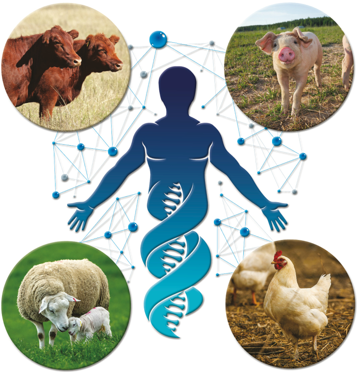
This issue of Animal Frontiers, “Farm animals are important biomedical models,” describes several examples in which cattle, sheep, pigs, or chickens provide an excellent physiological model for studies related to human health or disease (Figure 1). While previous reports have discussed the use of domestic animals as dual-purpose models that benefit agricultural and biomedical research (Ireland et al., 2008), this issue of Animal Frontiers provides additional and novel examples of the value of farm animals for biomedical research. Because farm animals are larger in size than laboratory species, scientists are able to collect larger volumes and more frequent samples of blood without significant changes in blood chemistry or substantive changes in blood volume as well as larger or more frequent tissue biopsies, thereby allowing the study of changes in hormones, metabolites, immune factors, or cellular components in the same animal over time. Many of these studies revealed that the physiology of humans is more closely related to the physiology of farm animals than to rodents. Finally, the human genome sequence is more similar to the genome sequences of cattle and pigs compared with rodents (Humphray et al., 2007; Tellam et al., 2009); thus, cattle and pigs may be better models for many human genetic diseases.
Figure 1.
Schematic depicting the use of cattle, pigs, sheep, and chickens as models for biomedical research.
The article by Proudfoot et al. (2019) discusses use of gene editing in pigs and the potential to use this technology in chickens to generate disease-resistant animals. Gene editing allows modification of a single gene or single nucleotide polymorphism without the addition of foreign DNA to the animal’s genome. Gene-edited pigs that are resistant to infection by important viral diseases, such as porcine reproductive and respiratory syndrome virus or transmissible gastroenteritis virus, have recently been produced. The avian egg has very different physiology compared with mammalian eggs; thus, gene editing in poultry has not yet been reported. However, use of chicken stem cells may provide a new method for successful gene editing in poultry. While the production of gene-edited farm animals holds tremendous potential for enhancing disease resistance, use of these animals is also likely to enhance our understanding of transmission of zoonotic diseases between animals and humans as well as our understanding of disease resistance in humans. As with any new technology, consumer acceptance and use of a science-based regulatory framework will determine whether or not the technology will be available to treat diseases in humans.
Wolf et al. (2019) describe production of genetically modified pigs as a source of cells, tissues, or organs for xenotransplantation. Pigs may be an important donor for human transplant tissues because many pig organs and tissues are similar to humans and pig cells can be genetically engineered to overcome issues associated with the risk of transmission of zoonotic pathogens or rejection of transplant tissues. To prevent hyperacute rejection of pig tissues in non-human primates, gene targeting or gene editing was used to knockout antigens (e.g., galactose-α1,3-galactose [αGal] or α-1,3-galactosyltransferase [GGTA1]) on pig cells that are not found on cells in humans and Old World monkeys and transgenic technology was used to express one or more human complement-regulatory proteins in pig cells. This technology holds significant promise for thousands of people waiting for transplants of pancreatic islets, neuronal cells, corneas, hearts, or kidneys.
Studies with farm animals in the areas of muscle development and metabolism are informative for animal production as well as human health as discussed by Zhao et al. (2019). The composition of muscle fibers affects growth efficiency and meat quality in farm animals and is associated with metabolic syndrome in humans. In skeletal muscle and brown adipose tissue, thermogenesis increases energy expenditure and heat production, which reduces feed efficiency in farm animals (an undesirable situation) and prevents obesity and metabolic diseases in humans (desirable conditions). While dysregulation of satellite cells and fibro/adipogenic progenitor cells leads to the favorable characteristic of increased intramuscular fat (or marbling) in cattle, this situation leads to muscle atrophy, fatty infiltration, and fibrosis in humans. Thus, understanding the cellular and molecular mechanisms that regulate skeletal muscle development and metabolism are essential for improving human health.
Abedal-Majed and Cupp (2019) describe livestock models to study infertility in humans. In most mammals, anovulation is a major cause of infertility. Pigs, sheep, and cattle have advantages to rodents as biomedical models and many studies conducted with these species have direct relevance to reproductive disorders in women. For example, polycystic ovarian syndrome (PCOS) is a multifactorial disorder that affects up to 20% of women of reproductive age (Di Pietro et al., 2015). Recently, a subpopulation of beef cows with excess intrafollicular androstenedione (A4; High A4 beef cows) and molecular phenotypes in ovarian theca cells was found to represent a naturally occurring, androgen excess animal model that is similar to women diagnosed with PCOS and subfertility. Thus, High A4 beef cows could be used to identify new cellular and molecular mechanisms regulating PCOS and/or develop potential treatments for PCOS in the future.
Intrauterine growth restriction (IUGR) is associated with increased mortality rates early in life and various metabolic disorders that may lead to reduced quality and duration of life in affected individuals in a variety of species. Beede et al. (2019) describe the use of pregnant sheep to study IUGR to advance our understanding of the mechanisms associated with adaptive fetal programming and metabolic dysfunction. Results indicate that adrenergic and inflammatory pathways are involved in regulation of skeletal muscle growth and glucose metabolism. Continued use of the pregnant sheep model may lead to prenatal interventions or postnatal treatments to improve outcomes in IUGR-born offspring.
Pregnant sheep and catheterized fetal sheep have been widely used to study normal and abnormal fetal development, as well as in utero treatment options for congenital birth defects under a variety of experimental conditions that could not be studied in human fetuses. Varcoe et al. (2019) summarize the most commonly used analgesics in fetal sheep studies and discuss the potential impacts of each drug on maternal pain and fetal well-being. Unlike ewes, it is not possible to directly assess effects of analgesia on fetal lambs. The authors also discuss various factors to consider when providing post-surgical anesthesia, such as route of administration, transport across the placenta, and metabolism of the drug by the placenta.
Ella et al. (2019) describe the use of various brain imaging technologies in farm animals, such as x-rays, magnetic resonance imaging (MRI), and computerized tomography scans. While these technologies have been used widely for brain imaging in human clinical studies, use of these technologies for brain imaging in farm animals has only recently been developed. A sheep model of Batten disease that shares many neuropathological features with humans has recently been described. The combined use of MRI imaging to monitor the brain in vivo and gene therapy with three different viral vectors in the ovine model of Batten disease provides an exciting opportunity for future clinical and translational studies with humans.
Food allergies, especially soy food allergies, are relatively common in humans and are a growing concern in companion animals. Radcliffe et al. (2019) discuss a subset of pigs that naturally develop allergies to soy. This pig model of soy protein-induced food allergenicity will be an important system for gaining new information regarding the genetic, genomic, biochemical, and physiological mechanisms associated with food allergenicity as well as development of potential intervention strategies that may be useful in humans.
Although zebrafish are not considered a farm animal, the importance of zebrafish in biomedical research cannot be discounted. Teame et al. (2019) identify several unique physiological characteristics of zebrafish, including external fertilization and development of nearly transparent embryos outside the uterus, that make zebrafish an interesting biomedical model. The authors also describe a standard diet for zebrafish larvae and adults, which should facilitate generation of consistent results in metabolic studies with zebrafish across laboratories throughout the world. Zebrafish may also be used to develop new therapies to prevent or treat important diseases in humans.
In conclusion, this issue of Animal Frontiers describes several novel uses of farm animals and zebrafish to gain a better understanding of the mechanisms underlying human diseases. The ability to collect frequent blood samples and(or) multiple tissue biopsies from farm animals; the similar physiology between farm animals and humans; the similar genome sequence between cattle, pigs, and humans; and the ability to genetically modify farm animals by gene targeting, gene editing, and(or) transgenesis are excellent advantages to the use of farm animals compared with traditional laboratory species when studying human diseases. Development of new technologies will likely allow further use of farm animals as additional biomedical models in the future.
About the Authors

Deb Hamernik is Associate Vice Chancellor for Research and Professor in the Department of Animal Science at the University of Nebraska-Lincoln. She administers funding for research seed grants and helps faculty form interdisciplinary research teams to enhance their competitiveness for extramural funding. Deb has been Editor-in-Chief of Animal Frontiers since July 2018 and is Past-President of the American Society of Animal Science.
Literature Cited
- Abedal-Majed M.A., and Cupp A.S.. . 2019. Livestock animals to study infertility in women. 2019. Anim. Front. 9(3):28–33. [DOI] [PMC free article] [PubMed] [Google Scholar]
- Beede K.A., Limesand S.W., Petersen J.L., and Yates D.T.. . 2019. Real supermodels wear wool: summarizing the impact of the pregnant sheep as an animal model for adaptive fetal programming. Anim. Front. 9(3):34–43. [DOI] [PMC free article] [PubMed] [Google Scholar]
- Di Pietro M., Parborell F., Irusta G., Pascuali N., Bas D., Bianchi M.S., Tesone M., and Abramovich D.. 2015. Metformin regulates ovarian angiogenesis and follicular development in a female polycystic ovary syndrome rat model. Endocrinology. 156:1453–1463. doi: 10.1210/en.2014-1765 [DOI] [PubMed] [Google Scholar]
- Ella A., Barrière D., Adriaenssen H., Palmer D.N., Melzer T.R., Mitchell N.L., and Keller M.. . 2019. The development of brain magnetic resonance approaches in large animal models for preclinical research. Anim. Front. 9(3):44–51. [DOI] [PMC free article] [PubMed] [Google Scholar]
- Humphray S.J., Scott C.E., Clark R., Marron B., Bender C., Camm N., Davis J., Jenks A., Noon A., Patel M., . et al. 2007. A high utility integrated map of the pig genome. Genome Biol. 8:R139. doi: 10.1186/gb-2007-8-7-r139 [DOI] [PMC free article] [PubMed] [Google Scholar]
- Ireland J.J., Roberts R.M., Palmer G.H., Bauman D.E., and Bazer F.W.. 2008. A commentary on domestic animals as dual-purpose models that benefit agricultural and biomedical research. J. Anim. Sci. 86:2797–2805. doi: 10.2527/jas.2008-1088. [DOI] [PubMed] [Google Scholar]
- Proudfoot C., Lillico S., and Tait-Burkard C.. . 2019. Genome editing for disease resistance in pigs and chickens. Anim. Front. 9(3):6–12. [DOI] [PMC free article] [PubMed] [Google Scholar]
- Radcliffe J.S., Brito L.F., Reddivari L., Schmidt M., Herman E.M. and Schinckel A.P.. . 2019. A swine model of soy protein induced food allergenicity: implications in human and swine nutrition. Anim. Front. 9(3):52–59. [DOI] [PMC free article] [PubMed] [Google Scholar]
- Teame T., Zhang Z., Ran C., Zhang H., Yang Y., Ding Q., Xie M., Gao C., Olsen R.E., Ye Y., . et al. 2019. The use of zebrafish (Danio rerio) as biomedical models. 2019. Anim. Front. 9(3):68–77. [DOI] [PMC free article] [PubMed] [Google Scholar]
- Tellam R.L., Lemay D.G., Van Tassell C.P., Lewin H.A., Worley K.C., and Elsik C.G.. 2009. Unlocking the bovine genome. BMC Genomics. 10:193. doi: 10.1186/1471-2164-10-193 [DOI] [PMC free article] [PubMed] [Google Scholar]
- Varcoe T.J., Darby J.R.T., Gatford K.L., Holman S.L., Cheung P., Berry M.J., Wiese M.D., and Morrison J.L.. . 2019. Considerations in selecting post-operative analgesia for pregnant sheep following fetal instrumentation surgery. Anim. Front. 9(3):60–67. [DOI] [PMC free article] [PubMed] [Google Scholar]
- Wolf E., Kemter E., Klymiuk N., and Reichart B.. . 2019. Genetically modified pigs as donors of cells, tissues and organs for xenotransplantation. Anim. Front. 9(3):13–20. [DOI] [PMC free article] [PubMed] [Google Scholar]
- Zhao L., Huang Y., and Du M.. . 2019. Farm animals for studying muscle development and metabolism: dual purposes for animal production and human health. Anim. Front. 9(3):21–27. [DOI] [PMC free article] [PubMed] [Google Scholar]


