Abstract
The development of methods to effectively capture N-glycopeptides from the complex biological samples is crucial to N-glycoproteome profiling. Herein, the hydrophilic chitosan–functionalized magnetic graphene nanocomposites (denoted as Fe3O4-GO@PDA-Chitosan) were designed and synthesized via a simple two-step modification (dopamine self-polymerization and Michael addition). The Fe3O4-GO@PDA-Chitosan nanocomposites exhibited good performances with low detection limit (0.4 fmol·μL−1), good selectivity (mixture of bovine serum albumin and horseradish peroxidase tryptic digests at a molar ration of 10:1), good repeatability (4 times), high binding capacity (75 mg·g−1). Moreover, Fe3O4-GO@PDA-Chitosan nanocomposites were further utilized to selectively enrich glycopeptides from human renal mesangial cell (HRMC, 200 μg) tryptic digest, and 393 N-linked glycopeptides, representing 195 different glycoproteins and 458 glycosylation sites were identified. This study provides a feasible strategy for the surface functionalized novel materials for isolation and enrichment of N-glycopeptides.
Subject terms: Chemical biology, Biomarkers, Chemistry, Materials science
Introduction
Protein N-glycosylation, as one of the most prevalent post-translational modifications (PTMs), can significantly change the structure, increase stability and subsequently endow new functions of protein1–3. Meanwhile, this modification plays determinant roles in physiological processes, such as molecular and cell recognition, signal transduction, immune response. Aberrant protein N-glycosylation is associated with human disease, such as rheumatoid arthritis, lupus erythematosus, Alzheimer’s disease (AD) and so on4–6 Therefore, the thorough analysis of protein N-glycosylation is beneficial for elucidating pathogenesis of many diseases and for the discovery of new clinical biomarkers and effective disease control7. In the recent years, mass spectrometry (MS) has been widely used in N-glycoproteme analysis due to its high sensitivity and high-throughput and capacity to analyze disease-associated glycoforms8. However, the low abundance of N-glycoproteins in biological samples, poor ionization of N-glycopeptides and the heterogeneity of glycan structures make the direct MS analysis extremely difficult8. Therefore, an enrichment step of N-glycopeptides prior to MS analysis is a prerequisite for the successful glycoproteomics analysis.
A variety of enrichment strategies towards glycoproteins/glycopeptides have been developed, such as boronic acid chemistry9–12, immobilized metal ion affinity chromatography (IMAC)13–15, molecularly imprinting method16–18, hydrophilic interaction liquid chromatography (HILIC)19–21 and so on. Among them, owning to good MS compatibility, excellent reproducibility, unbiased enrichment performance and simple operating process, HILIC approach, based on the hydrophilicity differences between glycopeptides and non-glycopeptides, is widely adopted in glycopeptides enrichment. Chitosan is a cationic polymer obtained by deacetylation of chitin, and has gained more attention as drug delivery carriers owning to its bio-safety, biocompatibility and biodegradability22. The polar groups (-OH, -NH2) endowed chitosan with selectivity towards glycopeptides through hydrogen bond between the glycan moieties and -OH/-NH2. Chitosan microspheres as adsorbent for isolation need an inconvenient and time-consuming centrifugation separation23. In recent years, magnetic separation has received extensive attention because of its better separation efficiency in contrast to the traditional approach. The combination of HILIC enrichment strategy and magnetic separation technology realized the rapid and efficient enrichment and separation of glycoproteins/glycopeptides. Li et al. fabricated Fe3O4@G6P magnetic microspheres for specific capture of N-linked glycopeptides24. The hydrophilic glucose-6-phosphate immobilized on the Fe3O4 nanoparticles endowed the microspheres with high selectivity and sensitivity. Fang et al. modified chitosan on magnetic Fe3O4 nanoparticles25. The HILIC microspheres showed the strong ability of fast magnetic separation and recognition toward glycopeptides. Notwithstanding these excellent reports, the synthesis of novel magnetic materials is still attracting extensive attentions aimed at optimizing the enrichment selectivity and sensitivity towards glycoproteins/glycopeptides. Magnetic Fe3O4 nanoparticle-decorated GO (Fe3O4-GO) has magnetic responsibility and large surface area, and has been widely used for glycoproteins/glycopeptides enrichment26,27. Although some efficient strategies (self-assembly, Au-S bond) were applied to fabricate hydrophilic materials, they suffered from tedious operation28,29. Based on these, the assembly of chitosan coated Fe3O4-GO nanocomposites by a convenient Michael addition reaction would be very attractive.
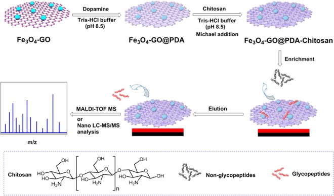
Herein, a new type of chitosan-functionalized hydrophilic magnetic nanocomposites, Fe3O4-GO@PDA-Chitosan (Fig. 1) was assembled via dopamine self-polymerization and Michael addition. Briefly, Fe3O4 NPs were interspersed on the surface of graphene oxide nanocomposites by solvothermal reaction. Polydopamine (PDA)-coated magnetic graphene was prepared via dopamine self-polymerization. Chitosan was grafted on the surface of Fe3O4-GO@PDA nanocomposites via Michael addition. The dopamine self-polymerization and Michael addition reaction occurred simply under mechanical agitation in Tris-HCl buffer. The polydopamine layer and chitosan on the surface of magnetic graphene could effectively enhance hydrophilic enrichment performance of magnetic nanocomposites for N-glycopeptides from the complex biological samples under an external magnetic field. This novel nanocomposite was applied to achieve good selectivity (mixture of bovine serum albumin and horseradish tryptic digests at a molar ration of 10:1), sensitivity (0.4 fmol·μL−1), binding capacity (75 mg·g−1) for N-glycopeptides enrichment.
Figure 1.
Schematic illustration of the procedure for preparation of Fe3O4-GO@PDAand Fe3O4-GO@PDA-Chitosan and N-glycopeptides enrichment.
Experimental Section
Materials
Chitosan (with a deacetylation, degree of 98%) was purchased from Aladdin (Shanghai, China). Horseradish peroxidase (HRP), immunoglobulin G (IgG), peptide-N4-(N-acetyl-β-D-glucosaminyl) asparagine amidase F (PNGase F), bovine serum albumin (BSA), and HPLC-grade acetonitrile (ACN) were purchased from Sigma-Aldrich (USA) (Beijing, China). Dithiothreitol (DTT), urea, ammonium bicarbonate (NH4HCO3) and iodoacetamide (IAA) were obtained purchased from Solarbio (China). Trifluoroacetic acid (TFA) was purchased from J&K (Beijing, China). 2,5-Dihydroxybenzoic acid (DHB) was obtained from TCI (Japan) (Shanghai, China). Trypsin was from Sangon Biotech Co. Ltd. (Shanghai, China). Graphene oxide (GO) was purchase from XF NANO (Nanjing, China). Dopamine chloride and tris(hydroxymethyl)aminomethane (Tris) were obtained from Tianjin Heowns Biotech Co. Ltd. (Tianjin, China). Iron (III) chloride hexahydrate (FeCl3·6H2O), sodium acetate (NaAc), ethylene glycol (EG), acetic acid, hydrogen peroxide (H2O2), ethanol, methanol were purchased from Concord Chemical Ltd. (Tianjin, China). Deionized water (18.25 MΩ cm) was prepared with a Milli-Q water purification system (Millipore, Milford, MA, USA).
Characterization
The morphology, structure and performance of the synthesized nanocomposites were evaluated according to a previous report30. Transmission electron microscope (TEM) images were obtained on a JEM-2100F (Japan) transmission electron microscope. Fourier transform infrared (FT-IR) spectra (4000-400 cm−1) in KBr were recorded using the BRUKER TENSOR 27 Fourier transform infrared spectrophotometer. The crystal structure of the nanocomposites was performed on a Rigaku (Japan) D/max/2500 v/pc with nickel-filtered Cu Kα source. The XRD patterns were collected in the range 3 < 2θ < 80° at a scan rate of 4.0°/min. The X-ray photoelectron spectra were obtained on a Thermo Fischer (USA) ESCALAB 250Xi X-ray photoelectron spectrometer (XPS) with an Mg Kα anode (15 kV, 400 W) at a takeoff angle of 45°. The source X-rays were not filtered and the instrument was calibrated against the C 1 s band at 285 eV. The magnetic properties were analyzed with a LDJ9600-1 (USA) vibrating sample magnetometer (VSM). The hydrophilicity of the nanocomposites was revealed with contact angle analyzer JY-82B (Dingsheng, China). Zeta potential of the nanocomposites was measured by Brook haven ZETAPALS/BI-200SM (USA) at room temperature. Thermogravimetric analysis (TGA) was carried out in nitrogen atmosphere at a heating rate of 10 °C·min−1 from room temperature to 800 °C (NETZCHSTA 449 F3, Germany). Matrix assisted laser desorption/ionization time-of-flight mass spectrometry (MALDI-TOF MS) were performed on a Bruker Auto flex III LRF200-CID instrument (Bruker Daltonics, Germany) with a pulsed nitrogen laser operating at 337 nm in linear positive ion mode and DHB (25 mg·mL−1, ACN/H2O/TFA, 80:19:1, v/v/v) was used as matrix.
Preparation of Fe3O4-GO nanocomposites
Fe3O4-GO NPs were synthesized with a solvothermalmethod29. Typically, GO (48.7 mg), NaAc (1.36 g) and FeCl3·6H2O (152 mg) were dissolved in EG (30 mL) under sonication for 2 h. The homogeneous solution was poured into a Teflon-lined stainless-steel autoclave (50 mL) and maintained at 180 °C for 16 h. With the help of an external magnetic field, the prepared Fe3O4-GO nanocomposites were separated from the reaction solvent and washed with deionized water and ethanol for several times, and dried in vacuum at 40 °C for 3 h.
Preparation of Fe3O4-GO@PDA nanocomposites
The Fe3O4-GO nanocomposites (40 mg) were dispersed in Tris buffer (100 mL, 10 mM, pH 8.5) under sonication, and dopamine chloride (30 mg) was added to the above solution. The mixture was stirred mechanically at 60 °C for 24 h. In an external magnetic field, the prepared Fe3O4-GO@PDA nanocomposites were separated from the reaction solvent and washed with deionized water and ethanol for several times, and dried in vacuum at 40 °C for 3 h.
Degradation of chitosan by hydrogen peroxide
5% H2O2 (100 mL) was added dropwise into the solution of Chitosan (10 g) in 2% acetic acid (200 mL). The mixture was stirred magnetically at 60 °C for 8 h. Then the reaction solution was cooled and passed through two layers of filter paper. The aqueous phase obtained was retained and the pH was adjusted to 10.0 with 1 N NaOH, set aside for 2 h, and again filtrated through two layers of filter paper. After concentration under reduced pressure, the solution was diluted with methanol, set aside for 5 h. The white precipitation was collected via filtration and washed with methanol. The white powder was obtained after drying under vacuum.
Preparation of Fe3O4-GO@PDA-Chitosan nanocomposites
Into the solution of chitosan (120 mg) in Tris-HCl buffer (120 mL, pH 8.5), the Fe3O4-GO@PDA-Chitosan nanocomposites (30 mg) was dispersed under sonication. The mixture was stirred mechanically at room temperature for 24 h. In an external magnetic field, the prepared Fe3O4-GO@PDA-Chitosan nanocomposites were separated from the reaction solvent and washed with deionized water and ethanol for several times, and dried in vacuum at 40 °C for 3 h.
Digestion of proteins
1 mg HRP (human IgG or BSA) was dissolved in 1 mL NH4HCO3 solution (50 mM, pH 8.3) under sonication and denatured by boiling for 15 min in water bath. And then, the denatured proteins were reduced with 3.1 mg DTT at 37 °C for 2 h in water bath and alkylated by 7.2 mg IAA using a shaking table at room temperature in the dark for 40 min. The mixture was incubated with trypsin at an enzyme-protein ratio of 1:25 (w/w) at 37 °C for 16 h. The tryptic digests were stored at −20 °C for later use.
Proteins extracted from human renal mesangial cells (HRMC) were precipitated by trichloroacetic acid and collected by centrifugation. The pellet was dissolved in 100 mM of NH4HCO3. The proteins underwent reduction, alkylation, and enzymolysis in sequence. The resulting digests were desalted using Sep-pak C18 cartridges (Water Ltd., Elstree, UK), evaporated to dryness, and stored at −20 °C for later use.
N-Glycopeptides enrichment under hydrophilic mode
Fe3O4-GO@PDA-Chitosan nancomposites (or Fe3O4-GO@PDA, 40 μg) were rinsed thrice with the loading buffer (ACN/H2O/TFA = 89:10.5:0.5, v/v/v), and then dispersed via ultrasonication in the above buffer (400 μL) containing a determined amount of HRP or IgG digests. After incubation at room temperature for 30 min, the nanocomposites were then washed thrice with the loading buffer. Finally, the N-glycopeptides captured by the Fe3O4-GO@PDA-Chitosan nancomposites (or Fe3O4-GO@PDA) were released by 2 × 13 μL elution buffer (ACN/H2O/TFA = 30:69.9:0.1, v/v/v). The supernatant was collected, lyophilized, re-dissolved in 4 μL elution buffer and analyzed by MALDI-TOF MS.
For the enrichment of N-glycopeptides from digests of human renal mesangial cells (HRMC, kindly donated by Dr. Mingzhen Li (Metabolic Diseases Hospital, Tianjin Medical University, Tianjin, China)), the operation was carried out according to a previous report30. Briefly, 200 μg of the sample was dissolved in 5 mL of loading buffer (ACN/H2O/TFA = 89:10.5:0.5, v/v/v), incubated with 20 mg of Fe3O4-GO@PDA-Chitosan nanocomposites for 1 h, and washed thrice with 2 mL of loading buffer. Then, the trapped glycopeptides were eluted twice with 400 μL of elution buffer, and the elution was evaporated to dryness. The obtained glycopeptides were redissolved in 10 mM of NH4HCO3, and glycan moieties were removed by 1000 unites of PNGase F. The mixture was desalted and enriched using Sep-pak C18 cartridges (Waters Ltd., Elstree, UK), evaporated to dryness, and redissolved prior to anlysis by nano LC-MS/MS.
Results and Discussion
Preparation and characterization of Fe3O4-GO@PDA-Chitosan nanocomposites
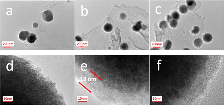
The morphology of Fe3O4-GO, Fe3O4-GO@PDA, Fe3O4-GO@PDA-Chitosan nanocomposites and the thickness of the grafted PDA layer were revealed by transmission electron microscopy (TEM) at the 200 or 10 nanometer scale. As shown in Fig. 2a,d, Fe3O4 nanoparticles were irregularly interspersed on the graphene nanosheets, indicating the successful assembly of Fe3O4-GO nanocomposites. By means of self-polymerization, the PDA layer was coated on the surface of Fe3O4-GO with the thickness of ~18 nm (Fig. 2b,e). The molecular weight of degraded chitosan was determined to be ~1471 g·mol−1 by Gel Permeation Chromatography, so the modification of degraded chitosan on the surface of Fe3O4-GO@PDA nanocomposites was not detected. When the chitosan shell was formed on the surface of Fe3O4-GO@PDA, the obtained Fe3O4-GO@PDA-Chitosan displayed an obvious core-shell structure (Fig. 2c,f).
Figure 2.
TEM images of Fe3O4-GO (a,d), Fe3O4-GO@PDA (b,e) and Fe3O4-GO@PDA-Chitosan (c,f) nanocomposites.
The Zeta potentials of the obtained nanocomposites were detected in acid aqueous solution (H2O/TFA = 99.5:0.5, v/v). The zeta potential of Fe3O4-GO@PDA was 25.70 mV (Fig. 3), which indicated that there were abundant phenolic group and amino groups onto the surface of PDA layer. And after graft of chitosan on the surface of PDA layer, the zeta potential increased to 29.01 mV due to the addition of amine and hydroxyl groups.
Figure 3.
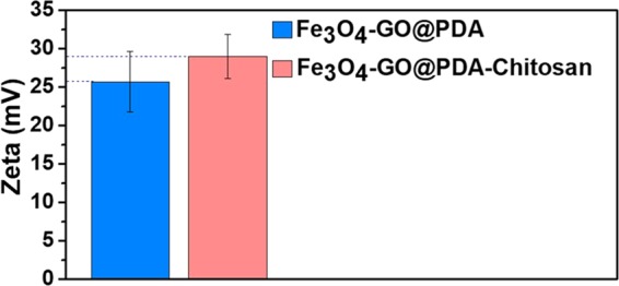
Zeta potential of Fe3O4-GO@PDA and Fe3O4-GO@PDA-Chitosan nanocomposites.
The magnetic properties of the obtained nanocomposites were measured by a vibrating sample magnetometer (VSM) at room temperature. The saturation magnetic (Ms) values of Fe3O4-GO, Fe3O4-GO@PDA and Fe3O4-GO@PDA-Chitosan nanocomposites were 34.06, 19.66, 16.21 emu·g−1, respectively (Fig. S1, Electronical Supporting Materials). Although the saturation magnetization value of Fe3O4-GO@PDA-Chitosan was reduced to 16.21 emu·g−1, the Fe3O4-GO@PDA-Chitosan nanocomposites can be quickly separated within 10 s under an external magnetic field (Fig. S1 inset, Electronical Supporting Materials) and re-dispersed quickly after removal of the magnetic field.
The crystalline phases of Fe3O4-GO, Fe3O4-GO@PDA and Fe3O4-GO@PDA-Chitosan nanocomposites were characterized by the XRD technique (Fig. S2, Electronical Supporting Materials). Six discernible diffraction peaks at 30.06°, 35.36°, 43.16°, 53.42°, 57.08°, 62.50° were index as (220), (311), (400), (422), (511) and (440) in the database of the Joint Committee on Powder Diffraction Standards (JCPDS card: 19-0629). This result indicated that the presence of Fe3O4 nanoparticles in the nanocomposites.
The thermogravimetric analysis (TGA) curves of Fe3O4-GO, Fe3O4-GO@PDA and Fe3O4-GO@PDA-Chitosan nanocomposites are shown in Fig. 4. It can be found that 13.9% weight loss occurred for Fe3O4-GO (Fig. 4a) corresponding to the content of GO. And there were 39.7 and 43.3% weight loss for Fe3O4-GO@PDA (Fig. 4b) and Fe3O4-GO@PDA-Chitosan (Fig. 4c), respectively. From the data of TGA curves, the amount of immobilized chitosan onto the Fe3O4-GO@PDA-Chitosan nanocomposites was calculated to 24.5 μmol·g−1.
Figure 4.
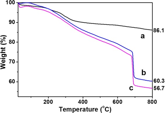
TGA curves of Fe3O4 (a), Fe3O4-GO@PDA (b) and Fe3O4-GO@PDA-Chitosan (c) nanocomposites.
The surface composition of the obtained nanocomposites (Fe3O4-GO, Fe3O4-GO@PDA, Fe3O4-GO@PDA-Chitosan) were characterized by XPS. As shown in Fig. 5, the peaks of Fe 2p, O 1 s, N 1 s and C 1 s were evidently observed. The contact angle of prepared nanocomposites (Fe3O4-GO, Fe3O4-GO@PDA, Fe3O4-GO@PDA-Chitosan) were measured with the powder tableting method (Fig. 6) to evaluate the relative surface hydrophilicity. The contact angle of Fe3O4-GO (a), Fe3O4-GO@PDA (b) and Fe3O4-GO@PDA-Chitosan (c) nanocomposites were 61.0°, 53.5°, and 45.7°, respectively. It indicated that the polydopamine layer coated on the surface of magnetic graphene played a role not only in facile synthesis procedure via Michael addition, but also in hydrophilicity raise; the modification of chitosan improved further the hydrophilicity of the nanocomposites.
Figure 5.
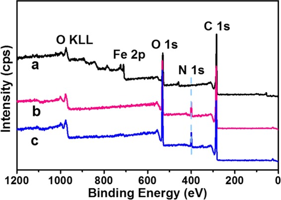
XPS spectrum of Fe3O4 (a), Fe3O4-GO@PDA (b) and Fe3O4-GO@PDA-Chitosan (c) nanocomposites.
Figure 6.
Water contact angles of Fe3O4 (a), Fe3O4-GO@PDA (b) and Fe3O4-GO@PDA-Chitosan (c) nanocomposites.
Glycopeptide enrichment from standard proteins by Fe3O4-GO@PDA-Chitosan nanocomposites
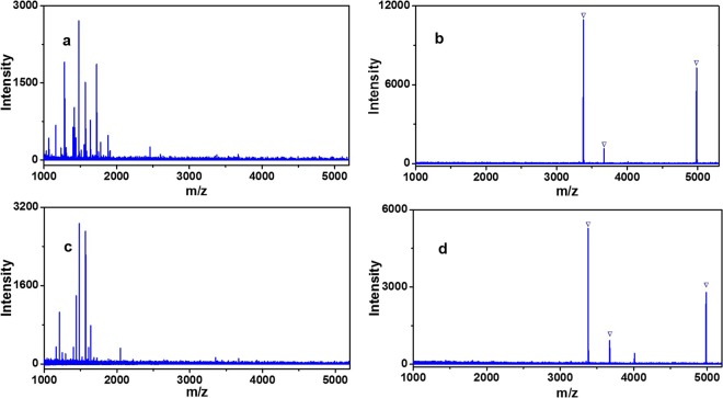
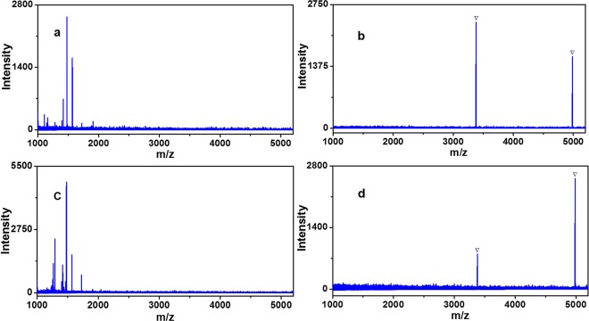
To select the optimal enrichment condition to glycopeptides, three different kinds of loading buffer (89% ACN/H2O, 0.1% TFA; 89% ACN/H2O, 0.5% TFA; 89% ACN/H2O, 1% TFA) were utilized for investigate the effect of capturing glycopeptides from HRP digestion by Fe3O4-GO@PDA-Chitosan nanocomposites and the results were displayed in Fig. S3 (Electronical Supporting Materials). The performance of Fe3O4-GO@PDA-Chitosan nanocomposites for enrichment of N-glycopeptides was the best in the loading buffer (89% ACN/H2O, 0.5% TFA). By taking advantage of the optimized enrichment condition, Fe3O4-GO@PDA-Chitosan nanocomposites were applied to capture N-glycopeptides from standard HRP tryptic digest (50 fmol·μL−1). As shown in Fig. 7a, the direct analysis of HRP digest (50 fmol·μL−1) without enrichment step, the signal peaks of low abundance of N-glycopeptides were completely suppressed, no target analyte was detected. After enrichment by Fe3O4-GO@PDA-Chitosan nanocomposites, the number and signal intensity of glycopeptides were distinctly enhanced, meanwhile, three N-glycopeptides were detected and non-glycopeptides were completely removed (Fig. 7b, detailed information is listed in Table S1, Electronical Supporting Materials). And no glycopeptides peaks were found in the supernatant (Fig. 7c). For comparison, three N-glycopeptides were also identified after enrichment with Fe3O4-GO@PDA nanocomposites, but the signal intensity was weaker and the one signal peak of non-glycopeptide appeared in MS spectra (Fig. 7d).
Figure 7.
MALDI-TOF mass analysis of tryptic digest of HRP (50 fmol·μL−1): (a) before enrichment, (b) eluent and (c) supernatant after enrichment with Fe3O4-GO@PDA-Chitosan nanocomposites; eluent after enrichment with (d) Fe3O4-GO@PDA nanocomposites. The peaks of glycopeptides are marked with  .
.
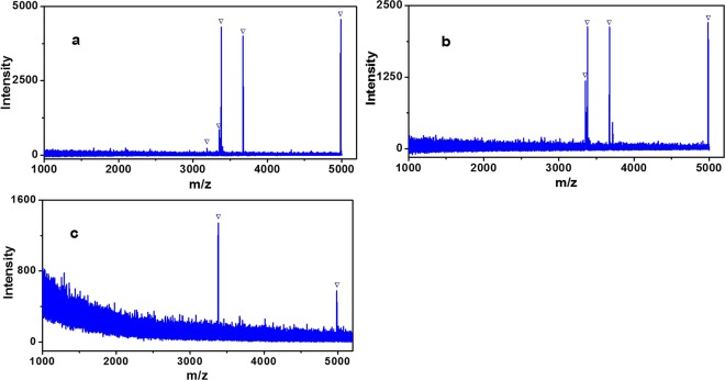
The detection sensitivity of Fe3O4-GO@PDA-Chitosan (or Fe3O4-GO@PDA) nanocomposites for N-glycopeptides enrichment was evaluated with lower concentrations of HRP tryptic digests. When the concentration of HRP digestion decreased to 0.5 fmol·μL−1, five N-glycopeptides were detected after enrichment with Fe3O4-GO@PDA-Chitosan nanocomposites, and four N-glycopeptides and one non-glycopeptide were detected after enrichment with Fe3O4-GO@PDA nanocomposites (Fig. 8a,b). Even at the concentration of 0.4 fmol·μL−1, two N-glycopeptides were still observed after treatment of Fe3O4-GO@PDA-Chitosan nanocomposites (Fig. 8c). The detection sensitivity of Fe3O4-GO@PDA-Chitosan nanocomposites was higher than some reported hydrophilic nanomaterials, such as Au NP-maltose/PDA/Fe3O4-RGO (2.5 fmol·μL−1)29, Fe3O4-DA-Maltose (1.25 fmol·μL−1)31, Fe3O4@mSiO2@G6P (0.5 fmol·μL−1)32, mMOF@Au@GSH (0.5 fmol·μL−1)20.
Figure 8.
MALDI-TOF mass analysis of tryptic digest of HRP (0.5 fmol·μL−1) after enrichment with Fe3O4-GO@PDA-Chitosan (a) and Fe3O4-GO@PDA nanocomposites (b); MALDI-TOF mass analysis of tryptic digest of HRP (0.4 fmol·μL−1) after enrichment with Fe3O4-GO@PDA-Chitosan(c) nanocomposites. The peaks of glycopeptides are marked with  .
.
The enrichment selectivity of Fe3O4-GO@PDA-Chitosan nanocomposites was further investigated, by evaluating the capture performance for N-glycopeptides from the mixture of HRP and BSA tryptic digest with different molar ratios. When the molar ratio of digests mixture of HRP and BSA was 1:1 or 1:10, the MS signals of N-glycopeptides were completely suppressed in direct analysis due to the strong signal suppression from an amount of non-glycopeptides (Fig. 9a,c). By contrast, after enrichment by Fe3O4-GO@PDA-Chitosan nanocomposites, two N-glycopeptides derived from HRP were detected with no non-glycopeptide signals (Fig. 9b). The molar ratio of HRP and BSA was further increased even 1:10, an overwhelming majority of non-glycopeptides were removed and the peaks of N-glycopeptides still absolutely dominated MS spectra (Fig. 9d). The enrichment selectivity of Fe3O4-GO@PDA-Chitosan nanocomposites was higher than some reported hydrophilic nanomaterials functionalized with saccharides, such as Au NP-maltose/PDA/Fe3O4-RGO29, Fe3O4-DA-Maltose31, MagG/Au/Glu33. The results indicated that Fe3O4-GO@PDA-Chitosan nanocomposites have the great potential in N-glycopeptides enrichment from complex biological sample.
Figure 9.
MALDI-TOF mass spectra of mixture of tryptic HRP and BSA before enrichment: (a) 1:1; (c) 1:10. MALDI-TOF MS spectra of the identified glycopeptides enriched by Fe3O4-GO@PDA-Chitosan nanocomposites from the tryptic mixture of HRP and BSA with the molar ratio of (b) 1:1; (d) 1:10. The peaks of glycopeptides are marked with  .
.
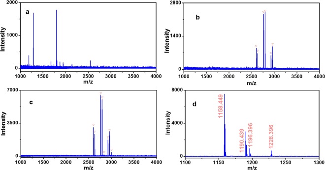
To demonstrate the unbiased enrichment performance of Fe3O4-GO@PDA-Chitosan nanocomposites towards different N-glycan, it was applied to capture N-glycopeptides from IgG digest, which contains different glycoforms from HRP. In direct MS analysis without enrichment, no N-glycopeptides were detected on account of the strong interference from non-glycopeptides (Fig. 10a). Seven N-glycopeptides were identified after enrichment with Fe3O4-GO@PDA nanocomposites (Fig. 10b). For comparison, fifteen N-glycopeptides were detected after enrichment by Fe3O4-GO@PDA-Chitosan nanocomposites (Fig. 10c, detailed information is listed in Table S2, Electronical Supporting Materials), and the signal intensity was stronger in MS spectra. The eluted glycopeptides from Fe3O4-GO@PDA-Chitosan nanocomposites enrichment were deglycosylated by PNGase F and two strong signals of deamidated peptides (at m/z 1158.4 (EEQFN#STFR), 1190.4 (EEQYN#STFR)) were detected (Fig. 10d), which demonstrated that the enriched peptide fragments were N-glycopeptides.
Figure 10.
MALDI-TOF mass spectra of tryptic digests of IgG (5 fmol·μL−1): direct analysis (a); analysis after enrichment with Fe3O4-GO@PDA (b) or Fe3O4-GO@PDA-Chitosan nanocomposites (c), and deglycosylation by PNGase F (d). The peaks of glycopeptides are marked with  .
.
Evaluation of binding capacity of Fe3O4-GO@PDA-Chitosan nanocomposites for N-glycopeptide enrichment
Different masses (20-100 μg) of Fe3O4-GO@PDA-Chitosan nanocomposites were dispersed with ultrasonication for 6 s in the loading buffer (400 μL, V(ACN)/V(H2O)/V(TFA) = 89:10.5:0.5) containing human IgG digest (3 μg). After the enrichment, the elution was analyzed with MALDI-TOF MS. When the MS signal intensity of six selected glycopeptides reach maximum, the total amount of glycopeptides were captured by the nanocomposites. The binding capacity was calculated by the mass ratio of human IgG (3 μg) and the Fe3O4-GO@PDA-Chitosan nanocomposites. As shown in Fig. 11, the N-glycopeptides from human IgG digest (3 μg) were almost bonded onto 40 μg of Fe3O4-GO@PDA-Chitosan nanocomposites, and the binding capacity of the nanocomposites was about 75 mg·g−1, Which was higher than some reported hydrophilic materials modified with saccharides such as Fe3O4@CS MCNCs (17.5 mg·g−1)25 and Fe3O4-DA-Maltose (43 mg·g−1)31. Meanwhile, the binding capacity of Fe3O4-GO@PDA nanocomposites was evaluated, the N-glycopeptides from human IgG digest (3 μg) were almost bonded onto 80 μg of Fe3O4-GO@PDA nanocomposites, and the binding capacity of the nanocomposites was about 37.5 mg·g−1 (Fig. S4, Electronical Supporting Materials).
Figure 11.
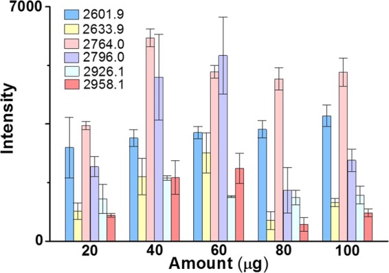
The intensity of six selected N-glycopeptides from tryptic digests of human IgG (3 μg) after enrichment by different amount of Fe3O4-GO@PDA-Chitosan nanocomposites.
The stability and reusability of Fe3O4-GO@PDA-Chitosan nanocomposites
To evaluate the stability and reusability of Fe3O4-GO@PDA-Chitosan nanocomposites, the material was firstly placed in a glass vial for three months at room temperature, and then applied to capture N-glycopeptides from HRP digest for four cycles. As shown in Fig. S5 (Electronical Supporting Materials), the enrichment performance of Fe3O4-GO@PDA-Chitosan nanocomposites in the first time and fourth time was all good, which indicates that the Fe3O4-GO@PDA-Chitosan nanocomposites own good stability and repeatability.
Practical application of Fe3O4-GO@PDA-Chitosan nanocomposites on glycopeptide enrichment
In order to test the practical application of Fe3O4-GO@PDA-Chitosan nanocomposites on glycopeptide enrichment in the complex samples, the Fe3O4-GO@PDA-Chitosan nanocomposites were applied to analyze the N-linked glycopeptides from the human renal mesangial cells (HRMC). HRMC serve as a filtration barrier of the kidney. The injury of mesangial cells could cause diabetic nephropathy, leading end-stage renal disease. Emerging evidence indicates that mesangial cells can be damaged by high glucose, however the mechanism is unclear. In this work, Fe3O4-GO@PDA-Chitosan nanocomposites were used to enrich the N-link glycopeptide from the HRMC which have been treated by high glucose and the glycopeptide enrichment was further identified by HPLC-MS/MS (The details of cell culture, protein extraction and MS/MS data analysis in the Electronical Supporting Materials). Finally, 393 N-linked glycopeptides, representing 195 different glycoproteins and 458 glycosylation sites were identified in HRMC (Table S3, Electronical Supporting Materials).
Conclusion
In summary, the novel chitosan-interspersed magnetic nanocomposites (Fe3O4-GO@PDA-Chitosan) were prepared successfully via a facile two-step method for selective enrichment of N-glycopeptides. Due to the enhanced hydrophilicity by the PDA layer and chitosan, the resulting nanocomposites exhibited good selectivity and sensitivity for N-glycopeptides enrichment from the complex biological samples. The combination of dopamine self-polymerization and Michael addition is a feasible strategy to assemble hydrophilic nanomaterials applied in N-glycopeptides enrichment.
Supplementary information
Acknowledgements
We gratefully appreciate the financial support by the Natural Science Foundation of Tianjin City (No 18JCQNJC09500) and the CAMS Innovation Fund for Medical Sciences (CIFMS, 2017-I2M-3-019 and 2016-I2M-3-022).
Author contributions
Langxing Chen and Changfen Bi conceived the experiments; Changfen Bi and Ye Yuan synthesized and characterized hydrophilic magnetic graphene nanocomposites, conducted the experiments and analyzed the results. Yuran Tu and Jiahui Wu completed the synthesis of degradated chitosan. YuLu Liang evaluated the stability and reusability of Fe3O4-GO@PDA-Chitosan nanocomposites. The manuscript was prepared by Changfen Bi, Langxing Chen, Yiliang Li, Xiwen He and Yukui Zhang. All authors edited the manuscript.
Competing interests
The authors declare no competing interests.
Footnotes
Publisher’s note Springer Nature remains neutral with regard to jurisdictional claims in published maps and institutional affiliations.
Contributor Information
Yiliang Li, Email: liyiliang75@163.com.
Langxing Chen, Email: lxchen@nankai.edu.cn.
Supplementary information
is available for this paper at 10.1038/s41598-019-56944-4.
References
- 1.Jürgen R. Protein N-glycosylation along the secretory pathway: relationship to organelle topography and function, protein quality control, and cell interactions. Chem. Rev. 2002;102:285–303. doi: 10.1021/cr000423j. [DOI] [PubMed] [Google Scholar]
- 2.Hart GW, Copeland RJ. Glycomics hits the big time. Cell. 2010;143:672–676. doi: 10.1016/j.cell.2010.11.008. [DOI] [PMC free article] [PubMed] [Google Scholar]
- 3.Jürgen R, et al. Protein N-glycosylation, protein folding, and protein quality control. Mol. Cells. 2010;30:497–506. doi: 10.1007/s10059-010-0159-z. [DOI] [PubMed] [Google Scholar]
- 4.Parekh RB, et al. Association of rheumatoid arthritis and primary osteoarthritis with changes in the glycosylation pattern of total serum IgG. Nature. 1985;316:452–457. doi: 10.1038/316452a0. [DOI] [PubMed] [Google Scholar]
- 5.Moran FP, et al. Interplay between protein glycosylation pathways in alzheimer’s disease. Sci. Adv. 2017;3:e1601576. doi: 10.1126/sciadv.1601576. [DOI] [PMC free article] [PubMed] [Google Scholar]
- 6.Green RS, et al. Mammalian N-glycan branching protects against innate immune self-recognition and inflammation in autoimmune disease pathogenesis. Immunity. 2007;27:308–320. doi: 10.1016/j.immuni.2007.06.008. [DOI] [PubMed] [Google Scholar]
- 7.Lan Y, et al. Serum glycoprotein-derived N- and O-linked glycans as cancer biomarkers. Am. J. Cancer Res. 2016;6:2390–2415. [PMC free article] [PubMed] [Google Scholar]
- 8.Alley JW, Jr., Mann FB, Novotny VM. High-sensitivity analytical approaches for the structural characterization of glycoproteins. Chem. Rev. 2013;113:2668–2732. doi: 10.1021/cr3003714. [DOI] [PMC free article] [PubMed] [Google Scholar]
- 9.Xiao HP, Chen WX, Smeekens MJ, Wu RH. An enrichment method based on synergistic and reversible covalent interactions for large-scale analysis of glycoproteins. Nat. Commun. 2018;9:1692. doi: 10.1038/s41467-018-04081-3. [DOI] [PMC free article] [PubMed] [Google Scholar]
- 10.Zhang XH, Wang JW, He XW, Chen LX, Zhang YK. Tailor-made boronic acid functionalized magnetic nanoparticles with a tunable polymer shell-assisted for the selective enrichment of glycoproteins/glycopeptides. ACS Appl. Mater. Interfaces. 2015;7:24576–24584. doi: 10.1021/acsami.5b06445. [DOI] [PubMed] [Google Scholar]
- 11.Wu Q, et al. 3-Carboxybenzoboroxole functionalized polyethylenimine modified magnetic graphene oxide nanocomposites for human plasma glycoproteins enrichment under physiological conditions. Anal. Chem. 2018;90:2671–2677. doi: 10.1021/acs.analchem.7b04451. [DOI] [PubMed] [Google Scholar]
- 12.Liu JX, et al. Boronic acid-functionalized particles with flexible three-dimensional polymer branch for highly specific recognition of glycoproteins. ACS Appl. Mater. Interfaces. 2016;8:9552–9556. doi: 10.1021/acsami.6b01829. [DOI] [PubMed] [Google Scholar]
- 13.Tan SY, Wang JD, Han Q, Liang QL, Ding MY. A porous graphene sorbent coated with titanium (IV)-functionalized polydopamine for selective lab-in-syringe extraction of phosphoproteins and phosphopeptides. Microchim. Acta. 2018;185:316. doi: 10.1007/s00604-018-2846-y. [DOI] [PubMed] [Google Scholar]
- 14.Zou XJ, Jie JZ, Yang B. Single-step enrichment of N-glycopeptides and phosphopeptides with novel multifunctional Ti4+-immobilized dendritic polyglycerol coated chitosan nanomaterials. Anal. Chem. 2017;89:7520–7526. doi: 10.1021/acs.analchem.7b01209. [DOI] [PubMed] [Google Scholar]
- 15.Hong YY, et al. Hydrophilic phytic acid-coated magnetic graphene for titanium (IV) immobilization as a novel hydrophilic interaction liquid chromatography-immobilized metal affinity chromatography. Anal. Chem. 2018;90:11008–11015. doi: 10.1021/acs.analchem.8b02614. [DOI] [PubMed] [Google Scholar]
- 16.Bie ZJ, Chen Y, Ye J, Wang SS, Liu Z. Boronate-affinity glycan-oriented surface imprinting: a new strategy to mimic lectins for the recognition of an intact glycoprotein and its characteristic fragments. Angew. Chem. Int. Ed. 2015;54:10211–10215. doi: 10.1002/anie.201503066. [DOI] [PubMed] [Google Scholar]
- 17.Sun XY, Ma R-T, Chen J, Shi Y-P. Magnetic boronate modified molecularly imprinted polymers on magnetite microspheres modified with porous TiO2 (Fe3O4@pTiO2@MIP) with enhanced adsorption capacity for glycoproteins and with wide operational pH range. Microchim. Acta. 2018;185:565. doi: 10.1007/s00604-018-3092-z. [DOI] [PubMed] [Google Scholar]
- 18.Luo J, et al. Double recognition and selective extraction of glycoprotein based on the molecular imprinted graphene oxide and boronate affinity. ACS Appl. Mater. Interfaces. 2017;9:7735–7744. doi: 10.1021/acsami.6b14733. [DOI] [PubMed] [Google Scholar]
- 19.Ma W, et al. Cysteine-functionalized metal-organic framework: facile synthesis and high efficient enrichment of N-linked glycopeptides in cell lysate. ACS Appl. Mater. Interfaces. 2017;9:19562–19568. doi: 10.1021/acsami.7b02853. [DOI] [PubMed] [Google Scholar]
- 20.Liu QJ, Deng CH, Sun NR. Hydrophilic tripeptide-functionalized magnetic metal-organic frameworks for the highly efficient enrichment of N-linked glycopeptides. Nanoscale. 2018;10:12149–12155. doi: 10.1039/C8NR03174F. [DOI] [PubMed] [Google Scholar]
- 21.Xia CS, et al. Two-dimensional MoS2-based zwitterionic hydrophilic interaction liquid chromatography material for the specific enrichment of glycopeptides. Anal. Chem. 2018;90:6651–6659. doi: 10.1021/acs.analchem.8b00461. [DOI] [PubMed] [Google Scholar]
- 22.Sangram KS, et al. Cationic polymers and their therapeutic potential. Chem. Soc. Rev. 2012;41:7147–7149. doi: 10.1039/c2cs35094g. [DOI] [PubMed] [Google Scholar]
- 23.He XM, et al. High strength and hydrophilic chitosan microspheres for the selective enrichment of N-glycopeptides. Anal. Chem. 2017;89:9712–9721. doi: 10.1021/acs.analchem.7b01283. [DOI] [PubMed] [Google Scholar]
- 24.Li YL, Wang JW, Sun NR, Deng C-H. Glucose-6-phosphate-functionalized magnetic microspheres as novel hydrophilic probe for specific capture of N-linked glycopeptides. Anal. Chem. 2017;89:11151–11158. doi: 10.1021/acs.analchem.7b03708. [DOI] [PubMed] [Google Scholar]
- 25.Fang CL, et al. One-pot synthesis of magnetic colloidal nanocrystal clusters coated with chitosan for selective enrichment of glycopeptides. Anal. Chim. Acta. 2014;841:99–105. doi: 10.1016/j.aca.2014.05.037. [DOI] [PubMed] [Google Scholar]
- 26.Wang JX, Li J, Yan GQ, Gao MX, Zhang XM. Preparation of a thickness-controlled Mg-MOFs-based magnetic graphene composite as a novel hydrophilic matrix for the effective identification of the glycopeptide in the human urine. Nanoscale. 2019;11:3701–3709. doi: 10.1039/C8NR10074H. [DOI] [PubMed] [Google Scholar]
- 27.Jiang B, Wu Q, Zhang LH, Zhang YK. Preparation and application of silver nanoparticle-functionalized magnetic graphene oxide nanocomposites. Nanoscale. 2017;9:1607–1615. doi: 10.1039/C6NR09260H. [DOI] [PubMed] [Google Scholar]
- 28.Wan H, et al. A dendrimer-assisted magnetic graphene-silica hydrophilic composite for efficient and selective enrichment of glycopeptides from the complex sample. Chem. Commun. 2015;51:9391–9394. doi: 10.1039/C5CC01980J. [DOI] [PubMed] [Google Scholar]
- 29.Bi CF, Jiang RD, He XW, Chen LX, Zhang YK. Synthesis of a hydrophilic maltose functionalized Au NP/PDA/Fe3O4-RGO magnetic nanocomposite for the highly specific enrichment of glycopeptides. RSC Adv. 2015;5:59408–59416. doi: 10.1039/C5RA06911D. [DOI] [Google Scholar]
- 30.Bi CF, et al. Maltose-functionalized hydrophilic magnetic nanoparticles with polymer brushes for highly selective enrichment of N-linked glycopeptides. ACS Omega. 2018;3:1572–1580. doi: 10.1021/acsomega.7b01788. [DOI] [PMC free article] [PubMed] [Google Scholar]
- 31.Bi CF, et al. Click synthesis of hydrophilic maltose-functionalized iron oxide magnetic nanoparticles based on dopamine anchors for highly selective enrichment of glycopeptides. ACS Appl. Mater. Interfaces. 2015;7:24670–24678. doi: 10.1021/acsami.5b06991. [DOI] [PubMed] [Google Scholar]
- 32.Li YL, Deng CH, Sun NR. Hydrophilic probe in mesoporous pore for selective enrichment of endogenous glycopeptides in biological samples. Anal. Chim. Acta. 2018;1024:84–92. doi: 10.1016/j.aca.2018.04.030. [DOI] [PubMed] [Google Scholar]
- 33.Feng XY, Deng CH, Gao MX, Yan GQ, Zhang XM. Novel synthesis of glucose functionalized magnetic graphene hydrophilic nanocomposites via facile thiolation for high-efficient enrichment of glycopeptides. Talanta. 2018;179:377–385. doi: 10.1016/j.talanta.2017.11.040. [DOI] [PubMed] [Google Scholar]
Associated Data
This section collects any data citations, data availability statements, or supplementary materials included in this article.


