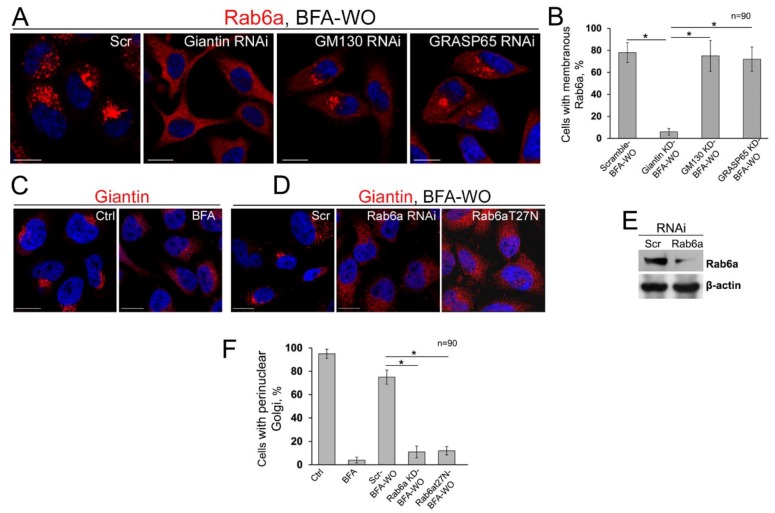
Figure 8.
The overlap of giantin and Rab6a during Golgi biogenesis. (A) Confocal immunofluorescence images of Rab6a in HeLa cells after 60 min of BFA-WO, pretreated with scramble, giantin, GM130, or GRASP65 siRNAs. All confocal images acquired with the same imaging parameters; bars, 10 μm. (B) Quantification of cells with membranous Rab6a in cells presented in (A); n = 90 cells from three independent experiments, results are expressed as a mean ± SD; * p < 0.001. (C) Giantin immunostaining in DMSO- and BFA-treated HeLa cells. (D) Giantin immunostaining in HeLa cells after 60 min of BFA-WO, transfected with scramble, Rab6a siRNAs, and dominant-negative (GDP-bound) Rab6a(T27N). (E) Rab6a W-B of lysates of HeLa cells treated with corresponding siRNAs; β-actin was a loading control. (F) Quantifications of cells with perinuclear Golgi in cells from (C,D); n = 90 cells from three independent experiments, results expressed as a mean ± SD; *, p < 0.001.

