Abstract
BACKGROUND:
Uncaria gambir (local name: gambir) is a plant native to Sumatera, Malaya and Borneo. This plant is potential as local wisdom for therapeutics. In Sumatera, gambir was used as a traditional treatment for fever, diarrhoea, diabetics and wound healing.
AIM:
To explore the efficacy of gambir extract on TNF alpha level, prostaglandin E2 level, lesson area, body weight, lipid profile and leptin level in Wistar rat-model gastritis.
METHODS:
This study was an experimental study, with a pre-post-test control group design. The subjects in this study were 30 male rats, 8 weeks old, weight 150-200 gram. Rats were administered with gambir extract at the dose of 20, 40 and 80 mg/kg BW/day for 3 days. Gambir was extracted by maceration methods. Statistical analysis was performed by SPSS 18.
RESULTS:
Gambir extract at the dose of 80 mg/kg BW exhibited the highest efficacy in reducing TNF alpha level, lesion area and increasing prostaglandin E2 level compared to gambir extract at doses of 20 mg/kg BW, 400 mg/kg BW, negative control, and positive control.
CONCLUSION:
Gambir extract was effective in reducing TNF alpha level, lesson area, and increasing prostaglandin E2 level in Wistar rat-model gastritis.
Keywords: Gambir extract, Uncaria gambir, TNF alpha, Prostaglandin E2, Gastritis
Introduction
Gastritis is an inflammatory condition that occurs in the gastric mucosa. The symptoms resulting from gastric mucosa inflammation are burning sensation and discomfort in the stomach. Gastritis is one of the serious health problems in the world [1]. The incidence of chronic gastritis increases with age. In Western countries, the prevalence of gastritis sufferers is almost 80% in the elderly population of sixty years old and predicted to become 100% in the population of seventy years old. Chemical irritants of gastric mucosae, such as alcohol and aspirin, induce chemical injury in gastric mucosa. It is one of the serious exogenous factors that induces gastric mucosa inflammation that will lead to gastritis [2]. Anti-gastritis drugs, such as antacids, PPI or H2 antagonists, are oral medications commonly used in the management of gastritis. The undesirable side effects of using oral anti-gastritis drugs are considerable, therefore exploring new drug compounds from natural ingredients is expected to be a solution to the discovery and development of new drugs with better efficacy and safety [3], [4], [5].
Indonesia is one of the countries with a rich biodiversity of medicinal plants. Gambir (Uncaria gambir) from the Rubiaceae family is one of Indonesia native medicinal plants [6]. Wide varieties of the study had been conducted to explore the potential effect of Gambir [7], [8], [9]. This study aimed to assess the potential of gambir extract in treating gastritis in vivo, which would further explore the effect of extracts on TNF-α and prostaglandin E2 so that new standardised herbs could be obtained in the management of gastritis.
Material and Methods
The study design was an experimental study, with post-test control group design. The study had been approved by the Bioethics Committee, Faculty of Medicine, Universitas Sriwijaya (189/kpt-fkunsri/rsmh/2017).
Gambir were provided by Indonesia Traditional Herbal Research Center, Tawangmangu, Central Java, Indonesia. Gambir was washed, dried and drilled, followed by maceration method by ethanol 96%, evaporated and gambir extract was obtained. Phytochemical analysis through Thin Layer Chromatography (TLC) was performed to obtain the component information of gambir extract.
Thirty rats (Eureka Research Laboratory, Indonesia) were used in this study. Inclusion criteria were male Wistar rats, eight weeks old, weight 150-200 gram and healthy. Rats were divided into 5 groups; every group consisted of 6 rats. Gastritis in rats was induced by administrating ethanol 96% for 3 days. Rats were treated, group 1: aqua dest 1 mL (negative control), group 2: ranitidine 20 mg/kg BW (positive control), group 3: gambir extract 20 mg/kg BW, group 4: gambir extract 40 mg/kg BW, and group 5: gambir extract 80 mg/kg BW. Treatment was carried out for 3 days.
Following the performed treatments, rats were killed by anaesthesia. Rats gastric organ was evacuated, washed and assayed for lesion area size by callipers, and the tissue was obtained for homogenisation by centrifugation 3000 rpm, at -4°C, for 20 minutes. The supernatant was collected and used to assay TNF alpha and prostaglandin E2 level using ELISA methods. The procedure of ELISA was based on the assay protocol on the manual (Cloud-Clone Corp®, Texas, USA). The sample solution was bottled using the capillary tube on Silica GF silent phase 254 which was activated by heating at 105°C-110°C for 1 hour then eluted with methanol: chloroform phase (1: 39) v/v. Chromatogram results were observed in UV254 nm. Spotting was detected by H2SO4 spray.
The statistical analysis was done by SPSS 18.0 (SPSS Inc., Chicago, Illinois, USA). Data were assessed for bivariate and multivariate analysis. T-test was used for bivariate analysis and pos hoc test for multivariate analysis.
Results
As shown in Table 1, gambir extract at the dose of 80 mg/kg BW was the most effective in reducing lesion area size, TNF alpha level and increasing prostaglandin E2 level compared to gambir extract at the dose of 20 mg/kg BW, 40 mg/kg BW, negative control and positive control. Gambir extract showed dose-dependent manner efficacy in reducing lesion area size, TNF alpha level and increasing prostaglandin E2 level. Gambir extracts at the dose of 80 mg/kg BW possesses higher efficacy to reduce lesion area size, TNF alpha level and increase prostaglandin E2 level compared to gambir extract at doses of 20 mg/kg BW and 40 mg/kg BW. The increasing doses of the extract were positively related to the efficacy to reduce lesion area size, TNF alpha level and increase prostaglandin E2 level, thus affecting the gastric mucosal lesion.
Table 1.
The Efficacy of Gambir Extract on Lesion Area, TNF Alpha and Prostaglandin E2
| Variable | Group | Mean ± SD | p-value* |
|---|---|---|---|
| Lession area (mm) | Negative control | 2.72 ± 0,83 | 0.01 |
| Positive control | 0.42 ± 0,13 | - | |
| Extract 20 mg/kg BW | 0.58 ± 0.22 | 0.01 | |
| Extract 40 mg/kg BW | 0.43 ± 0.24 | 0.56 | |
| Extract 80 mg/kg BW | 0.29 ± 0.10 | 0.02 | |
| TNF alpha (pg/mL) | Negative control | 212 ± 10.09 | 0.01 |
| Positive control | 170 ± 13,76 | - | |
| Extract 50 mg/kg BW | 198 ± 20.53 | 0.01 | |
| Extract 100 mg/kg BW | 169 ± 10.78 | 0.43 | |
| Extract 200 mg/kg BW | 149 ± 10.84 | 0.01 | |
| Prostaglandin E2 (pg/mL) | Negative control | 28.78 ± 7.87 | 0.01 |
| Positive control | 60,65 ± 11.09 | - | |
| Extract 50 mg/kg BW | 49.23 ± 7.34 | 0.01 | |
| Extract 100 mg/kg BW | 61.23 ± 5.87 | 0.34 | |
| Extract 200 mg/kg BW | 73.13 ± 6.35 | 0.02 |
Paired T test; VS positive control; p = 0.05.
Phytochemical Analysis
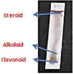
Qualitative test of phytochemical component performed on gambir extract exhibited that gambir contained an alkaloid, steroid/ternoid (essential oil), and flavonoid components.
Figure 1.

Thin Layer Chromatography Analysis of Gambir Extract
Discussion
Gambir extract showed dose-dependent manner efficacy in reducing lesion area size, TNF alpha level and increasing prostaglandin E2 level. Gambir extracts at the dose of 80 mg/kg BW possesses higher efficacy to reduce lesion area size, TNF alpha level and increase prostaglandin E2 level compared to gambir extract at doses of 20 mg/kg BW and 40 mg/kg BW. The increasing doses of the extract were positively related to the efficacy to reduce lesion area size, TNF alpha level and increase prostaglandin E2 level, thus affecting the gastric mucosal lesion in rats [10], [11]. Based on the phytochemical analysis, gambir extract contained an alkaloid, steroid and flavonoid. Quercetin (one of flavonoid) supplementation was reported to reduce stress oxidative in hypertensive patients [12]. Its antioxidant activity may also suppress the elevation of oxidant, MDA in diet-induced obesity rat models [13], [14]. Quercetin was reported to downregulate the expression of NfKB, TNF alpha and other proinflammatory cytokines. Flavonoid had the potential to reduce the damaged of the gastric mucosal membrane by inhibiting apoptosis and reducing anti-inflammatory activity. Flavonoid had the efficacy to inhibit apoptosis in 3T3-L1 preadipocytes by decreasing the mitochondria membrane potential, downregulating expression of B-cell lymphoma 2 (Bcl-2) and poly (ADP-ribose) polymerase (PARP), and activating Bcl-2 homologous antagonist/killer (Bak), Bcl-2-associated X protein (Bax), and cysteine-dependent aspartate-directed proteases 3 (caspase 3) [15], [16]. Gambir extract was the potential to inhibit inflammation in the gastric mucosal membrane by decreasing the inflammatory process through increased prostaglandin E2. Prostaglandin E2 plays a pivotal role to protect gastric mucosal membrane [17].
Prostaglandin E2 is a prostaglandin with complex biological activity, which is involved in GI-RI inflammation [18]. PGE2 synthase is the last key enzyme in the synthesis of prostaglandin E2. Prostaglandin E2 is the prostaglandin with the highest abundance and greatest distribution in the human body. It serves a major role in inflammation as a pain and fever mediator during inflammation. Additionally, it may induce vasodilation and microvascular leakage [19]. As a type of unsaturated fatty acid, prostaglandin E2 is predominantly composed of 20 carbon atoms. It has the basic structure of one five-carbon ring and two side chains. Arachidonic acid is synthesised into prostaglandin E2 under the catalysis of cyclooxygenase (COX) and prostaglandin synthase [20]. Prostaglandin E2 escapes through facilitated diffusion and binds to E-prostanoid 1 – 4 in an autocrine or paracrine manner. In this way, it may alter the levels of intracellular second messengers, and send signals to cells, causing a series of physiological or pathophysiological changes [18].
Cyclooxygenase prevents the conversion of arachidonic acid into prostaglandin E2. Therefore, it may quickly alleviate active substance-induced inflammation. As a result, it may reduce the excitability of the peripheral and central pain-sensing conduction system [21]. Thus, COX-2 serves anti-inflammatory and analgesic functions. Prostaglandins may promote the secretion of gastric fluid and bicarbonate. In this manner, they can protect the gastric mucosal barrier, promote the renewal of gastric mucosal cells and improve mucosal blood flow. Furthermore, COX-2 may stimulate the active transport process of cells, activate adenylate cyclase and stabilise the lysosome. It may also maintain the level of mucosal thiol compounds and stimulate surface-active phospholipids in gastric mucosa [22].
TNF-α, the major pro-inflammatory cytokine released from the migrated macrophages, plays an important role in the pathogenesis of gastric ulcers through stimulation of intercellular adhesion molecule (ICAM)-1 expression on vascular endothelial cells which increases leukocyte adhesion to the endothelial surface on post-capillary venules and promotes transendothelial migration of leukocytes to inflammatory sites [23]. TNF-α also increases intracellular oxidative stress and up-regulation of cytokine-induced neutrophil chemoattractant (CINC)-1 mRNA and protein in gastric epithelial cells [24].
In conclusion, the gambir extract exhibited efficacy in increasing gastric mucosal integrity through reducing lesion area size, TNF alpha level and increasing prostaglandin E2 level.
Footnotes
Funding: This research did not receive any financial support
Competing Interests: The authors have declared that no competing interests exist
References
- 1.Kumar V, Abbas AK, Fausto N, Aster JC. Robbins and Cotran Pathologic Basis of Disease. 8th edition. Philadelphia, USA: Saunders Elsevier; 2011. [Google Scholar]
- 2.Liu X, Chen Z, Mao N, Xie Y. The protective of hydrogen on stress-induced gastric ulceration. Int Immunopharmacol. 2012;13(2):197–203. doi: 10.1016/j.intimp.2012.04.004. https://doi.org/10.1016/j.intimp.2012.04.004 PMid:22543062. [DOI] [PubMed] [Google Scholar]
- 3.Choi JI, Raghavendran HRB, Sung N, Kim JH, Chun BS, Ahn DH, et al. Effect of nigella sativa on aspirin-induced stomach ulceration in rats. Chemico-Biological Interactions. 2010;183(1):249–54. doi: 10.1016/j.cbi.2009.09.015. https://doi.org/10.1016/j.cbi.2009.09.015 PMid:19788892. [DOI] [PubMed] [Google Scholar]
- 4.Thong-Ngam D, Choochuay S, Patumraj S, Chayanupatkul M, Klaikaew N. Curcumin prevents indomethacin-induced gastropathy in rats. World J Gastroenterol. 2012;18(13):1479–84. doi: 10.3748/wjg.v18.i13.1479. https://doi.org/10.3748/wjg.v18.i13.1479 PMid:22509079 PMCid:PMC3319943. [DOI] [PMC free article] [PubMed] [Google Scholar]
- 5.Kumar A, Chomwal R, Kumar P, Sawal R. Anti-inflammatory and wound healing activity of Curcuma aromatica salisb extract and its formulation. J Chem Pharm Res. 2009;1(1):304–10. [Google Scholar]
- 6.Hussin MH, Kassim MJ. The corrosion inhibition and adsorption behavior of Uncaria gambir extract on mild steel in 1 M HCl. Mater Chem Phys. 2011;125(3):461–8. https://doi.org/10.1016/j.matchemphys.2010.10.032. [Google Scholar]
- 7.Amir M, Mujeeb M, Khan A, Ashraf K, Sharma D, Aqil M. Phytochemical analysis and in vitro antioxidant activity of Uncaria gambir. Int J Green Pharm. 2012;6(1):67–72. https://doi.org/10.4103/0973-8258.97136. [Google Scholar]
- 8.Anggraini T, Akihiro T, Yoshino T, Itani T. Antioxidative activity and catechin content of four kinds of Uncaria gambir extracts from West Sumatra Indonesia. Afr J Biochem Res. 2011;5(1):33–8. [Google Scholar]
- 9.Widiyarti G, Sundowo A, Hanafi M. The free radical scavenging and anti-hyperglycemic activities of various gambirs available in Indonesian market. Makara Sains. 2011;15(2):129–34. https://doi.org/10.7454/mss.v15i2.1062. [Google Scholar]
- 10.Yoshikawa T, Naito Y, Ueda S, Oyamada H, Takemura T, Yoshida N, Sugino S, Kondo M. Role of oxygen-derived free radicals in the pathogenesis of gastric mucosal lesions in rats. J Clin Gastroenterol. 1990;12(1):S65–71. doi: 10.1097/00004836-199001001-00012. https://doi.org/10.1097/00004836-199001001-00012 PMid:2212551. [DOI] [PubMed] [Google Scholar]
- 11.Naito Y, Yoshikawa T, Matsuyama K, Yagi N, Arai M, Nakamura Y, et al. Effects of oxygen radical scavengers on the quality of gastric ulcer healing in rats. J Clin Gastroenterol. 1995;21(1):S82–6. [PubMed] [Google Scholar]
- 12.Eseberri I, Miranda J, Lasa A, Churruca I, Portillo MP. Doses of quercetin in the range of serum concentrations exert delipidating effects in 3T3-L1 preadipocytes by acting on different stages of adipogenesis, but not in mature adipocytes. Oxid Med Cell Longev. 2015;2015:480943. doi: 10.1155/2015/480943. https://doi.org/10.1155/2015/480943 PMid:26180590 PMCid:PMC4477249. [DOI] [PMC free article] [PubMed] [Google Scholar]
- 13.Kleemann R, Verschuren L, Morrison M, Zadelaar S, van Erk M, Wielinga PY, et al. Anti-inflammatory, anti-proliferative and anti-atherosclerotic effects of quercetin in human in vitro and in vivo models. Atherosclerosis. 2011;218(1):44–52. doi: 10.1016/j.atherosclerosis.2011.04.023. https://doi.org/10.1016/j.atherosclerosis.2011.04.023 PMid:21601209. [DOI] [PubMed] [Google Scholar]
- 14.Yang JY, Della-Fera MA, Rayalam S, Ambati S, Hartzell DL, Park HJ, et al. Enhanced inhibition of adipogenesis and induction of apoptosis in 3T3-L1 adipocytes with combinations of resveratrol and quercetin. Life Sci. 2008;82:1032–9. doi: 10.1016/j.lfs.2008.03.003. https://doi.org/10.1016/j.lfs.2008.03.003 PMid:18433793. [DOI] [PubMed] [Google Scholar]
- 15.Hsu CL, Yen GC. Induction of cell apoptosis in 3T3-L1 pre-adipocytes by flavonoids is associated with their antioxidant activity. Mol Nutr Food Res. 2006;50:1072–9. doi: 10.1002/mnfr.200600040. https://doi.org/10.1002/mnfr.200600040 PMid:17039455. [DOI] [PubMed] [Google Scholar]
- 16.Yamamoto Y, Oue E. Antihypertensive effect of quercetin in rats fed with a high-fat high-sucrose diet. Biosci Biotechnol Biochem. 2006;70(4):933–9. doi: 10.1271/bbb.70.933. https://doi.org/10.1271/bbb.70.933 PMid:16636461. [DOI] [PubMed] [Google Scholar]
- 17.Villegas I, La Casa C, de La Lastra CA, Motilva V, Herrer'ıas JM, Mart'ın MJ. Mucosal damage induced by preferential COX-1 and COX-2 inhibitors:role of prostaglandins and inflammatory response. Life Sci. 2004;74(7):873–84. doi: 10.1016/j.lfs.2003.07.021. https://doi.org/10.1016/j.lfs.2003.07.021 PMid:14659976. [DOI] [PubMed] [Google Scholar]
- 18.Del Carmen S, de Moreno de LeBlanc A, LeBlanc JG. Development of a potential probiotic yoghurt using selected anti-inflammatory lactic acid bacteria for prevention of colitis and carcinogenesis in mice. J Appl Microbiol. 2016;121:821–830. doi: 10.1111/jam.13213. https://doi.org/10.1111/jam.13213 PMid:27341191. [DOI] [PubMed] [Google Scholar]
- 19.Negroni A, Prete E, Vitali R, Cesi V, Aloi M, Civitelli F, Cucchiara S, Stronati L. Endoplasmic reticulum stress and unfolded protein response are involved in paediatric inflammatory bowel disease. Dig Liver Dis. 2014;46:788–794. doi: 10.1016/j.dld.2014.05.013. https://doi.org/10.1016/j.dld.2014.05.013 PMid:24953208. [DOI] [PubMed] [Google Scholar]
- 20.Roblin X, Boschetti G, Williet N, Nancey S, Marotte H, Berger A, Phelip JM, Peyrin-Biroulet L, Colombel JF, Del Tedesco E. Azathioprine dose reduction in inflammatory bowel disease patients on combination therapy:An open-label, prospective and randomised clinical trial. Aliment Pharmacol Ther. 2017;46:142–149. doi: 10.1111/apt.14106. https://doi.org/10.1111/apt.14106 PMid:28449228. [DOI] [PubMed] [Google Scholar]
- 21.Brzozowski T, Konturek PC, Konturek SJ, Sliwowski Z, Drozdowicz D, Stachura J, Pajdo R, Hahn EG. Role of prostaglandins generated by cyclooxygenase-1 and cyclooxygenase-2 in healing of ischemia-reperfusion-induced gastric lesions. Eur J Pharmacol. 1999;385:47–61. doi: 10.1016/s0014-2999(99)00681-0. https://doi.org/10.1016/S0014-2999(99)00681-0. [DOI] [PubMed] [Google Scholar]
- 22.Tlaskalová-Hogenová H, Stěpánková R, Kozáková H, Hudcovic T, Vannucci L, Tučková L, Rossmann P, Hrnčíř T, Kverka M, Zákostelská Z, et al. The role of gut microbiota (commensal bacteria) and the mucosal barrier in the pathogenesis of inflammatory and autoimmune diseases and cancer:Contribution of germ-free and gnotobiotic animal models of human diseases. Cell Mol Immunol. 2011;8:110–120. doi: 10.1038/cmi.2010.67. https://doi.org/10.1038/cmi.2010.67 PMid:21278760 PMCid:PMC4003137. [DOI] [PMC free article] [PubMed] [Google Scholar]
- 23.Kast RE. Tumor necrosis factor has positive and negative self regulatory feed back cycles centered around cAMP. Int J Immunopharmacol. 2000;22(11):1001–1006. doi: 10.1016/s0192-0561(00)00046-1. https://doi.org/10.1016/S0192-0561(00)00046-1. [DOI] [PubMed] [Google Scholar]
- 24.Konturek PC, Duda A, Brzozowski T, Konturek SJ, Kwiecien S, Drozdowicz D, et al. Activation of genes for superoxide dismutase, interleukin-1beta, tumor necrosis factor-alpha, and intercellular adhesion molecule-1 during healing of ischemia-reperfusion-induced gastric injury. Scand J Gastroenterol. 2000;35(5):452–63. doi: 10.1080/003655200750023697. https://doi.org/10.1080/003655200750023697 PMid:10868446. [DOI] [PubMed] [Google Scholar]


