. 2019 Dec 19;8:e50279. doi: 10.7554/eLife.50279

Figure 7. Importance of the common activation pathway in pathophysiological and biological contexts.

(a) Comparison of disease-associated mutations in the common activation pathway (further decomposed into layers 1–4), ligand-binding pocket, G-protein-coupling region and other regions. Red line denotes the mean value. (b) Mapping of disease-associated mutations in class A GPCRs to the common activation pathway. (c) Key roles of the residues constituting the common activation pathway have been reported in numerous experimental studies on class A GPCRs. Two hundred seventy two (272) CAMs/CIMs from 41 receptors were mined from the literature for the 14 hub residues (i.e. residues that have more than one edges in the pathway).

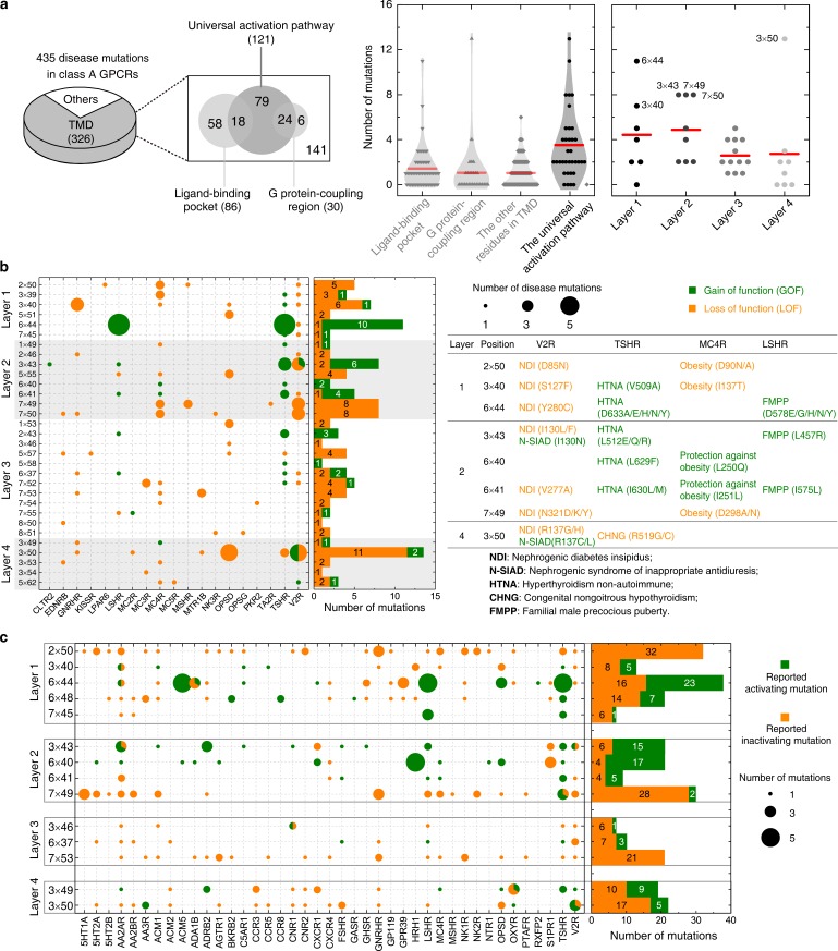
Figure 7—source data 1. Constitutively activating/inactivating mutations for the 14 hub residues in the common activation pathway.

Figure 7.

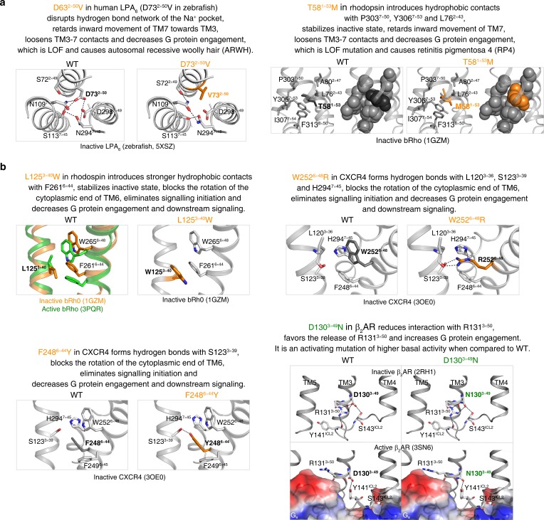
Figure 7—figure supplement 1. The common activation pathway can be used to mechanistically interpret disease-associated mutations and CAMs/CIMs.

Figure 7—figure supplement 1.

(a) Pathway-guided mechanistic interpretations of two disease mutations. (b) Pathway-guided mechanistic interpretations of four CAMs/CIMs.
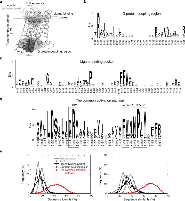
Figure 7—figure supplement 2. Residues in the common activation pathway are more conserved than other functional regions of GPCR.

Figure 7—figure supplement 2.

(a) Illustration of different functional regions of GPCR. (b-d) Sequence pattern of the G protein-coupling region (b), ligand-binding pocket (c) and the common activation pathway (d). (e) Distribution of sequence identity (left) and similarity (right) for functional regions across 286 non-olfactory class A receptors.