

Abstract
Objective
To assess the changes in alar base width in patients who underwent Lefort 1 osteotomy with anterior maxillary osteotomy (AMO), where conventional alar cinch suturing was done to control the alar base widening.
Materials and Methods
This study was conducted on pre-operative and post-operative photographs of 100 female patients aged between 18 and 30 years who underwent Lefort 1 osteotomy with AMO. The derived values were then compared and analysed using t test.
Results
The mean alar base width pre-operatively was 14.11 mm and post-operatively was 15.28 mm. The mean increase in alar base width was 1.176 mm. The result indicated a definitive change between pre-operative and post-operative alar base widths with mean increase in width of 1.176 mm (P = 0.000), which is clinically not very significant.
Conclusion
The effect of Lefort 1 osteotomy on the alar base can be well controlled by the conventional alar cinch suturing without any additional anchorage on the ANS with predictable results.
Keywords: Alar, Lefort, Osteotomy
Introduction
Lefort 1 osteotomy is a versatile procedure and a work horse of orthognathic surgery used to correct mid-face deformities in all three dimensions. Lefort 1 osteotomy and subsequent repositioning of the maxilla invariably lead to widening of the alar base depending on the skeletal movement performed and is due to the detachment of the subnasal soft tissue and muscles during the osteotomy which may not be reattached appropriately at the correct location [1]. Various methods have been described in the literature to control this widening. Alar base cinch suturing, modified cinch suturing by anchoring the sutures over the anterior nasal spine or deepening and creation of the pyriform floor contour are few techniques used alone or in combination to control the alar base flare in cases of Lefort 1 osteotomy with each method having its own advantages and disadvantages.
Presented here is the result of photographic analysis done on 100 female patients who underwent Lefort 1 osteotomy with anterior maxillary osteotomy (AMO) to assess the changes in alar base width pre-operatively and post-operatively, where conventional alar cinch suturing was done to control the alar base widening.
Methodology
This study was conducted on pre-operative and post-operative photographs of 100 female patients aged between 18 and 30 years who underwent Lefort 1 osteotomy with AMO.
The pre-operative and post-operative alar base widths were measured on the patient’s photograph using a divider and a scale. The intercanthal distance was measured from one medial canthus to the other, and the alar width was measured at the base of the alae. The photographs were taken in a studio set-up, with the patient 6 feet away from the camera in a sitting posture with head positioned naturally/neutrally (FH plane parallel to the floor). The camera was at the level of nose. No positioning device was used during photography. Since the pre-operative and post-operative alar base widths were not recorded earlier, we did a retrospective, photographic study to analyse the changes in the alar base following Lefort 1 AMO with cinch sutures. To standardise the values, these measurements were equated to the intercanthal distance which remains unchanged on both pre-operative and post-operative photographs for each patient. This was then equated to the mean alar base width (32 mm) in Indian population (Fig. 1). The derived values were then compared and analysed using t test.
Fig. 1.

Methodology
Statistical Analysis
The Student’s t test was used to compare the differences between the pre-operative and post-operative alar base widths. SPSS version 17.0 software (SPSS Inc., Chicago, IL, USA) was used for all analyses. Significance was set at P < 0.05.
Result
One hundred female patients who underwent a maxillary Le Fort I osteotomy with AMO aged between 18 and 30 years were studied. The mean age of the patients was 24.2 years.
The mean alar base width pre-operatively was 14.11 mm and post-operatively was 15.28 mm. The mean increase in alar base width was 1.176 mm. The result indicated a definitive change between pre-operative and post-operative alar base widths with mean increase in width of 1.176 mm (P = 0.000), which is clinically not very significant (Table 1, Fig. 2).
Table 1.
Mean pre-operative and post-operative alar base widths
| Alar base | Mean | SD | SE of mean | Mean difference | t | P value |
|---|---|---|---|---|---|---|
| Pre-op alar base | 14.11 | 2.45 | 0.24 | − 1.176 | − 5.665 | 0.000* |
| Post-op alar base | 15.28 | 2.35 | 0.24 |
P value less than 0.05 indicates that the results are statistically significant
Fig. 2.

Comparison of mean pre-operative and post-operative alar base widths
The average movement of maxilla was 7.29 mm (measurements were made on the pre-operative and post-operative standardised cephalographs). Range was 5–15 mm. The amount of maxillary vertical impaction and the corresponding change in alar base are shown in Table 2. (–) value in the alar base width change indicates the widening of the alar base.
Table 2.
Maxillary movement versus alar base change
| Sl no. | Maxillary movement (mm) | Alar base difference (mm) | Sl no. | Maxillary movement (mm) | Alar base difference (mm) |
|---|---|---|---|---|---|
| 1 | 5 | − 0.3 | 51 | 7 | − 1 |
| 2 | 5 | 0 | 52 | 7 | − 2.2 |
| 3 | 5 | − 0.8 | 53 | 7 | 1.3 |
| 4 | 5 | − 3.6 | 54 | 7 | − 0.9 |
| 5 | 5 | − 0.9 | 55 | 7 | 0 |
| 6 | 5 | − 1.2 | 56 | 7 | 0 |
| 7 | 5 | − 0.5 | 57 | 7 | − 2.5 |
| 8 | 5 | − 0.7 | 58 | 7 | 0.9 |
| 9 | 5 | − 2.7 | 59 | 7 | − 0.5 |
| 10 | 5 | − 1.4 | 60 | 7 | − 2.2 |
| 11 | 5 | − 2.5 | 61 | 7 | 2.9 |
| 12 | 5 | − 1.5 | 62 | 8 | − 3.9 |
| 13 | 5 | 1 | 63 | 8 | − 0.3 |
| 14 | 5 | 0.7 | 64 | 8 | − 0.52 |
| 15 | 5 | − 2.1 | 65 | 8 | − 2.6 |
| 16 | 5 | 1.5 | 66 | 8 | − 2.6 |
| 17 | 5 | 0 | 67 | 8 | − 1.3 |
| 18 | 5 | − 5.3 | 68 | 8 | − 1.4 |
| 19 | 5 | − 1.4 | 69 | 8 | − 1.3 |
| 20 | 5 | − 1.9 | 70 | 8 | − 1.9 |
| 21 | 5 | − 0.2 | 71 | 8 | − 0.9 |
| 22 | 5 | − 1.6 | 72 | 8 | − 2.2 |
| 23 | 5 | − 2.3 | 73 | 8 | − 3.5 |
| 24 | 5 | 2.5 | 74 | 8 | 0.4 |
| 25 | 5 | − 3.5 | 75 | 8 | 1.2 |
| 26 | 5 | − 1.2 | 76 | 8 | − 1.1 |
| 27 | 5 | 0.4 | 77 | 8 | − 1.8 |
| 28 | 5 | 4.1 | 78 | 9 | − 3.1 |
| 29 | 6 | 0 | 79 | 9 | − 2.3 |
| 30 | 6 | − 2.1 | 80 | 9 | − 1.2 |
| 31 | 6 | − 1.2 | 81 | 9 | − 2.6 |
| 32 | 6 | 0.4 | 82 | 9 | − 2.5 |
| 33 | 6 | − 3 | 83 | 9 | − 2.6 |
| 34 | 6 | − 0.6 | 84 | 9 | − 2.1 |
| 35 | 6 | − 3.3 | 85 | 9 | − 3 |
| 36 | 6 | 0.9 | 86 | 9 | − 1.8 |
| 37 | 6 | − 2.7 | 87 | 10 | 0 |
| 38 | 6 | − 2.1 | 88 | 10 | − 1.8 |
| 39 | 6 | − 0.9 | 89 | 10 | − 1 |
| 40 | 7 | − 1.4 | 90 | 10 | 1 |
| 41 | 7 | − 1.7 | 91 | 10 | − 4.4 |
| 42 | 7 | 3.6 | 92 | 10 | − 2.6 |
| 43 | 7 | − 2.3 | 93 | 10 | 2.7 |
| 44 | 7 | − 0.2 | 94 | 11 | − 1.8 |
| 45 | 7 | − 4.8 | 95 | 11 | − 1.4 |
| 46 | 7 | − 0.6 | 96 | 11 | − 2.9 |
| 47 | 7 | − 3.3 | 97 | 12 | − 4 |
| 48 | 7 | 0 | 98 | 15 | − 1.8 |
| 49 | 7 | − 0.5 | 99 | 15 | − 0.9 |
| 50 | 7 | − 0.9 | 100 | 15 | − 3.4 |
Since cinch sutures were used, there was no significant correlation between the amount of maxillary movement and the corresponding change in the alar base.
Discussion
Lefort 1 osteotomy with AMO is indicated mainly for dentoalveolar protrusion or for correction of anterior open bite. Movement of the skeletal unit brings about significant changes in the associated soft tissues which depend on the type and amount of movement, the incision, its closure, etc. [2].
Alar base cinch suturing, modified cinch suturing or deepening and creation of the pyriform floor contour are used alone or in combination to control the alar base flare in cases of Lefort 1 osteotomy. The alar cinch suturing was first described by the plastic surgeons for nasal recontouring which was then incorporated into maxillary orthognathic surgery [3]. In the classic alar cinching technique, a single suture is passed bilaterally across the alar-facial groove soft tissue muscle complex and tightened adequately. Various modifications of this method have been described. One of the modifications consists of anchoring the suture through the anterior nasal spine. A trans-septal modified alar cinch suturing technique with combined extraoral skin reinsertion and anterior nasal spine anchorage has also been described by some authors [4, 5]. There is a conflict of opinion among the authors regarding the most ideal technique with each author highlighting their method as more favourable. Some authors have reported that the conventional alar cinch suturing has a limited effect in reducing the nasal flaring compared with anterior nasal spine anchoring, and few have reported that the alar base widening is even more in patients with conventional cinch sutures than in patients without them [5–7].
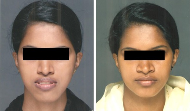
In all 100 cases in this study, alar cinch suturing was done by the conventional method without anchoring it to the anterior nasal spine. The advantage as observed by the first author (and the chief Maxillofacial Surgeon of all 100 cases) being that, since patient has a nasoendotracheal tube in situ, the exact nasal contour on the side of the tube is difficult to ascertain and possibilities of having that side widened is high. If anchored to the anterior nasal spine, then post-operative alterations are not feasible. When done in the conventional way, without any anchorage to the ANS, post-operative manipulations can be done to make minor contour adjustments and make the defect, if any, inconspicuous. In all 100 patients, the alar base widening was well controlled with the conventional cinching technique (Figs. 3a, b ;4a, b; 5a, b; 6a, b ;7a, b).
Fig. 3.

Case 1: a pre-op, b post-op
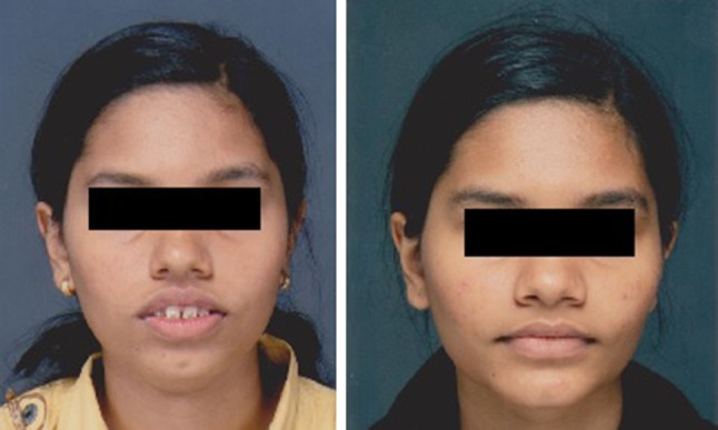
Fig. 4.
Case 2: a pre-op, b post-op
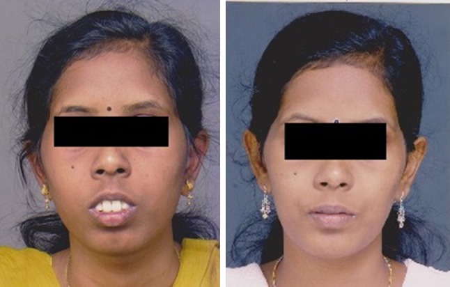
Fig. 5.

Case 3: a pre-op, b post-op
Fig. 6.
Case 4: a pre-op, b post-op
Fig. 7.
Case 5: a pre-op, b post-op
In this case series, all 100 patients underwent a Lefort 1 osteotomy with AMO with on table extraction of the first premolars followed by a conventional alar cinch suturing and V–Y closure of the lip. All the cases were operated by a single surgeon. Post-operative photographs were captured 6 months post-surgery. The pre-operative and post-operative alar base widths were measured and equated to the intercanthal width, and the derived values were compared.
A significant change in the alar base width was noted with an average of 1.176 mm, which is clinically insignificant.
Conclusion
The effect of Lefort 1 osteotomy on the alar base can be well controlled by the conventional alar cinch suturing without any additional anchorage on the ANS with predictable results.
Compliance with Ethical Standards
Conflict of interest
No potential conflict of interest relevant to this article was reported.
Footnotes
Publisher's Note
Springer Nature remains neutral with regard to jurisdictional claims in published maps and institutional affiliations.
Contributor Information
Varghese Mani, Email: Varghese.mani@gmail.com.
Prasanth Panicker, Email: drprasanthpanicker555@gmail.com.
Archana Shenoy, Email: archana95shenoy@gmail.com.
Ashford Lidiya George, Email: drlidiyamaxfac@gmail.com.
Tojan Chacko, Email: tojanchacko@gmail.com.
References
- 1.Westermark AH, Bystedt H, Von Konow L, Sällström KO. Nasolabial morphology after Le Fort I osteotomies. Effect of alar base suture. Int J Oral Maxillofac Surg. 1991;20:25–30. doi: 10.1016/S0901-5027(05)80690-3. [DOI] [PubMed] [Google Scholar]
- 2.O’Ryan FS, Schendel SA. Nasal anatomy and maxillary surgery. Part II: unfavorable nasolabial esthetics following the Le Fort I osteotomy. Int J Adult Orthodon Orthognath Surg. 1989;4:75–84. [PubMed] [Google Scholar]
- 3.Collins PC, Epker BN. The alar base cinch: a technique for prevention of alar base flaring secondary to maxillary surgery. Oral Surg Oral Med Oral Pathol. 1982;53:549–553. doi: 10.1016/0030-4220(82)90338-3. [DOI] [PubMed] [Google Scholar]
- 4.Nirvikalpa N, Narayanan Wahab A, Ramadorai A. Comparison between the classical and a modified transseptal technique of alar cinching for Le Fort I osteotomies: a prospective randomized controlled trial. Int J Oral Maxillofac Surg. 2013;42:49–54. doi: 10.1016/j.ijom.2012.05.027. [DOI] [PubMed] [Google Scholar]
- 5.Yen CY, Kuo CL, Liu IH, Su WC, Jiang HR, Huang IG, Liu SY, Lee SY. Modified alar base cinch suture fixation at the bilateral lower border of the piriform rim after a maxillary Le Fort I osteotomy. Int J Oral Maxillofac Surg. 2016;45(11):1459–1463. doi: 10.1016/j.ijom.2016.06.002. [DOI] [PubMed] [Google Scholar]
- 6.Howley C, Nayeem A, Lee R, Cox S. Use of the alar base cinch suture in Le Fort I osteotomy. Br J Oral Maxillofac Surg. 2011;49:127–130. doi: 10.1016/j.bjoms.2010.02.009. [DOI] [PubMed] [Google Scholar]
- 7.Khamashta-Ledezma L, Naini FB. Prospective assessment of maxillary advancement effects: maxillary incisor exposure, and upper lip and nasal changes. Am J Orthod Dentofacial Orthop. 2015;147:454–464. doi: 10.1016/j.ajodo.2014.11.028. [DOI] [PubMed] [Google Scholar]