Figure 2.
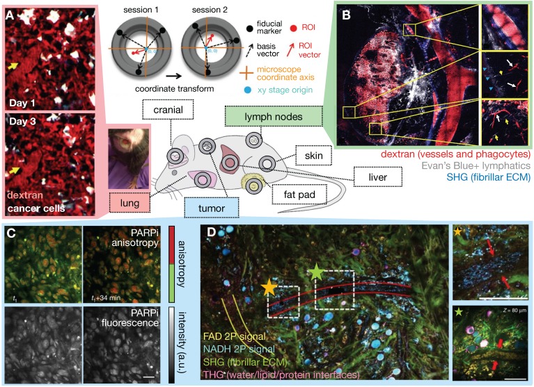
Intravital microscopy (IVM) developments aid EPR assessment. The availability of surgically implanted imaging windows at various anatomical sites allows tissue stabilization and longitudinal IVM of orthotopic disease sites over days and weeks (center). (A) Microcartography performed using fiducial marks etched on imaging windows enables precise localization across imaging sessions. Here, lung tumor vasculature was followed over multiple days. Yellow arrows indicate the location of the same microvessel branch point each day. A photograph of a long-term lung window is shown in a freely moving mouse (Adapted with permission from 26, copyright 2018 Springer Nature). (B) IVM mosaicking combines large scale and zoomed-in views of the TME. Here, a 10 x 10 mosaic covers a 4 x 4 mm lymph node area. Subcapsular sinuses are magnified at right, and shadows of erythrocytes (white arrows) and lymphatic capillaries (yellow arrows) are visible (Adapted with permission from 27, copyright 2017 Elsevier). (C) Real-time target engagement of the PARP inhibitor olaparib (PARPi) can be visualized in vivo using anisotropy imaging. High anisotropy (red) indicates the fluorescently-labeled PARPi has bound to a protein, which is localized in the nuclei of cancer cells (Adapted with permission from 199, copyright 2014 Springer Nature). (D) Multiphoton label-free IVM of a large tumor field (1.5 × 1.5 mm2) highlights cellular and ECM structures simultaneously (Adapted with permission from 40, copyright 2018 Springer Nature).

