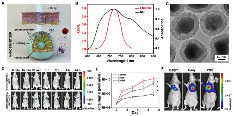
Figure 17.
(A) Schematic illustration for the design and mechanism of LGO:Cr@mSiO2 mediated X-PDT. (B) The absorption spectrum of 2,3-naphthalocyanine (NC) (black) and the XEOL spectrum of LGO:Cr (red). (C) TEM image of LGO:Cr@mSiO2. (D) In vivo imaging based on X-ray excited persistent luminescence signals and assessed in H1299 lung tumor models. (E) Tumor growth assessed by monitoring BLI signal changes at different time points. (F) Representative bioluminescence imaging for the three treatment groups taken on day 7. Adapted with permission from Ref 62. Copyright 2017 Royal Society of Chemistry.

