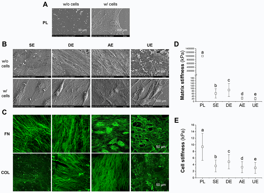
Fig. 1.

Effect of biochemical and biophysical properties of dECMs on expanded cells’ morphology and stiffness. Scanning electron microscopy (SEM) was used to observe SDSC morphology during expansion on Plastic (PL, A) and dECMs (B), including SECM (SE), DECM (DE), AECM (AE), and UECM (UE). Scale bars: 30 μm for dECMs and 300 μm for expanded cells on different culture substrates. Immunostaining was used to identify typical matrix protein expression including fibronectin (FN) and type I collagen (COL) in dECMs (C). Scale bar: 50 μm. Atomic force microscopy (AFM) was used to measure stiffness of both cell substrates (D) and expanded SDSCs (E). Data are shown as average ± standard deviation (SD) for n=1881-2001 (D) and n=1778-1981 (E). Groups not connected by the same letter are significantly different (P < 0.05).
