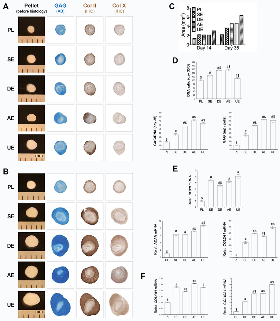
Fig. 4.

dECM expanded SDSCs exhibited enhanced chondrogenic potential. After expansion on different culture substrates for one passage, expanded cells were chondrogenically induced in a pellet culture system. Pellet samples were collected for data analysis at two time points: day 14 (earlier stage) (A/E/F) and day 35 (late stage) (B/C/D). Before histological staining, pellet size was measured with a scale bar as mm; Alcian blue (AB) was used to stain sulfated GAGs and immunohistochemistry staining (IHC) was used to detect type II collagen (Col II) and type X collagen (Col X) (A/B). Both day 14 and day 35 pellet sizes were quantified by ImageJ (C). Biochemical analysis was used for DNA and GAG amounts in pellets; cell viability in chondrogenic induction was evaluated using DNA ratio (DNA amount adjusted by that at day 0); a ratio of GAG to DNA indicated chondrogenic index (D). Real-time PCR was used to evaluate expression of chondrogenic marker genes (SOX9, ACAN, and COL2A1) (E) and hypertrophic genes (COL1A1 and COL10A1) (F) in pellets after chondrogenic induction. Data are shown as average ± standard deviation (SD) for n=4. P < 0.05 indicates a statistically significant difference compared with the PL group (#) and/or the SE group ($).
