Abstract
Adrenocortical carcinoma (ACC) is a rare and highly aggressive malignant neoplasm which can produce intravascular extension into the inferior vena cava (IVC) and can rarely extend into the right atrium. We describe the 18F Fluorodeoxyglucose Positron Emission Tomography-Computed Tomography findings of a 57-year-old man diagnosed with ACC with IVC thrombus extending up to the right atrium.
Keywords: Adrenocortical carcinoma, FDG, inferior vena cava, PET-computed tomography, thrombus
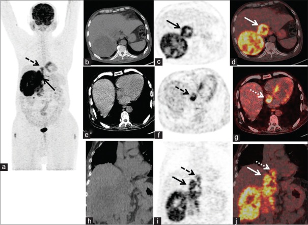
A 57-year-old man presented to surgery outpatient department with intermittent abdominal pain in the right hypochondrium for 3 months. Contrast-enhanced computed tomography (CT) of the abdomen was advised which showed a heterogeneous mass with few necrotic areas measuring ~12.2 cm × 11.8 cm × 13 cm in the right suprarenal region with hypodense area in the intrahepatic inferior vena cava (IVC). On suspicion of adrenocortical carcinoma (ACC), biochemical tests were done, which showed elevated serum cortisol and Dihydroepiandosterone DHEA levels, suggestive of secretory activity of the tumor. Considering surgery as the curative treatment option, the patient was referred for 18F-FDG-PET-CT scan to rule out any distant metastasis. PET-CT findings revealed a large FDG avid heterogenous right suprarenal mass [Figure 1a-j]. Right adrenal was not visualized separately. FDG avid IVC thrombus(Standard uptake volume SUVmax~7.6) extending up to the right atrium was also seen [Figure 1e-j dashed black and white arrows]. Fine-needle aspiration cytology of the mass was consistent with features of ACC. The patient underwent right adrenalectomy with IVC thrombectomy, and the histopathology was consistent with ACC.
Figure 1.
(a) MIP of PET-computed tomography image showing FDG uptake in the right hypochondrium of the abdomen. (b) Axial computed tomography image showing right suprarenal mass and hypodense lesion in inferior vena cava showing increased FDG uptake in PET (c, solid black arrow) and fused PET-computed tomography image (d, solid white arrow). (e) Hypodense lesion in the right atrium showing FDG uptake in the PET (f, dashed black arrow) and fused PET-computed tomography (g, dashed white arrow). (h-j) Coronal images showing FDG avid tumor thrombus extending from the inferior vena cava (solid black and white arrows) to the right atrium (dashed black and white arrows)
ACC is a rare and aggressive neoplasm with a very poor 5-year survival rate of 15%–44% in a series reported in the literature.[1,2,3] These neoplasms tend to grow very rapidly with common sites of metastases being liver, lung, and local invasion into kidneys, renal veins, and IVC.[4] Few case reports have highlighted the pattern of FDG uptake in IVC thrombus in case of ACC.[5,6] Sharma et al. in their series of 24 patients have demonstrated that avidity of FDG quantitatively assessed by SUVmax can differentiate between a benign and malignant tumor thrombus.[7] Most of the malignant thrombus had a SUVmax of >6.0, and in our case also, SUVmax of 7.6 was suggestive of a malignant tumor thrombus. Our case reiterates the fact that 18F-FDG-PET-CT can be helpful in the detection of primary tumor with local venous and visceral invasion as well as distant metastases in cases of ACC.
Declaration of patient consent
The authors certify that they have obtained all appropriate patient consent forms. In the form the patient(s) has/have given his/her/their consent for his/her/their images and other clinical information to be reported in the journal. The patients understand that their names and initials will not be published and due efforts will be made to conceal their identity, but anonymity cannot be guaranteed.
Financial support and sponsorship
Nil.
Conflicts of interest
There are no conflicts of interest.
References
- 1.Ng L, Libertino JM. Adrenocortical carcinoma: Diagnosis, evaluation and treatment. J Urol. 2003;169:5–11. doi: 10.1016/S0022-5347(05)64023-2. [DOI] [PubMed] [Google Scholar]
- 2.Ayala-Ramirez M, Jasim S, Feng L, Ejaz S, Deniz F, Busaidy N, et al. Adrenocortical carcinoma: Clinical outcomes and prognosis of 330 patients at a tertiary care center. Eur J Endocrinol. 2013;169:891–9. doi: 10.1530/EJE-13-0519. [DOI] [PMC free article] [PubMed] [Google Scholar]
- 3.Annamaria P, Silvia P, Bernardo C, Alessandro de L, Antonino M, Antonio B, et al. Adrenocortical carcinoma with inferior vena cava, left renal vein and right atrium tumor thrombus extension. Int J Surg Case Rep. 2015;15:137–9. doi: 10.1016/j.ijscr.2015.07.008. [DOI] [PMC free article] [PubMed] [Google Scholar]
- 4.Francis IR, Smid A, Gross MD, Shapiro B, Naylor B, Glazer GM, et al. Adrenal masses in oncologic patients: Functional and morphologic evaluation. Radiology. 1988;166:353–6. doi: 10.1148/radiology.166.2.3336710. [DOI] [PubMed] [Google Scholar]
- 5.Chiche L, Dousset B, Kieffer E, Chapuis Y. Adrenocortical carcinoma extending into the inferior vena cava: Presentation of a 15-patient series and review of the literature. Surgery. 2006;139:15–27. doi: 10.1016/j.surg.2005.05.014. [DOI] [PubMed] [Google Scholar]
- 6.Senthil R, Mittal BR, Kashyap R, Bhattacharya A, Radotra BD, Bhansali A, et al. 18F FDG PET/CT demonstration of IVC and right atrial involvement in adrenocortical carcinoma. Jpn J Radiol. 2012;30:281–3. doi: 10.1007/s11604-011-0037-4. [DOI] [PubMed] [Google Scholar]
- 7.Sharma P, Kumar R, Jeph S, Karunanithi S, Naswa N, Gupta A. 18F-FDG PET-CT in the diagnosis of tumor thrombus: Can it be differentiated from benign thrombus? Nucl Med Commun. 2011;32:782–8. doi: 10.1097/MNM.0b013e32834774c8. [DOI] [PubMed] [Google Scholar]


