Figure 5.
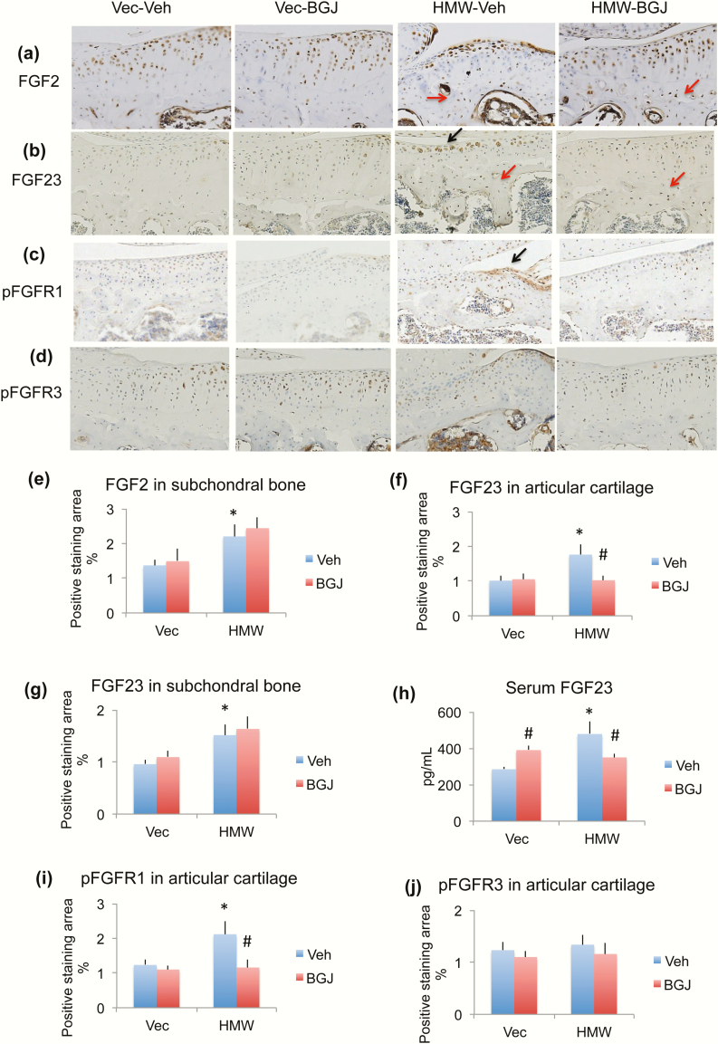
Effect of BGJ on OA regulatory proteins in knees of VectorTg and HMWTg mice. Representative immunohistochemical staining images for (a) FGF2, (b) FGF23, (c) pFGFR1, and (d) pFGFR3. Quantitative analysis of percentage of positive staining area for (e) FGF2 in subchondral bone, (f) FGF23 in articular cartilage, (g) FGF23 in subchondral bone, (i) pFGFR1 in articular cartilage, and (j) pFGFR3 in articular cartilage. (h) Serum FGF23 level. (a and e) FGF2 immunohistochemical staining showed that FGF2 expression in cartilage was similar among 4 groups. However, FGF2 expression in the osteocytes of subchondral bone was markedly increased in the HMWTg vehicle-treated group; BGJ treatment did not alter FGF2 expression in either VectorTg mice or HMWTg mice, n = 4–5 males/group. (b, f, and g) FGF23 protein expression was significantly increased in chondrocyte of articular cartilage and osteocytes of subchondral bone in knees from HMWTg vehicle-treated mice compared with VectorTg vehicle-treated mice. BGJ treatment significantly rescued abnormal FGF23 expression in articular cartilage, but not in subchondral bone, n = 7–9 males/group. (h) Serum FGF23 level was significantly increased in HMWTg vehicle-treated mice compared with VectorTg vehicle-treated mice. BGJ treatment significantly increased FGF23 serum level in VectorTg mice, but significantly decreased serum FGF23 level in HMWTg mice, n = 7–9 males/group. (c and i) phosphorylated FGFR1 (pFGFR1) was significantly increased in HMWTg-vehicle group compared with VectorTg-vehicle group, which was rescued with BGJ treatment, n = 7–9 males/group. (d and j) pFGFR3 protein expression was similar among all 4 groups, n = 4–5 males/group. *compared with VectorTg-vehicle group P < 0.05; #compared with corresponding vehicle group P < 0.05 by 2-way ANOVA.

