Article title: Reciprocal control of miR-197 and IL-6/STAT3 pathway reveals miR-197 as potential therapeutic target for hepatocellular carcinoma.
Author: Wang H, Su X, Yang M, Chen T, Hou J, Li N, Cao X.
Journal: OncoImmunology.
DOI: https://doi.org/10.1080/2162402X.2015.1031440
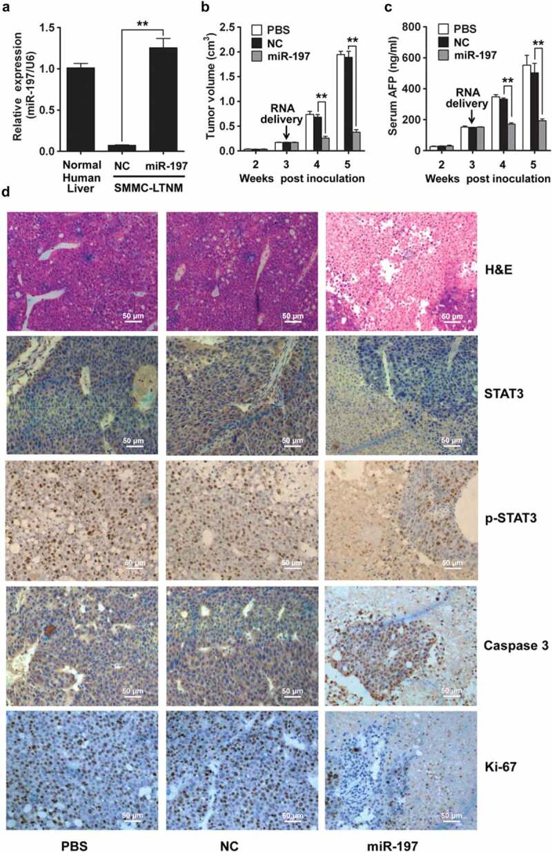
In the published version of Figure 5D, the data for PBS group in STAT3 staining panel and the PBS, NC and miR-197 groups in p-STAT3 staining panel were inadevertently presented with incorrect images during figure assembly for manuscript submission. The corrected Figure 5 is shown as below. The figure legend is correct as published and is also shown for reference. The data were not manipulated in any way, the errors have no bearings on the interpretation of the results, nor do they influence the conclusions of the work.
Figure 5.

miR-197 suppresses HCC growth in vivo.
(A) qRT-PCR analysis of miR-197 expression in normal human liver tissue, or SMMC-LTNM tumor tissue two weeks after intratumoral injection of cholesterol-conjugated miR-197 mimics (miR-197) or negative control (NC).(B–C) Two weeks after subcutaneous inoculation of SMMC-LTNM tumor cells, HCC-bearing nude mice were treated by intratumoral injection of cholesterol-conjugated miR-197, NC or PBS. Tumor volume (B), serum AFP levels (C) were shown as indicated in (C).(D) H&E staining and detection of STAT3, p-STAT3, Caspase 3 and Ki-67 by IHC in HCC tissues was performed two weeks after intratumoral injection of cholesterol-conjugated miR-197, NC or PBS. Scale bars, 50 μm.Data are shown as mean ± s.d. (n = 3) of one representative experiment. Similar results were obtained in three independent experiments. **p < 0.01.
We apologize for any inconvenience it may have caused.


