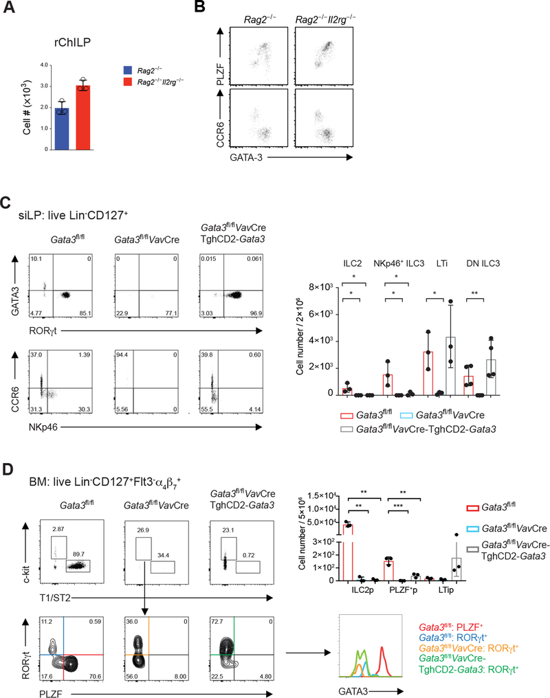
Figure 7. Dose-Dependent GATA3-Mediated Transcriptional Regulation Dictates the Development of ILCs Prior to Cytokine Signals.
(A) The total rChILP cell numbers in the Rag2−/− and Rag2−/−Il2rg−/− mice were counted and plotted (mean ± s.d.; n = 3).
(B) The GATA3high or PLZF+ non-LTi ILC progenitors and GATA3low or CCR6+ LTi progenitors from the Rag2−/−Il2rg−/− mice were analyzed in comparison with these populations in the Rag2−/− mice.
(C) Flow cytometry analysis of ILCs found in the siLP of the Gata3fl/flVavCre-TghCD2-Gata3 mice. Dead cells were excluded by a fixable viability dye, and the Lin−CD127+ ILCs were further gated for analyses. The total numbers of indicated populations were counted and plotted (mean ± s.d.; n = 3; *P < 0.05, **P < 0.01, Student’s t-test).
(D) Flow cytometry analysis of rChILPs in the bone marrow of the Gata3fl/flVavCre-TghCD2-Gata3 mice. The plots were first gated on the live Lin−CD127+Flt-3−α4β7+ population. T1/ST2−c-Kit+ cells were further analyzed for the expression of RORγt and PLZF. The degree of GATA3 expression by different subsets was further compared. The total numbers of indicated populations were counted and plotted (mean ± s.d.; n = 3; **P < 0.01, ***P < 0.001, Student’s t-test).
Data are representative of three independent experiments (A-D) or a combination of three independent experiments (C-D).
See also Figure S7.

