



Abstract
Malignant infantile osteopetrosis (MIOP) is a rare inherited bone metabolism disorder characterized by increased bone mineral density (BMD) and abnormal hematopoiesis. Hematopoietic stem cell transplantation (HSCT) is currently the only curative therapy for MIOP. However, a higher risk of secondary malignancy occurs in children previously exposed to cytotoxic drugs. Here we report a rare case of a 3-year-old female patient with MiT family translocation renal cell carcinoma (MiTF tRCC), who is a survivor of HSCT for MIOP 2 years earlier. The patient had a complete resection of the tumor. Microscopically, we detected diffusely and papillary-like arranged tumor cells whose cytoplasm was bright and clear. Immunohistochemistry showed tumor cells diffusely expressed TFE3, and fluorescence in situ hybridization (FISH) demonstrated disruption of the TFE3 locus, confirming the diagnosis of Xp11 translocation RCC, the subtype of MiTF tRCC. This case supports the view that chemotherapy exposure is a risk factor for MiTF tRCC and indicates the possible association of HSCT with MiTF tRCC.
Keywords: Malignant infantile osteopetrosis, hematopoietic stem cell transplantation, MiT family translocation renal cell carcinoma, Xp11 translocation renal cell carcinoma, chemotherapy
Introduction
Malignant infantile osteopetrosis (MIOP) is a rare inherited bone metabolism disorder that has an incidence of 1:250,000 in the general population [1]. Autosomal recessive is the inheritance pattern of MIOP, and most cases are associated with mutations in the TCIRG1 gene. The failure in bone resorption due to reduced or dysregulated activity of osteoclasts results in generalized osteosclerosis [1]. MIOP has a mortality rate of 70% by the age of 6 years and nearly 100% by the end of the first decade. The only potentially curative therapy is hematopoietic stem cell transplantation (HSCT) [2]. However, the conditioning regimen of HSCT, covering DNA topoisomerase II inhibitors or alkylating agents, may increase the risk of secondary MiT family translocation renal cell carcinoma (MiTF tRCC).
MiTF tRCC is a rare childhood and adult tumor, comprising Xp11 translocation RCC and t(6;11) RCC. The Xp11 tRCC was first officially recognized in the 2004 WHO renal tumor classification, and it harbors gene fusions involving TFE3 with one of multiple reported genes including ASPSCR1 (ASPL), PRCC, NonO, SFPQ, CLTC, PARP14, LUC7L3, DVL2, KHSRP, and RBM10 [3,4]. Although RCCs account for <5% of renal tumors in children, Xp11 tRCC likely constitutes approximately 50% of these cases [3]. It has similar clinical manifestations compared to other RCCs [5], and pediatric patients usually have a better prognosis than adults [3]. Surgery is the most common therapeutic strategy for this tumor [5].
Here we report a rare case of a 3-year-old female patient with MiTF tRCC, who was a survivor of HSCT for MIOP 2 years earlier.
Case report
A 3-year-old female was diagnosed with MIOP at 7 months of age. She was referred to our hospital with the chief complaint of gross motor retardation. A physical examination revealed frontal bossing, mental retardation, moderate hearing loss in the left ear, and nystagmus. A blood routine revealed anemia (Hb 89.2 g/L) and thrombocytopenia (91*109/L). The blood biochemistry showed elevated α-hydroxybutyrate dehydrogenase (α-HBDH, 301 IU/L), lactate dehydrogenase (LDH, 391 IU/L), creatine kinase isoenzyme (CK-MB, 41.9 IU/L), and reduced serum calcium (1.21 mmol/L). Blood gas analysis revealed a significant decrease in blood oxygen saturation (73.2%) and oxygen partial pressure (39.3 mmHg).
The extensively increased bone mineral density (BMD) and medullary cavity stenosis were detectable on radiographs (Figure 1B and 1C) with a ‘bone-in-bone’ appearance of the vertebrae (Figure 1D). An abdominal ultrasound disclosed pronounced hepatosplenomegaly. Magnetic resonance imaging (MRI) of the brain showed a thin corpus callosum and a plump lateral ventricle (Figure 1A). Genetic detection identified two heterozygous mutations in the TCIRG1 gene on chr11p6: a nonsense mutation (C.2008C>T) and a frameshift mutation (C.1188: lack of C), confirming the diagnosis of MIOP.
Figure 1.

Radiological findings of MIOP. A. MRI showed thin corpus callosum and plump lateral ventricle. B. The pelvis X-ray revealed uneven bone density increased, periosteal reaction could be seen on both sides of the sacrum and femur. C. The left lower extremity X-ray revealed extensively increased BMD and medullary cavity stenosis. D. Whole spine lateral X-ray showed skull bones, vertebrae, pelvis had increased BMD. The vertebral bodies showed a ‘bone-in-bone’ appearance, and the anterior ribs were widened with reduced uneven bone density.
The patient underwent an HLA haploidentical HSCT at 10 months of age at Beijing Children’s Hospital, conditioned with fludarabine, busulfan and cyclophosphamide. Anti-thymocyte globulin (ATG) was used for GvHD prophylaxis, which, however, was switched to basiliximab considering the patient’s allergy to ATG. She received donated bone marrow and peripheral blood stem cell transfusions, consisting of 24.63×10⁸/kg of mononuclear cells and 10.22×10⁶/kg of CD34+ cells. Neutrophils and platelets were successfully engrafted on day +11 and day +29 respectively. No appearance of aGvHD was observed after transplantation, and immunosuppressant tacrolimus has been used for the long term.
After 6 months of transplantation, the blood routine showed no evidence of anemia (Hb120 g/L), and the platelet count (191*1012/L) returned to normal. The blood biochemistry revealed that HBDH (248 IU/L), LDH (291 IU/L) and CK-MB (27 IU/L) were markedly decreased than before, and the patient’s serum calcium level (2.57 mmol/L) was in the normal range. No cGvHD was observed in the following days.
During regular follow-up, she remained entirely well. But 2 years after the transplantation, abdominal ultrasonography indicated an occupation of the right kidney; there was no abdominal pain, fever or hematuria. A routine reexamination showed a progressive enlargement of the kidney mass. Ultrasonography of the abdomen revealed a 25×28×22 mm middle echogenic nodule in the middle of the right kidney with a clear boundary. The MRI reported a solid mass of approximately 26×29×22 mm in the middle of the right kidney, which was slightly enhanced by the enhancement scan (Figure 2A and 2B). She underwent tumor enucleation, having a complete resection with a margin of 0.2 cm from the edge of the tumor. No adjuvant treatment was delivered, and the patient remained free of disease at 3 months’ follow-up.
Figure 2.
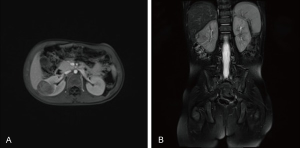
A. MR on fat-suppressed T1WI sequence revealed a 26×29×22 mm solid mass with isointensity signal, low signal and patchy high signal in the middle of the right kidney. B. MR on fat-suppressed T2WI sequence revealed isointensity signal and high signal.
Pathologic manifestations
The surgical specimen demonstrated a well encapsulated tumor measuring 30×27×25 mm. The tumor section was grayish red, yellow and soft, with focal areas of necrosis and calcification (Figure 3A).
Figure 3.

Pathological findings of the tumor. A. Gross appearance demonstrated a well encapsulated tumor measuring 3.0×2.7×2.5 cm. The section was grayish red, yellow and soft, with focal areas of necrosis and calcification. B-D. The microscopic examination revealed papillary-like arranged tumor cells whose cytoplasm was bright and clear, and nucleolus was obvious (HE staining, ×100, ×200, ×400). E. Tumor cells with focal areas of hemorrhage and necrosis (HE staining, ×100). F. The tumor cells broke through the capsule in some areas, invading the kidney tissue (HE staining, ×100).
The microscopic examination revealed diffusely arranged tumor cells and a papillary-like structure (Figure 3B), with focal areas of hemorrhage and necrosis (Figure 3E). The cytoplasms of the tumor cells were bright and clear, and the nuclei were obvious (Figure 3C and 3D). The tumor cells broke through the capsule in some areas, which could be observed in the kidney tissue (Figure 3F). But none was found in the perirenal fat.
Immunohistochemistry showed the tumor cells were positive for TFE3 (Figure 4A), PAX8 (Figure 4B), CD10 (membranous, Figure 4C), PAX5 (Figure 4D), PAX2 (weak, Figure 4E), CD68 (sparse, Figure 4F) and vimentin (scarce, Figure 4L), and were negative for MelonA (Figure 4I), HMB45 (Figure 4J) and WT1 (Figure 4K). The cell proliferation marker, Ki67, showed a proliferation rate of 5% (Figure 4G), and P53 showed a weak positive rate of 40% (Figure 4H). These markers were accordance with the characteristics of Xp11 tRCC. Fluorescence in situ hybridization (FISH) demonstrated a disruption of the TFE3 locus consistent with an Xp11 translocation (Figure 5), supporting the diagnosis of Xp11 tRCC, the subtype of MiTF tRCC.
Figure 4.

Immunohistochemical manifestations of the tumor cells. A. TFE3+. B. PAX8+. C. CD10+, membranous. D. PAX5+. E. PAX2+, weak. F. CD68+, sparse. G. Ki67 5%+. H. P53 40%+, weak. I. MelonA-. J. HMB45-. K. WT1-. L. Vimentin+, scarce. (A-G, ×200; H-L, ×100).
Figure 5.

Fluorescence in situ hybridization. Red arrow, a break-apart TFE3 probe showed disruption of the TFE3 gene with separation of green and red signals. Green arrow, normal gene: green and red signal merged together.
Discussion
MIOP is a rare genetic metabolism disorder. The diagnosis of MIOP mainly depends on typical imaging findings. In addition, the Osteopetrosis Working Group also recommends genetic testing, which can provide key information on clinical performance as well as prognosis and play an important role in decision for treatment [6].
However, malignancy after MIOP is extremely rare. So far, only one study reported that a 3-month-old male patient was diagnosed as MIOP and acute myeloid leukemia (AML), type M3 simultaneously with the manifestations of chest infection, growth retardation and hepatosplenomegaly. The author speculated that AML was associated with a concomitant gene mutation, but this was not confirmed genetically [7]. Our case is the first reported case of MiTF tRCC that occurred after MIOP. Due to the low number of reported cases, we cannot ascertain whether there is a possible genetic association between MIOP and MiTF tRCC.
The category of MiTF tRCC is composed of Xp11 tRCC and t(6;11) RCC, and Xp11 tRCC takes up majority of those cases. Xp11 tRCC harbors a chromosome translocation involving the Xp11 breakpoint, resulting in gene fusions involving the TFE3 gene [8]. The most common subtypes are the ASPSCR1-TFE3 RCC due to t(X;17)(p11;q25) translocation [9], and the PRCC-TFE3 RCC due to t(X;1)(p11;q21) translocation [10].
Microscopically, Xp11 tRCC is similar to clear cell RCC or papillary RCC [11]. The most distinctive histologic morphology is papillary architecture and epithelioid clear cells. In typical cases, abundant psammoma bodies can be found [8]. Xp11 tRCC can also resemble other renal neoplasms, including extensive cystic change simulating multilocular cystic RCC, sarcomatoid transformation, oncocytic areas mimicking oncocytoma, trabecular architecture mimicking a carcinoid tumor, colonization of renal pelvic urothelium mimicking urothelial carcinoma [12], etc. The broad morphological spectrum of Xp11 tRCC makes it difficult for pathologists to distinguish it from other tumor types both in adults and children only by the morphology of HE staining.
Therefore, immunohistochemistry is a common and essential method for differential diagnosis. Owning to the overexpression of TFE3, strong nuclear TFE3 immunoreactivity is the most sensitive and specific immunohistochemical marker for Xp11 tRCC. The Xp11 RCC underexpresses epithelial markers, such as cytokeratins and EMA. In contrast, this tumor consistently expresses CD10 and RCC markers like PAX2 and PAX8, similar to both clear cell RCC and papillary RCC, while vimentin is often negative. Occasionally, Xp11 tRCC may express melanocytic markers, such as Melan A and HMB45, particularly in typical cases. Besides, cathepsin-K labels approximately 60% of Xp11 tRCC, and almost all t(6;11) RCC, but no other common RCC subtypes [8].
In the past, TFE3 break-apart FISH was regarded as the best method to diagnose Xp11 tRCC. However, Argani et al. [4] reported 3 cases of Xp11 tRCC with RBM10-TFE3 gene fusion. TFE3 rearrangement was not detected by conventional TFE3 break-apart FISH, but was suggested by TFE3 IHC, which highlights the combination of TFE3 IHC and TFE3 FISH in diagnosis. In this case, the HE staining showed typical morphology, except for the psammoma bodies. Immunohistochemically, the tumor cells were positive for TFE3, CD10, PAX8, PAX5, PAX2 and vimentin (scarce), and negative for MelonA, HMB45 and WT1. Finally, the TFE3 break-apart FISH validated the diagnosis of Xp11 tRCC.
MiTF tRCC occurring as a second malignancy is even more unusual. Exposure to chemotherapy is currently the only known risk factor of MiTF tRCC, although its mechanism is not yet clear. Argani et al. [13] reported that 15% (6 of 39) of translocation RCCs were associated with previous exposure to cytotoxic chemotherapy. The indications for the antecedent chemotherapy included acute promyelocytic leukemia, acute myeloid leukemia with t(9;11), bilateral Wilms’ tumor, systemic lupus erythematosus, and conditioning regimen of bone marrow transplant for Hurler’s syndrome. And they suggested that MiTF tRCC should be added to the list of possible chemotherapy-associated secondary malignancies in children. Malouf et al. [14] also reported that 7% (4 of 54) of patients with MiTF tRCC had received chemotherapy for germ cell tumors, nephroblastoma, rhabdomyosarcoma, and medulloblastoma, respectively. Additionally, other translocation RCCs arising in the setting of prior chemotherapy have been described, including neuroblastoma [14,15], primitive neuroectodermal tumor [16,17], Ewing sarcoma [16], rhabdomyosarcoma [18], acute lymphoblastic leukemia [17], and cardiac leiomyosarcoma [19]. With the exception of cases with unknown chemotherapy regimens, DNA topoisomerase II inhibitors and/or alkylating agents were used in all of the above cases. Although they have differing mechanisms of action, both cytotoxic agents break DNA, which may initiate repair or recombination mechanisms that permit a chromosome translocation to occur, increasing the risk of translocation malignancy [8]. Our case is the same as the above cases. The patient received a conditioning regimen of alkylating agents covering busulfan and cyclophosphamide of HSCT, which further confirmed the recognized association of MiTF tRCC with prior cytotoxic chemotherapy.
In addition to chemotherapy, HSCT may also be associated with secondary malignancy. Several large studies have demonstrated that childhood survivors of HSCT represent a particularly high risk of secondary solid malignancies, reporting cumulative incidence up to 6.7% at 15 years of follow-up [20]. Risk factors include radiation and chemotherapy exposure, cGvHD, use of immunosuppressants, viruses [20,21], etc. However, reports of secondary MiTF tRCC after HSCT are scarce. Argani et al. [13] reported a 6-year-old female patient with t(6;11) RCC who underwent matched unrelated cord blood transplantation preceded by conditioning with high-dose cyclophosphamide, total body irradiation, and ATG at 6 months of age. Al-Mashaikhi et al. [22] reported a case of a 17-year-old male patient with Xp11 tRCC who received an autologous HSCT at the age of 3 due to neuroblastoma after 4 cycles of chemotherapy and radiotherapy. The conditioning regimen was carboplatin, etoposide and cyclophosphamide. In this case, we speculate that long-term use of immunosuppressant tacrolimus probably increased the risk of secondary translocation RCC. Because tacrolimus enhances the overexpression of transforming growth factor-β (TGF-β) and promotes progression and metastasis of tumors [23], the tumor in this case showed progressive enlargement.
In summary, to our knowledge, this is the first reported case of MiTF tRCC occurring after MIOP, which supports the view that chemotherapy exposure is a risk factor for MiTF tRCC and indicates the possible association of HSCT with MiTF tRCC. However, for lack of enough cases, the clear relationship between HSCT and MiTF tRCC still needs more evidence to confirm.
Disclosure of conflict of interest
None.
References
- 1.Sobacchi C, Schulz A, Coxon FP, Villa A, Helfrich MH. Osteopetrosis: genetics, treatment and new insights into osteoclast function. Nat Rev Endocrinol. 2013;9:522–536. doi: 10.1038/nrendo.2013.137. [DOI] [PubMed] [Google Scholar]
- 2.Eapen M, Davies SM, Ramsay NK, Orchard PJ. Hematopoietic stem cell transplantation for infantile osteopetrosis. Blood. 2015;2:270–276. doi: 10.1038/sj.bmt.1701474. [DOI] [PubMed] [Google Scholar]
- 3.Srigley JR, Delahunt B, Eble JN, Egevad L, Epstein JI, Grignon D, Hes O, Moch H, Montironi R, Tickoo SK, Zhou M, Argani P. The international society of urological pathology (ISUP) vancouver classification of renal neoplasia. Am J Surg Pathol. 2013;37:1469–1489. doi: 10.1097/PAS.0b013e318299f2d1. [DOI] [PubMed] [Google Scholar]
- 4.Argani P, Zhang L, Reuter VE, Tickoo SK, Antonescu CR. RBM10-TFE3 renal cell carcinoma: a potential diagnostic pitfall due to cryptic intrachromosomal xp11.2 inversion resulting in false-negative TFE3 FISH. Am J Surg Pathol. 2017;41:655–662. doi: 10.1097/PAS.0000000000000835. [DOI] [PMC free article] [PubMed] [Google Scholar]
- 5.Liu N, Wang Z, Gan W, Xiong L, Miao B, Chen X, Guo H, Li D. Renal cell carcinoma associated with xp11.2 translocation/TFE3 gene fusions: clinical features, treatments and prognosis. PLoS One. 2016;11:e166897. doi: 10.1371/journal.pone.0166897. [DOI] [PMC free article] [PubMed] [Google Scholar]
- 6.Wu CC, Econs MJ, DiMeglio LA, Insogna KL, Levine MA, Orchard PJ, Miller WP, Petryk A, Rush ET, Shoback DM, Ward LM, Polgreen LE. Diagnosis and management of osteopetrosis: consensus guidelines from the osteopetrosis working group. J Clin Endocrinol Metab. 2017;102:3111–3123. doi: 10.1210/jc.2017-01127. [DOI] [PubMed] [Google Scholar]
- 7.Prasad R, Jaiswal BP, Mishra OP, Singh UK. Association of possible osteopetrosis with acute myeloid leukaemia in a child. BMJ Case Rep. 2013:2013. doi: 10.1136/bcr-2013-009765. [DOI] [PMC free article] [PubMed] [Google Scholar]
- 8.Argani P. MiT family translocation renal cell carcinoma. Semin Diagn Pathol. 2015;32:103–113. doi: 10.1053/j.semdp.2015.02.003. [DOI] [PubMed] [Google Scholar]
- 9.Argani P, Antonescu CR, Illei PB, Lui MY, Timmons CF, Newbury R, Reuter VE, Garvin AJ, Perez-Atayde AR, Fletcher JA, Beckwith JB, Bridge JA, Ladanyi M. Primary renal neoplasms with the ASPL-TFE3 gene fusion of alveolar soft part sarcoma: a distinctive tumor entity previously included among renal cell carcinomas of children and adolescents. Am J Pathol. 2001;159:179–192. doi: 10.1016/S0002-9440(10)61684-7. [DOI] [PMC free article] [PubMed] [Google Scholar]
- 10.Argani P, Antonescu CR, Couturier J, Fournet J, Sciot R, Debiec-Rychter M, Hutchinson B, Reuter VE, Boccon-Gibod L, Timmons C, Hafez N, Ladanyi M. PRCC-TFE3 renal carcinomas: morphologic, immunohistochemical, ultrastructural, and molecular analysis of an entity associated with the t(X;1)(p11.2;q21) Am J Surg Pathol. 2002;26:1553–1566. doi: 10.1097/00000478-200212000-00003. [DOI] [PubMed] [Google Scholar]
- 11.Hirobe M, Masumori N, Tanaka T, Kitamura H, Tonooka A, Hasegawa T, Tsukamoto T. Clinicopathological characteristics of Xp11.2 translocation renal cell carcinoma in adolescents and adults: diagnosis using immunostaining of transcription factor E3 and fluorescence in situ hybridization analysis. Int J Urol. 2016;23:140–145. doi: 10.1111/iju.13007. [DOI] [PubMed] [Google Scholar]
- 12.Green WM, Yonescu R, Morsberger L, Morris K, Netto GJ, Epstein JI, Illei PB, Allaf M, Ladanyi M, Griffin CA, Argani P. Utilization of a TFE3 break-apart FISH assay in a renal tumor consultation service. Am J Surg Pathol. 2013;37:1150–1163. doi: 10.1097/PAS.0b013e31828a69ae. [DOI] [PubMed] [Google Scholar]
- 13.Argani P, Lae M, Ballard ET, Amin M, Manivel C, Hutchinson B, Reuter VE, Ladanyi M. Translocation carcinomas of the kidney after chemotherapy in childhood. J. Clin. Oncol. 2006;24:1529–1534. doi: 10.1200/JCO.2005.04.4693. [DOI] [PubMed] [Google Scholar]
- 14.Malouf GG, Camparo P, Molinie V, Dedet G, Oudard S, Schleiermacher G, Theodore C, Dutcher J, Billemont B, Bompas E, Guillot A, Boccon-Gibod L, Couturier J, Escudier B. Transcription factor E3 and transcription factor EB renal cell carcinomas: clinical features, biological behavior and prognostic factors. J Urol. 2011;185:24–29. doi: 10.1016/j.juro.2010.08.092. [DOI] [PubMed] [Google Scholar]
- 15.Ramphal R, Pappo A, Zielenska M, Grant R, Ngan BY. Pediatric renal cell carcinoma: clinical, pathologic, and molecular abnormalities associated with the members of the mit transcription factor family. Am J Clin Pathol. 2006;126:349–364. doi: 10.1309/98YE9E442AR7LX2X. [DOI] [PubMed] [Google Scholar]
- 16.Rais-Bahrami S, Drabick JJ, De Marzo AM, Hicks J, Ho C, Caroe AE, Argani P. Xp11 translocation renal cell carcinoma: delayed but massive and lethal metastases of a chemotherapy-associated secondary malignancy. Urology. 2007;70:173–178. doi: 10.1016/j.urology.2007.03.037. [DOI] [PubMed] [Google Scholar]
- 17.Schafernak KT, Yang XJ, Hsueh W, Leestma JL, Stagl J, Goldman S. Pediatric renal cell carcinoma as second malignancy: reports of two cases and a review of the literature. Can J Urol. 2007;14:3739–3744. [PubMed] [Google Scholar]
- 18.Brassesco MS, Valera ET, Bonilha TA, Scrideli CA, Carvalho de Oliveira J, Pezuk JA, Barros Silva GE, Costa RS, Tone LG. Secondary PSF/TFE3-associated renal cell carcinoma in a child treated for genitourinary rhabdomyosarcoma. Cancer Genet. 2011;204:108–110. doi: 10.1016/j.cancergencyto.2010.10.006. [DOI] [PubMed] [Google Scholar]
- 19.Dhall D, Al-Ahmadie HA, Dhall G, Shen-Schwarz S, Tickoo SK. Pediatric renal cell carcinoma with oncocytoid features occurring in a child after chemotherapy for cardiac leiomyosarcoma. Urology. 2007;70:113–178. doi: 10.1016/j.urology.2007.03.055. [DOI] [PubMed] [Google Scholar]
- 20.Lowe T, Bhatia S, Somlo G. Second malignancies after allogeneic hematopoietic cell transplantation. Biol Blood Marrow Transplant. 2007;13:1121–1134. doi: 10.1016/j.bbmt.2007.07.002. [DOI] [PubMed] [Google Scholar]
- 21.Danylesko I, Shimoni A. Second malignancies after hematopoietic stem cell transplantation. Curr Treat Options Oncol. 2018;19:9. doi: 10.1007/s11864-018-0528-y. [DOI] [PubMed] [Google Scholar]
- 22.Al-Mashaikhi N, Yang J, Terry J, Barr R. Renal cell carcinoma with Xp 11.2 translocation as a second tumor in a long-term survivor of advanced neuroblastoma. Pediatr Hematol Oncol. 2015;32:215–222. doi: 10.3109/08880018.2014.979457. [DOI] [PubMed] [Google Scholar]
- 23.Maluccio M, Sharma V, Lagman M, Vyas S, Yang H, Li B, Suthanthiran M. Tacrolimus enhances transforming growth factor-beta1 expression and promotes tumor progression. Transplantation. 2003;76:597–602. doi: 10.1097/01.TP.0000081399.75231.3B. [DOI] [PubMed] [Google Scholar]