Abstract
Long non-coding RNAs (lncRNAs) have been implicated in tumor development and progression. The lncRNA HERC2P3, located on human chromosome 15q11.1-q11.2, is one of pseudogenes of HERC2 (an E3 ubiquitin protein ligase). Its role and expression are still unclear in cancer. In the present study, we investigated the effects of HERC2P3 on gastric cancer cell growth and migration via CCK-8 assays and Transwell assays in vitro and tumor-bearing mouse model in vivo. The results demonstrated that HERC2P3 silencing inhibited cell growth and migration, although it only had a weak effect on cell growth. Western blot analysis revealed that Akt phosphorylation level could be reduced when HERC2P3 was knocked down, indicating Akt signaling may be involved in the HERC2P3-mediated tumor development. In addition, we analyzed the expression of HERC2P3 through quantitative RT-PCR in 30 paired gastric cancer samples and found HERC2P3 was up-regulated in gastric adenocarcinoma tissues compared with corresponding non-tumor tissues. Taken together, our results demonstrate that the abrogation of HERC2P3 could suppress tumor cell growth and migration, with important implication for validating HERC2P3 as a potential target for human gastric cancer therapy.
Keywords: lncRNA, HERC2P3, cell proliferation, cell migration, gastric cancer
Introduction
Gastric cancer (GC), one of the common malignant tumors, remains the second leading cause of cancer related death [1]. Numerous efforts have been made to improve the therapy of GC, however, the 5-year overall survival (OS) rate of GC patients is still lower than 25% due to most patients present with advanced disease [2,3]. Many studies have demonstrated that the most important method to prolong overall survival is early diagnosis. Therefore, revealing a novel mechanism of GC development and promoting effective markers of GC are urgently required.
The ENCODE project revealed that more than 98% of the human genome are non-coding RNA (ncRNA). Long non-coding RNA (lncRNA) with a length more than 200 nucleotides is a subcategory of non-coding RNA. Although the majority of the rest transcribes into noncoding transcripts lncRNA is considered as a transcription noise with no biological functions initially, recently years lncRNA has been implicated in numerous cellular and carcinogenesis processes, including cell differentiation, apoptosis, embryonic development and disease process [4-6]. Notably, many studies have proved lncRNA may function as an oncogene or tumor suppressor gene [7,8]. H19, a well-known lncRNA, is associated with embryonic stem cell differentiation and increasing evidence indicates that H19 plays an oncogenic role in bladder and hepatocellular carcinoma and breast cancer [6,9,10]. Another noted lncRNA, MEG3 was aberrantly expressed in many human cancers and inhibited cell proliferation in cancer cells [11]. These studies are exciting and suggest that lncRNAs contribute differently to human malignancy, however, a great number of lncRNAs were still to be characterized.
In the present study, we focused on an lncRNA HERC2P3 (Hect domain and RLD 2 pseudogene 3), which is located on chromosome 15q11.1-q11.2, including 26 exons [12]. The role of HERC2P3 in cancer is still unknown. Our study demonstrated that HERC2P3 plays a critical role in the cell growth and migration in human GC cells. Furthermore, HERC2P3 is frequently up-regulated at RNA level in 30 paired GC samples. Our data suggest that HERC2P3 may serve as a potential biomarker for diagnosis and treatment of gastric cancer.
Materials and methods
Cell culture
The human gastric cancer cell lines AGS and SGC7901 were purchased from the Shanghai Cell Bank of the Chinese Academy of Sciences (Shanghai, China). AGS and SGC7901 cell lines were cultured in a humidified atmosphere at 37°C with 5% CO2 and cultured in MEM medium supplemented with 10% fetal bovine serum and 100 μg/ml penicillin/streptomycin.
Transwell assays
Cell migration assays were conducted using the Transwell chamber (Corning, USA). 3×10⁴ cells were planted into the upper part of a Transwell chamber with 400 μl MEM medium, while 800 μl MEM medium with 10% FBS were added into the bottom. Then the chambers were maintained in the incubator (37°C and 5% CO2) for 48 h. Then the cells were stained with 0.5% crystal violet and photographed. The cells were counted in 5 random sections under microscope. The experiment was repeated three times.
Cell proliferation assay
After transfection, AGS and SGC7901 cells were seeded in a 96-well plate at a concentration of 3×10³ per well. Cells were tested for proliferation per 24 h using Cell Counting Kit-8 (CCK-8, Dojindo Laboratories, Kumamoto, Japan), 10 μl of Cell Counting Kit-8 reagent was added to each well for 1 h at 37°C and then the absorbance of 450 mm wavelength was read. All experiments were performed independently at least three times in triplicate.
RNA interferences, lentivirus construction and transfection
The siRNAs and lentivirus used in the experiment were obtained from GenePharma, Shanghai, China. The sequences of siRNA are siHERC2P3-1 (5-GCUGUUCAGAACAUUCGAA-3), siHERC2P3-2 (5-CACCAAGACUUUAUGCGGA-3) and siNC (5-UUCUCCGAACGUGUCACGUdTdT-3). The cell transfection with siRNAs was conducted using Lipofectamine 3000 (Invitrogen) according to the manufacturer’s constructions. The lentivirus LV-shHERC2P3 was packaged using the siHERC2P3-2 sequences mentioned above. Stably infected cell lines were isolated by puromycin selection.
RNA extraction and quantitative real-time PCR
Total RNA was extracted from frozen tissues or cultured cells using TRIZOL reagent (Invitrogen, Carlsbad, Calif). For real-time PCR, the isolated RNA was reverse transcribed to cDNA using a Reverse Transcription Kit. Real-time PCR was performed with SYBR Green Kit (Roche, Switzerland). The results were normalized to the expression of β-actin. The primers were as follows: HERC2P3-QF: 5-GCGATCAGGATAGGCCTCCT-3, HERC2P3-QR: 5-GTTCGGCTTCAGAGT-CGTCC-3, ß-actin-qF: 5-CCTGGCACCCAGCACAATG-3, ß-actin-qR: 5-GGGCCGGACTCGTCATACT-3.
Animal experiments
Nude mice (Four-week-old, male) were obtained from the Animal Center of the Chinese Academy of Science (Shanghai, China) and raised under specific pathogen free conditions. 1.5×10⁶ SGC7901 cells transfected with LV-shHERC2P3 or a LV-NC were injected through tail vein. After 6 weeks, the mice were sacrificed and its lungs were collected and photographed. The nodes on the surface were counted. For tumor growth, 2×10⁶ cells were resuspended in 150 µl PBS and subcutaneously injected into the right flank of each nude mouse. After 4 weeks, the mice were sacrificed and the tumor was collected and weighted.
Gastric cancer tissue samples
The tissue samples were gained from the gastric cancer patients who had surgery at Shanghai East Hospital, all the samples were snap-frozen in liquid nitrogen and stored at -80°C prior to RNA extraction. All the patients were provided with written informed consent under the approval of Human Resources Ethics Committee of Shanghai East Hospital.
Western blot
Total proteins were extracted in RIPA buffer, and the protein concentration was determined using the BCA protein assay kit (TaKaRa, Japan). To detect the expression of Akt (1:1000, Cell Signaling Technology, #9272), phospho-Akt (Ser473) (1:1000, Cell Signaling Technology, #4051), Slug (Slug (1:500, Cell Signaling Technology, #9585)), Cyclin D1 (1:1000, Cell Signaling Technology, #2978) and β-actin (1:500, Santa Cruz Biotechnology, sc-58673), we loaded 10 μl of whole-cell protein lysates on SDS-PAGE and performed Western blotting. Detection of proteins was achieved by using the Odyssey Infared Imaging System (Li-COR, USA) according to the manufacturer’s instructions.
Statistical analysis
Each experiment was repeated two or three times or more as mentioned. Data are presented as mean ± s.d., unless stated otherwise. Data processing was analyzed through Student’s t-test: *P<0.05, **P<0.01. Only p-value less than 0.05 were considered statistically significant.
Results
Silencing of HERC2P3 inhibited cell growth in GC cells
To explore the function of HERC2P3 in gastric cancer, firstly, two specific siRNAs against HERC2P3 and the negative control si-NC were transfected into AGS and SGC7901 cells, respectively. As shown in Figure 1A, the expression of HERC2P3 was decreased in the two cells after transfection with si-HERC2P3-1 and si-HERC2P3-2. CCK8 assays were performed and the results indicated that SGC7901 cells transfected with si-HERC2P3s exhibited a slower growth rate than control group (Figure 1B). However, AGS cells did not show the similar result, implying that the effect of HERC2P3 on cell growth is dependent on cell type. These results suggest that HERC2P3 knockdown may decrease tumor cell growth capacity.
Figure 1.
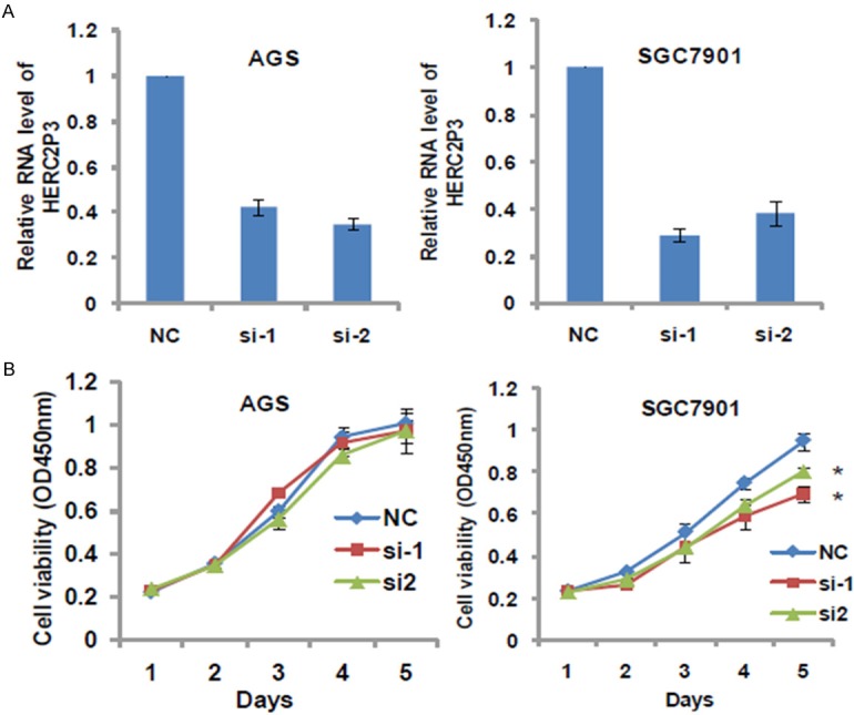
Knocking down HERC2P3 inhibited the cell growth in gastric cancer cells. A. HERC2P3 expression was detected in AGS and SGC7901 cells after transfection with si-HERC2P3. B. The cell grow rates were determined by performing CCK8 proliferation assays. HERC2P3 depletion inhibited the proliferation of SGC7901 cells.
HERC2P3 depletion restrained cell migration in GC cells
Next, Transwell assay was used to determine the effect of HERC2P3 knockdown on cell migration. After 24 hours of suspending the cells in the upper chamber, it was found that the cells transiently transfected with si-HERC2P3s migrated to the lower chamber less than those transiently transfected with si-NC, indicating that knockdown of HERC2P3 strongly suppressed cell migration in AGS and SGC7901 cells (Figure 2A, 2B). These findings suggest that HERC2P3 play an important role on GC cell migration in vitro.
Figure 2.
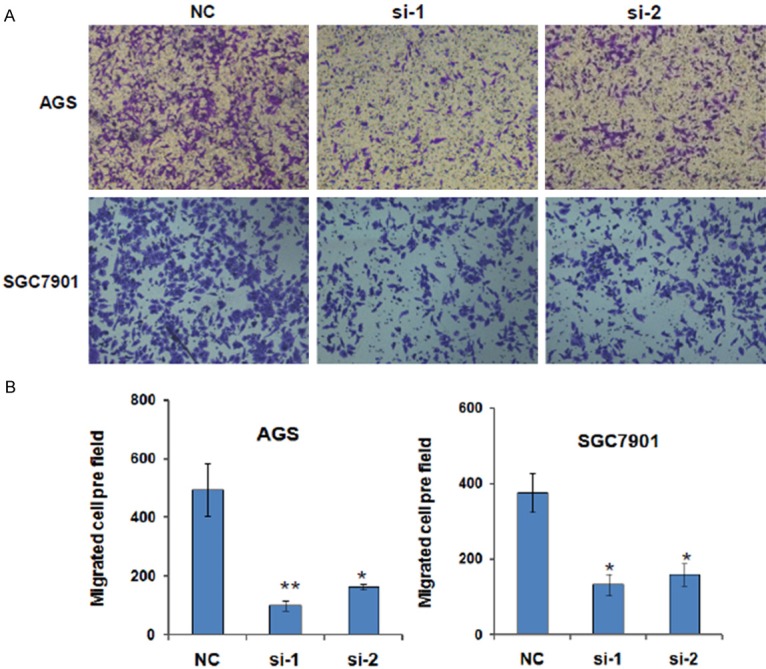
HERC2P3 depletion inhibited the cell migration in gastric cancer cells. A. HERC2P3 suppression by siRNA decreased AGS and SGC7901 cell motility by Transwell test. B. The migrated cell number of AGS and SGC7901 cells decreased after knocking down HERC2P3.
HERC2P3 was decreased in SGC7901 /LV-shHERC2P3 cells
As HERC2P3 depletion inhibited GC cell proliferation and migration, we constructed a stable cell line by infecting SGC7901 cells with LV-shHERC2P3 lentivirus. After puromycin selected, more than 90% of the SGC7901 cells strongly expressed GFP fluorescence (Figure 3A), revealing in these cells HERC2P3 was successfully knocked down. Moreover, the mRNA level of HERC2P3 was further verified by qRT-PCR. The resulting data showed that HERC2P3 was significantly downregulated in the SGC7901/LV-shHERC2P3 cells (Figure 3B).
Figure 3.
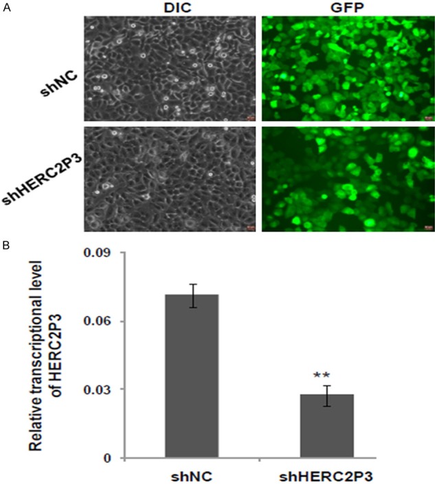
LV-shHERC2P3 was transfected effectively in SGC7901 cells. A. Represent images of SGC7901 cells infected with lV-shHERC2P3 and lV-shNC for 96 h. B. LV-shHERC2P3 transduction obviously inhibited HERC2P3 mRNA levels as determined by qRT-PCR.
Silencing of HERC2P3 suppresses the tumorigenicity and metastasis of GC cells in vivo
Based on our findings in vitro, we performed tumor xenograft assays to examine whether silencing HERC2P3 could decrease the tumorigenicity in vivo. The results demonstrated that low HERC2P3 expression had a slight suppressive effect on the growth of SGC7901 xenograft tumors in nude mice (n=5) (Figure 4A), consistent with the in vitro result. Moreover, we also used SGC7901 stable cell line to analyze the effect of HERC2P3 on tumor metastasis. Knockdown of HERC2P3 in SGC7901 cells by using lenti-viral shRNA significantly reduced the nodule number formed on the lung surface from mice receiving SGC7901-LVshHERC2P cells than that formed from mice receiving SGC7901-LVshNC cells (Figure 4B), indicating HERC2P3 promotes GC cell metastasis in vivo.
Figure 4.
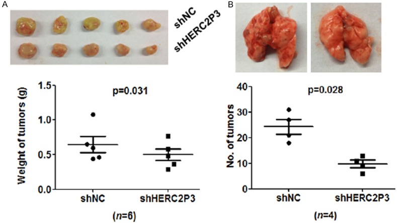
Knocking down HERC2P3 inhibits gastric cancer cell metastasis in vivo. A. The tumors from SGC7901/LV-shHERC2P3 group were averagely lighter than shNC group. B. The numbers of nodes on the lungs’ surface from shHERC2P3 group were less than the negative control group.
Knockdown of HERC2P3 could reduce AKT phosphorylation
Our above studies have demonstrated that silencing HERC2P3 decreased cell growth and migration in vivo and in vitro. Subsequently we attempted to explore the underlying mechanisms by western blot analysis. AGS and SGC7901 cells were transfected with si-HERC2P3 or si-NC and then the proteins were extracted. The western blot results showed that the phosphorylated AKT (p-Akt) was decreased in si-HERC2P3 transfected cells than that in si-NC transfected cells (Figure 6). Furthermore, silencing HERC2P3 could reduce the expression of cyclin D1 and Slug in SGC7901 cells whereas no significant difference was observed in AGS cells (Figure 6). This phenomenon might be in accordance with cell proliferation experiment, reflecting the cell type specific. These results strongly suggest that Akt signaling may mediate the regulation of HERC2P3 on tumor cell proliferation and migration.
Figure 6.
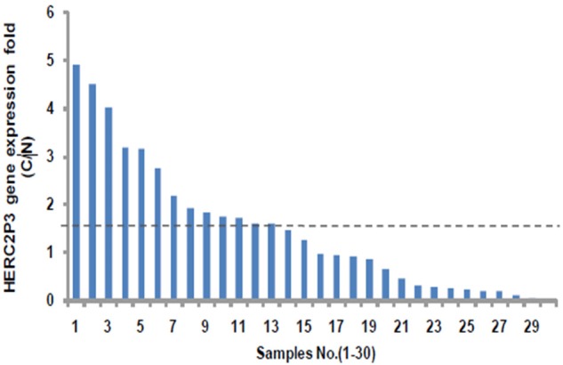
The expression level of HERC2P3 in 30 paired gastric cancer samples. The values “≥1.5” means up-regulation. The values “≤0.66” means down-regulation. The values “<1.5 and >0.66” means not significant variation.
HERC2P3 is frequently up-regulated at gene level in 30 paired GC samples
To determine the expression pattern of HERC2P3 in the human GC tissues, we examined HERC2P3 expression in 30 paired of GC tissues by quantitative RT-PCR. Among 30 paired samples, 13 paired samples were found to be at least 1.5 fold overexpressed, 9 samples were down-regulated and 8 samples were not significantly variable (Figure 5). The data suggest that HERC2P3 is largely overexpressed in GC.
Figure 5.
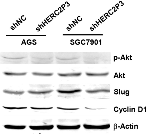
Knocking down HERC2P3 downregulated pAKT expression in vivo. The tumor extracts from each group were subjected to a Western blot analysis to detect pAKT, AKT, Slug, cyclin D and β-actin.
Discussion
LncRNAs have been identified as being linked to human disease and exerting specific functions. Recent years, many researchers have also proved that lncRNAs exhibits its correlation with the occurrence, development and metastasis of cancer [13]. LncRNAs are of great importance in the cancer field and play different biological and physical roles in normal individuals. Several GC related lncRNAs including H19 [14], AC130710 [15], FER1L4 [16], MRUL [17] and UCA1 [18] are explored. With the technological development and thousands of genomic sequences analysis, we are more likely to find possible therapeutic targets and new prognostic markers of lncRNAs. Although there have been multiple studies of the lncRNA, this is the first study that describes the correlation between GC and HERC2P3.
The human HERC gene family has six members. HERC2 is one of HERC family which is highly homologous and comprises the “large” HERC subfamily. HERC2 resides in the 15q11-q13 region which is highly conserved, encoding a putative giant protein of 528 kD, comprises 93 exons. HERC2 has been reported that HERC2 shows a predictive role in advanced NSCLC [19]. HERC2P3 is one of the pseudogene of HERC2 and little is known about its function in the occurrence of disease and cancer development.
In the present study, we tried to figure out the function of HERC2P3 in gastric cancer development. Firstly, we found that knockdown of HERC2P3 inhibited cell growth and migra-tion in human gastric cancer cell lines in vitro. Then we got the similar results through tumor xenograft formation in nude mice. Next, we detected the expression of HERC2P3 in human gastric cancer specimens by qRT-PCR. Our data showed that HERC2P3 is upregulated in most of gastric cancer tissues compared with adjacent non-cancer tissues. Many studies have confirmed that TNM stages, tumor size and distal metastasis are independent prognosis factor in GC [20,21]. However, in our study, not any correlation between HERC2P3 and clincopathological factors was found, which may be due to the small amount of samples. Our findings demonstrate the roles of HERC2P3 in gastric cancer cells, suggesting HERC2P3 may offer a new therapeutic target for this disease. Mechanistically, western blot analysis indicated that silencing HERC2P3 decreased the p-AKT level in both AGS and SGC7901 cells, knocking down HERC2P3 also reduced the level of cyclin D1 in SGC7901 cells. AKT, a serine/threonine protein kinase, is a critical downstream target of PI3K, which plays an important role in cell growth modulation, angiogenesis, migration, and metabolism [22-24]. The phosphorylation of AKT can promote cell proliferation and cell metastasis. Our data showed a possible way how LncRNA HERC2P3 works on gastric cancer cells. The findings that p-AKT and cyclin D1 are regulated by HERC2P3 support HERC2P3 involvement in cancer cell proliferation and migration from molecular level.
In conclusion, we identified HERC2P3 as a new lncRNA involved in the gastric cancer development. It is overexpressed in patients with gastric cancer and may play a significant oncogenetic role in gastric cancer. Further studies are needed to expound the detailed clinic relevance and underlying mechanism of which HERC2P3 contributes to gastric cancer.
Acknowledgements
This work was supported by grants from the Outstanding Young Foundation of Pudong Health Bureau of Shanghai (No. PWRq2015-09) and Science and Technology Development Foundation (No. PKJ2015-Y56) of Pudong New District, Shanghai.
Disclosure of conflict of interest
None.
References
- 1.Orditura M, Galizia G, Sforza V, Gambardella V, Fabozzi A, Laterza MM, Andreozzi F, Ventriglia J, Savastano B, Mabilia A, Lieto E, Ciardiello F, De Vita F. Treatment of gastric cancer. World J Gastroenterol. 2014;20:1635–1649. doi: 10.3748/wjg.v20.i7.1635. [DOI] [PMC free article] [PubMed] [Google Scholar]
- 2.Duraes C, Almeida GM, Seruca R, Oliveira C, Carneiro F. Biomarkers for gastric cancer: prognostic, predictive or targets of therapy? Virchows Arch. 2014;464:367–378. doi: 10.1007/s00428-013-1533-y. [DOI] [PubMed] [Google Scholar]
- 3.Karim-Kos HE, de Vries E, Soerjomataram I, Lemmens V, Siesling S, Coebergh JW. Recent trends of cancer in europe: a combined approach of incidence, survival and mortality for 17 cancer sites since the 1990s. Eur J Cancer. 2008;44:1345–1389. doi: 10.1016/j.ejca.2007.12.015. [DOI] [PubMed] [Google Scholar]
- 4.Gutschner T, Diederichs S. The hallmarks of cancer: a long non-coding RNA point of view. RNA Biol. 2012;9:703–719. doi: 10.4161/rna.20481. [DOI] [PMC free article] [PubMed] [Google Scholar]
- 5.Lai MC, Yang Z, Zhou L, Zhu QQ, Xie HY, Zhang F, Wu LM, Chen LM, Zheng SS. Long noncoding RNA MALAT-1 overexpression predicts tumor recurrence of hepatocellular carcinoma after liver transplantation. Med Oncol. 2012;29:1810–1816. doi: 10.1007/s12032-011-0004-z. [DOI] [PubMed] [Google Scholar]
- 6.Matouk IJ, Mezan S, Mizrahi A, Ohana P, Abu-Lail R, Fellig Y, Degroot N, Galun E, Hochberg A. The oncofetal H19 RNA connection: hypoxia, p53 and cancer. Biochim Biophys Acta. 2010;1803:443–451. doi: 10.1016/j.bbamcr.2010.01.010. [DOI] [PubMed] [Google Scholar]
- 7.Lee NK, Lee JH, Ivan C, Ling H, Zhang X, Park CH, Calin GA, Lee SK. MALAT1 promoted invasiveness of gastric adenocarcinoma. BMC Cancer. 2017;17:46. doi: 10.1186/s12885-016-2988-4. [DOI] [PMC free article] [PubMed] [Google Scholar]
- 8.Xue D, Zhou C, Lu H, Xu R, Xu X, He X. LncRNA GAS5 inhibits proliferation and progression of prostate cancer by targeting miR-103 through AKT/mTOR signaling pathway. Tumour Biol. 2016 doi: 10.1007/s13277-016-5429-8. [Epub ahead of print] [DOI] [PubMed] [Google Scholar]
- 9.Li S, Yu Z, Chen SS, Li F, Lei CY, Chen XX, Bao JM, Luo Y, Lin GZ, Pang SY, Tan WL. The YAP1 oncogene contributes to bladder cancer cell proliferation and migration by regulating the H19 long noncoding RNA. Urol Oncol. 2015;33:427, e1–10. doi: 10.1016/j.urolonc.2015.06.003. [DOI] [PubMed] [Google Scholar]
- 10.Vennin C, Spruyt N, Dahmani F, Julien S, Bertucci F, Finetti P, Chassat T, Bourette RP, Le Bourhis X, Adriaenssens E. H19 non coding RNA-derived miR-675 enhances tumorigenesis and metastasis of breast cancer cells by downregulating c-Cbl and Cbl-b. Oncotarget. 2015;6:29209–29223. doi: 10.18632/oncotarget.4976. [DOI] [PMC free article] [PubMed] [Google Scholar]
- 11.Zhou Y, Zhang X, Klibanski A. MEG3 noncoding RNA: a tumor suppressor. J Mol Endocrinol. 2012;48:R45–53. doi: 10.1530/JME-12-0008. [DOI] [PMC free article] [PubMed] [Google Scholar]
- 12.Strausberg RL, Feingold EA, Grouse LH, Derge JG, Klausner RD, Collins FS, Wagner L, Shenmen CM, Schuler GD, Altschul SF, Zeeberg B, Buetow KH, Schaefer CF, Bhat NK, Hopkins RF, Jordan H, Moore T, Max SI, Wang J, Hsieh F, Diatchenko L, Marusina K, Farmer AA, Rubin GM, Hong L, Stapleton M, Soares MB, Bonaldo MF, Casavant TL, Scheetz TE, Brownstein MJ, Usdin TB, Toshiyuki S, Carninci P, Prange C, Raha SS, Loquellano NA, Peters GJ, Abramson RD, Mullahy SJ, Bosak SA, McEwan PJ, McKernan KJ, Malek JA, Gunaratne PH, Richards S, Worley KC, Hale S, Garcia AM, Gay LJ, Hulyk SW, Villalon DK, Muzny DM, Sodergren EJ, Lu X, Gibbs RA, Fahey J, Helton E, Ketteman M, Madan A, Rodrigues S, Sanchez A, Whiting M, Madan A, Young AC, Shevchenko Y, Bouffard GG, Blakesley RW, Touchman JW, Green ED, Dickson MC, Rodriguez AC, Grimwood J, Schmutz J, Myers RM, Butterfield YS, Krzywinski MI, Skalska U, Smailus DE, Schnerch A, Schein JE, Jones SJ, Marra MA Mammalian Gene Collection Program Team. Generation and initial analysis of more than 15,000 full-length human and mouse cDNA sequences. Proc Natl Acad Sci U S A. 2002;99:16899–16903. doi: 10.1073/pnas.242603899. [DOI] [PMC free article] [PubMed] [Google Scholar]
- 13.Esteller M. Non-coding RNAs in human disease. Nat Rev Genet. 2011;12:861–874. doi: 10.1038/nrg3074. [DOI] [PubMed] [Google Scholar]
- 14.Zhang EB, Han L, Yin DD, Kong R, De W, Chen J. c-Myc-induced, long, noncoding H19 affects cell proliferation and predicts a poor prognosis in patients with gastric cancer. Med Oncol. 2014;31:914. doi: 10.1007/s12032-014-0914-7. [DOI] [PubMed] [Google Scholar]
- 15.Xu C, Shao Y, Xia T, Yang Y, Dai J, Luo L, Zhang X, Sun W, Song H, Xiao B, Guo J. lncRNAAC130710 targeting by miR-129-5p is upregulated in gastric cancer and associates with poor prognosis. Tumour Biol. 2014;35:9701–9706. doi: 10.1007/s13277-014-2274-5. [DOI] [PubMed] [Google Scholar]
- 16.Liu Z, Shao Y, Tan L, Shi H, Chen S, Guo J. Clinical significance of the low expression of FER1L4 in gastric cancer patients. Tumour Biol. 2014;35:9613–9617. doi: 10.1007/s13277-014-2259-4. [DOI] [PubMed] [Google Scholar]
- 17.Wang Y, Zhang D, Wu K, Zhao Q, Nie Y, Fan D. Long noncoding RNA MRUL promotes ABCB1 expression in multidrug-resistant gastric cancer cell sublines. Mol Cell Biol. 2014;34:3182–3193. doi: 10.1128/MCB.01580-13. [DOI] [PMC free article] [PubMed] [Google Scholar]
- 18.Gao J, Cao R, Mu H. Long non-coding RNA UCA1 may be a novel diagnostic and predictive biomarker in plasma for early gastric cancer. Int J Clin Exp Pathol. 2015;8:12936–12942. [PMC free article] [PubMed] [Google Scholar]
- 19.Bonanno L, Costa C, Majem M, Sanchez JJ, Rodriguez I, Gimenez-Capitan A, Molina-Vila MA, Vergnenegre A, Massuti B, Favaretto A, Rugge M, Pallares C, Taron M, Rosell R. Combinatory effect of BRCA1 and HERC2 expression on outcome in advanced non-small-cell lung cancer. BMC Cancer. 2016;16:312. doi: 10.1186/s12885-016-2339-5. [DOI] [PMC free article] [PubMed] [Google Scholar]
- 20.Adachi Y, Oshiro T, Mori M, Maehara Y, Sugimachi K. Tumor size as a simple prognostic indicator for gastric carcinoma. Ann Surg Oncol. 1997;4:137–140. doi: 10.1007/BF02303796. [DOI] [PubMed] [Google Scholar]
- 21.Wang W, Li YF, Sun XW, Chen YB, Li W, Xu DZ, Guan XX, Huang CY, Zhan YQ, Zhou ZW. Prognosis of 980 patients with gastric cancer after surgical resection. Chin J Cancer. 2010;29:923–930. doi: 10.5732/cjc.010.10290. [DOI] [PubMed] [Google Scholar]
- 22.Porta C, Paglino C, Mosca A. Targeting PI3K/Akt/mTOR signaling in cancer. Front Oncol. 2014;4:64. doi: 10.3389/fonc.2014.00064. [DOI] [PMC free article] [PubMed] [Google Scholar]
- 23.King D, Yeomanson D, Bryant HE. PI3King the lock: targeting the PI3K/Akt/mTOR pathway as a novel therapeutic strategy in neuroblastoma. J Pediatr Hematol Oncol. 2015;37:245–251. doi: 10.1097/MPH.0000000000000329. [DOI] [PubMed] [Google Scholar]
- 24.Johnson SM, Gulhati P, Rampy BA, Han Y, Rychahou PG, Doan HQ, Weiss HL, Evers BM. Novel expression patterns of PI3K/Akt/mTOR signaling pathway components in colorectal cancer. J Am Coll Surg. 2010;210:767–776. 776–778. doi: 10.1016/j.jamcollsurg.2009.12.008. [DOI] [PMC free article] [PubMed] [Google Scholar]


