Abstract
Emodin exhibits anti-proliferative effects in numerous cancer cell lines via different mechanisms. The present study aimed to study the effects and underlying molecular mechanisms of emodin on bladder cancer cells. We treated two bladder cancer cell lines, T24 and 5637, with different concentrations of emodin (20, 40 and 80 µmol/L) or DMSO (Control). We analyzed the biological effects of emodin on cell growth, invasion, as well as the mRNA and protein expression of Notch1, Jagged1, VEGF, VEGFR2 and MMP2 using Cell Counting Kit-8, transwell, reverse transcription-quantitative PCR and western blot assays. Emodin repressed cell growth, invasion and Notch1 expression in a concentration-dependent fashion. And Notch1 over-expression assay showed that the anti-proliferative and anti-invasive roles of emodin, along with the down-regulated effects on the expression of Notch1, Jagged1, VEGF, VEGFR2 and MMP2 were partially rescued by Notch1 over-expression. In conclusion, Emodin might suppress the progression of bladder cancer via inhibiting the expression of Notch1.
Keywords: Emodin, Notch1, bladder cancer, proliferation, invasion
Introduction
Bladder cancer is the second most common malignancy of the urogenital system. Transitional cell carcinoma (TCC), also known as urothelial carcinoma, begins from abnormalities of the urothelial cells lining of the bladder. TCC accounts for the vast majority of bladder cancer [1,2]. The majority of bladder cancer patients present with superficial tumors, while 20% to 40% of patients either present with or develop invasive disease. Transurethral resection of bladder tumor and immunotherapy are often applied to treat and prevent superficial tumor recurrence. And for muscle invasive disease, the radical ablative surgery and a combination of radiation and chemotherapy have been identified as effective clinical treatments [3]. The recovery rate of bladder cancer depends on the depth of the tumor invading into the bladder wall. Whereas, in order to improve the overall survival of invasive bladder cancer, new treatment options are urgently needed.
Emodin (6-methyl-1, 3, 8-trihydroxyanthraquinone) is a purgative and active component isolated from the herb. It is also produced by many species of fungi, such as Aspergillus, Pyrenochaeta and Pestalotiopsis [4]. In China, emodin is traditionally used for the treatment of skin burns, gallstone, hepatitis, inflammation and osteomyelitis [5,6], as it processes antimicrobial, antifungal, antiviral, anti-inflammatory, antioxidant and chemopreventive activities [7,8]. Early studies have demonstrated that emodin is able to inhibit tyrosine kinase activity of Her-2/Neu in breast and lung cancer [9,10]. In recent years, more and more evidences have indicated the anti-proliferative effects of emodin against varieties of human cancer cells. For example, emodin inhibits the growth of prostate cancer cells via down-regulating androgen receptor [11] or via the Notch signaling pathway [12]. In cervical cancer cell, emodin is also reported to induce cell apoptosis through activation of caspase9 [13]. Emodin can induce lung adenocarcinoma cell apoptosis via a reactive oxygen species-dependent mitochondrial signaling pathway [14]. Although emodin shows its inhibitory action on many cancer cells through different mechanisms, its effects on bladder cancer are still not clear.
In this study, we analyzed the biological effects of emodin on the proliferation and invasion of two bladder cancer cell lines, T24 and 5637. We also examined the relative protein expression which might be regulated by emodin in both bladder cancer cell lines. Taken together, the present study proved that emodin repressed the proliferation and invasion of bladder cancer cells through down-regulating the expression of Notch1.
Materials and methods
Cell culture
The bladder cancer cells, T24 and 5637, were purchased from American Type Culture Collection (ATCC). Cells were cultured in Dulbecco’s Modified Eagle’s Medium (DMEM) supplemented with 10% fetal bovine serum (FBS, Gibico) plus 1% antibiotics (100 U/ml penicillin and 100 µg/ml streptomycin), and maintained at 37°C in a 5% CO2 incubator.
Concentration screening
T24 and 5637 cells were cultured in DMEM medium added with 0 µmol/L (DMSO), 20 µmol/L, 40 µmol/L and 80 µmol/L emodin, respectively. Cell Counting Kit-8 (CCK-8), transwell, reverse transcription-quantitative PCR (qRT-PCR) and western blot assays were performed to select the appropriate concentration of emodin.
Notch1 over-expression
Full-length human Notch1 cDNA was synthesized and cloned into pCDNA3.1(+) (Addgene). pCDNA3.1(+) was served as negative control. The constructs were transfected into bladder cancer cells using Lipofectamine 2000 (Invitrogen) per the manufacture’s introductions.
Over-expression assay
The bladder cancer cells were divided into five groups. And the experimental design was as following: 1. Control group: T24 and 5637 cells without any treatments; 2. Negative control (NC) group: T24 and 5637 cells were transfected with control vector and cultured in DMEM medium added with DMSO; 3. Notch1 group: T24 and 5637 cells were transfected with full-length cDNA of Notch1 and cultured in DMEM medium added with DMSO; 4. NC + Emodin group: T24 and 5637 cells were transfected with control vector and cultured in DMEM medium added with 80 µmol/L emodin; 5. Notch1 + Emodin group: T24 and 5637 cells were transfected with full-length cDNA of Notch1 and cultured in DMEM medium added with 80 µmol/L emodin.
CCK-8 assay
CCK-8 assay was used to analyze the proliferation of bladder cancer cells. T24 and 5637 cells were seeded in 96-well plates and cell growth was measured with commercial CCK-8 Assay Kit (Sigma) at 0 h, 24 h, 48 h and 72 h. Absorbance excited at 450 nm of reacted cells was examined to valuate cell proliferation.
Transwell assay
Transwell assay was performed to evaluate the invasiveness of bladder cancer cells. T24 and 5637 cells were serum starved for 24 h after medication or transfection. Then, the cells were seeded into upper chamber of a 24-well transwell chamber (Trueline) which was coated with 50 µl Matrigel (1:2 dilution, BD Biosciences) containing a polycarbonate filter. The lower chamber was filled with 0.75 ml DMEM culture medium. After 24 h of incubation, the non-migrated cells were scraped from the upper chamber, and the adherent cells were stained with crystal violet and photographed.
Reverse transcription-quantitative PCR (qRT-PCR)
Total RNA was extracted using the TRIzol reagent (Invitrogen) at 48 h after medication or transfection. The reverse transcription was carried out using cDNA Synthesis Kit (Fermentas). Real-time PCR was performed using a standard SYBR Green PCR kit. The cycle conditions were 10 min at 95°C, 40 cycles of 15 s at 95°C and 45 s at 60°C, 15 s at 95°C, 1 min at 60°C followed by 15 s at 95°C and 15 s at 60°C. And the data were analyzed using ABI Prism 7300 SDS software. All the processes were according to the instructions of manufactures. GADPH was served as an internal control. The primer sequences were as follow: Notch1 (NM_017617.3): Primer F 5’ GACGCACAAGGTGTCTTC 3’, Primer R 5’ TTGCCCAGGTCATCTACG 3’; VEGF (NM_001025366.2), Primer F 5’ ATTTCTGGGATTCCTGTAG 3’, Primer R 5’ CAGTGAAGACACCAATAAC 3’; VEGFR2 (NM_002253.2), Primer F 5’ CTCAGCAGGATGGCAAAG 3’, Primer R 5’ ACTGTCCGTCTGGTTGTC 3’; Jagged1 (NM_000214.2), Primer F 5’ CTTCACGGGAACATACTG 3’, Primer R 5’ GCACTTGTAGGAGTTGAC 3’; MMP-2 (NM_004530.4), Primer F 5’ TTGACGGTAAGGACGGACTC 3’, Primer R 5’ GGCGTTCCCATACTTCACAC 3’; GAPDH (NM_001256799.1), Primer F 5’ CACCCACTCCTCCACCTTTG 3’, Primer R 5’ CCACCACCCTGTTGCTGTAG 3’.
Western blot assay
At 48 h post treatments, bladder cancer cells were harvested and subjected to SDS-PAGE. After transferred to a nitrocellulose membrane, the membranes were blocked with 5% skim milk and then incubated with primary antibodies. After incubation with secondary antibodies, the blots were visualized by the enhanced chemiluminescence system. The antibody list was as follows: Notch1 (1:1000, #3608, Cell Signaling Technology), VEGF (1:1000, Ab46154, Abcam), VEGFR2 (1:1000, AF6281, Affinity), Jagged1 (1:1000, Ab109536, Abcam), MMP2 (1:1000, Ab92536, Abcam), GAPDH (1:2000, #5174, Cell Signaling Technology).
Statistical analysis
Data analysis was performed by GraphPad Prism software (San Diego, CA). Values were presented as means ± SD. Statistical significance was determined by two-tailed Student’s t-test. P<0.05 was considered to be statistically significant.
Results
Emodin inhibited proliferation and invasion of bladder cancer cells concentration dependently
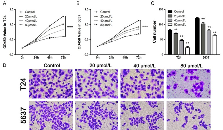
To evaluate the effects of emodin on cell proliferation and invasion of bladder cancer cells, CCK-8 and transwell assays were conducted in T24 and 5637 cells respectively. As shown in Figure 1A and 1B, optical density (OD) value was measured at 0 h, 24 h, 48 h and 72 h after the addition of control medium and different concentrations of emodin. Emodin showed a remarkable concentration-dependent inhibitory effects on the proliferation of both bladder cancer cells. Then we also estimated the invasion cell number of two bladder cancer cells at 48 h post medication. As expected, cell invasive capacity was also inhibited by emodin in a concentration-dependent fashion (Figure 1C and 1D). These data suggested that emodin treatment (20-80 µmol/L) significantly suppressed the proliferation and invasion of bladder cancer cells in a concentration-dependent manner.
Figure 1.

Emodin inhibited cell growth and invasion of bladder cancer cell in a dose-dependent fashion. A and B: Cell proliferation of two cell lines was examined at 0 h, 24 h, 48 h and 72 h after emodin addition (n=3). C and D: Different concentrations of emodin inhibited cell invasion in two bladder cancer cells. Data were shown as mean ± S.D., *P<0.05, **P<0.01, ***P<0.001.
Emodin inhibited the expression of Notch1 in bladder cancer cells
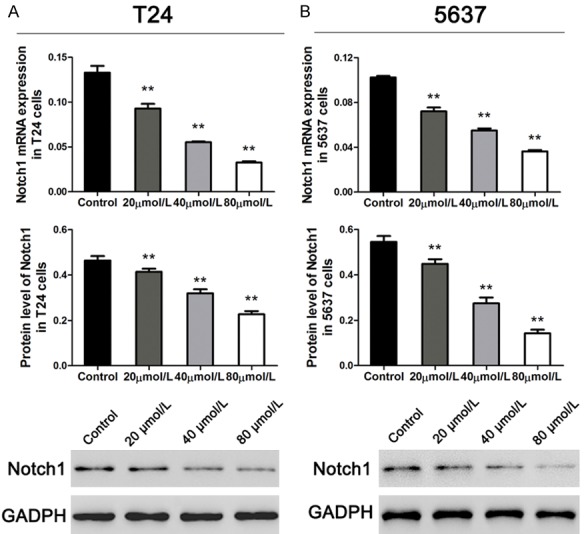
We examined the mRNA expression and protein levels of Notch1 in T24 and 5637 cells using qRT-PCR and western blot at 48 h after different concentrations of emodin medication. As shown in Figure 2, the expression of Notch1 was significantly depressed by emodin addition in both T24 and 5637 cells. And the inhibitory effect of emodin on Notch1 expression was concentration dependent. Therefore, we selected the dose of 80 µmol/L to perform further assays.
Figure 2.

Emodin inhibited Notch1 expression concentration-dependently in bladder cancer cells. A and B: mRNA and protein expression of Notch1 in T24 and 5637 cell lines (n=3). Data were shown as mean ± S.D., *P<0.05, **P<0.01, ***P<0.001.
Emodin affected cell proliferation and invasion via regulating Notch1 in bladder cancer cells
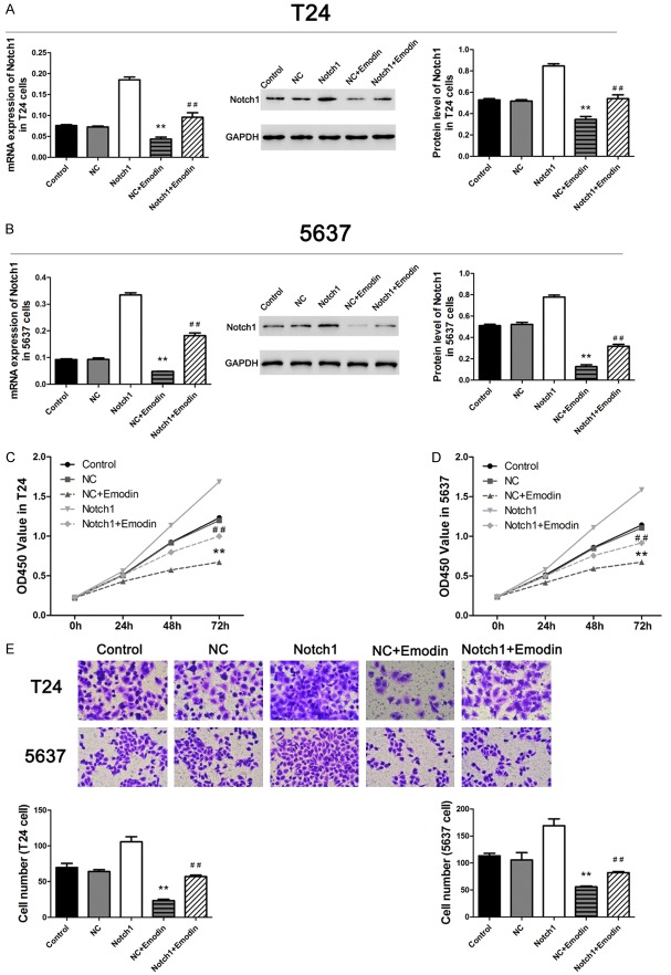
In over-expression assay, we transfected Notch1 cDNA into T24 and 5637 cells to increase the expression of Notch1 and treated bladder cancer cells with 80 µmol/L emodin as described above. As illustrated in Figure 3A and 3B, we found that the expression of Notch1 was significantly increased post transfection. Whereas, emodin treatment obviously inhibited the expression of Notch1 in two bladder cancer cells. And the effects of cDNA and emodin addition on cell proliferation and invasion showed the same trend (as shown in Figure 3C-E). These results indicated that emodin might suppress cell proliferation and invasion via regulating the expression of Notch1 in bladder cancer cells.
Figure 3.
Emodin affected cell proliferation and invasion via down regulating Notch1 in two bladder cancer cell lines. A and B: Expression of Notch1 was inhibited by emodin in both NC cells and over-expressed cells (n=3). C and D: Cell proliferation was inhibited in emodin medication groups (n=3). E: Cell invasion was significantly depressed in emodin medication groups (n=3). Data were shown as mean ± S.D., *P<0.05, **P<0.01 (compared with negative controls); #P<0.05, ##P<0.01 (compared with Notch1 groups).
Emodin reduced the expression of related proteins via inhibiting Notch1 in bladder cancer cells
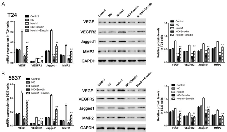
To further confirm the biological effects of emodin medication, we then examined the endogenous expression of four proteins, which were involved in proliferation and invasion processes, using qRT-PCR and western blot at 48 h post cDNA transfected and emodin addition. As shown in Figure 4, the mRNA and protein levels of Jagged1, VEGF, VEGFR2 and MMP2 were obviously inhibited after treated with emodin, and such effects were partially rescued in Notch1 over-expression groups. Taken together, these results indicated that emodin might inhibit cell growth and invasion relating proteins via suppressing Notch1 in bladder cancer cells.
Figure 4.

Emodin suppressed the expression of invasion-related proteins through down regulating Notch1. A and B: mRNA and protein level of VEGF, VEGFR2, Jagged1 and MMP-2 were inhibited in emdoin treated groups (n=3). Data were shown as mean ± S.D., *P<0.05, **P<0.01 (compared with negative controls); #P<0.05, ##P<0.01 (compared with Notch1 groups).
Discussion
Except of the anti-proliferative effect of emodin, many studies have focused on the invasion inhibitory effect of emodin on human cancer cells, and different mechanisms have been proposed. In squamous cell carcinoma and breast cancer cells, emodin is proved inhibiting cell invasion via suppressing AP-1 and nuclear factor-κB (NF-κB) signaling pathways [15]. Emodin also inhibits CXCL12-induced migration and invasion of prostate and lung cancer cells by blocking the expression of CXC chemokine receptor-4 [16]. Aloe-emodin, a structurally similar compound of emodin, has been reported to inhibit invasion in nasopharyngeal carcinoma cell by the down-regulation of MMP-2 through the p38-NF-κB-dependent pathway [17]. In bladder cancer cells, aloe-emodin has also been suggested inhibiting proliferation via activating the p53 dependent apoptotic pathway [18].
In the current study, we found that the proliferation and invasion of bladder cancer cells was dose-dependently inhibited by emodin exposure. Our previous study has showed that emodin inhibits the growth of prostate cancer cells via the Notch signaling pathway [12]. Notch signaling pathway, a highly conserved cell signaling system, is actively involved in embryonic development, cell differentiation, cell-cell communication, cell proliferation and apoptosis [19]. Aberrantly activated Notch signaling contributes to the tumorigenesis of a variety of human cancers [20]. Notch inhibitors, especially γ-secretase inhibitors, have been regarded as targeted therapeutic agents [21,22]. Here, we found the inhibitory effect of emodin on Notch1 expression. And over-expression assay showed that Notch1 overexpression partially rescued the inhibitory effects of emodin on bladder cancer cell proliferation and invasion. Therefore, the related data suggested that emodin might inhibit the growth and invasive capacity of bladder cancer cell through down-regulating Notch1.
We then examined the relative protein expression in bladder cancer cells, containing VEGF, VEGFR2, Jagged1 and MMP-2. VEGF and VEGFR2 are identified as metastasis-related genes. A previous study suggested that VEGF expression was an independent prognostic factor of recurrence and metastasis of bladder cancer [23]. Down-regulation of Jagged1, a ligand of Notch1, obviously inhibits cell invasion and migration in prostate cancer cells [24,25]. MMP-2 is also involved in the metastasis of human cancers, including breast, gastric and bladder cancer [26-28]. In this study, the data showed that over-expressed Notch1 significantly up-regulated the expression of these four proteins. And the down-regulation of Notch1 induced by emodin medication caused the inhibition of VEGF, VEGFR2, Jagged1 and MMP-2 expression in bladder cancer cells. These results indicated that emodin inhibited cell invasion by related proteins through down-regulating Notch1.
Summarily, our study revealed that emodin inhibited cell proliferation and invasion of bladder cancer cells in a concentration-dependent way via the down-regulation of Notch1 expression. And the proteins related to cell invasion were also inhibited by emodin through regulating the expression of Notch1.
Acknowledgements
This study was supported by Natural Science Foundation of Zhejiang province (Y2111329 and LY17H050002), Science and Technology Plan Projects of Zhejiang Province (2014C37016), Chinese Medicine Science and Technology Plan Projects of Zhejiang Province (2016ZB099, 2013ZA107 and 2011ZB099), Medicine and health science and technology plan projects of Zhejiang Province (2011KYB066 and 2015KYB295) and Science and Technology Plan Projects of Hangzhou Zhejiang Province (2017A05, 20110833B05 and 20110733Q12).
Disclosure of conflict of interest
None.
References
- 1.Heney NM, Ahmed S, Flanagan MJ, Frable W, Corder MP, Hafermann MD, Hawkins IR. Superficial bladder cancer: progression and recurrence. J Urol. 1983;130:1083–1086. doi: 10.1016/s0022-5347(17)51695-x. [DOI] [PubMed] [Google Scholar]
- 2.Vale CL. Adjuvant chemotherapy in invasive bladder cancer: a systematic review and metaanalysis of individual patient data Advanced Bladder Cancer (ABC) meta-analysis collaboration. Eur Urol. 2005;48:189. doi: 10.1016/j.eururo.2005.04.005. [DOI] [PubMed] [Google Scholar]
- 3.Stein JP, Lieskovsky G, Cote R, Groshen S, Feng AC, Boyd S, Skinner E, Bochner B, Thangathurai D, Mikhail M. Radical cystectomy in the treatment of invasive bladder cancer: longterm results in 1,054 patients. J. Clin. Oncol. 2001;19:666–675. doi: 10.1200/JCO.2001.19.3.666. [DOI] [PubMed] [Google Scholar]
- 4.Tsai TH. Analytical approaches for traditional chinese medicines exhibiting antineoplastic activity. J Chromatogr B Biomed Sci Appl. 2001;764:27. doi: 10.1016/s0378-4347(01)00277-8. [DOI] [PubMed] [Google Scholar]
- 5.Efferth T, Li PC, Konkimalla VS, Kaina B. From traditional Chinese medicine to rational cancer therapy. Trends Mol Med. 2007;13:353–361. doi: 10.1016/j.molmed.2007.07.001. [DOI] [PubMed] [Google Scholar]
- 6.Wang H, Dong Y, Xiu ZL. Microwave-assisted aqueous two-phase extraction of piceid, resveratrol and emodin from Polygonum cuspidatum by ethanol/ammonium sulphate systems. Biotechnol Lett. 2008;30:2079–84. doi: 10.1007/s10529-008-9815-1. [DOI] [PubMed] [Google Scholar]
- 7.Chang CH, Lin CC, Yang JJ, Namba T, Hattori M. Anti-inflammatory effects of emodin from ventilago leiocarpa. Am J Chin Med. 2012;24:139–142. doi: 10.1142/S0192415X96000189. [DOI] [PubMed] [Google Scholar]
- 8.Jayasuriya H, Koonchanok NM, Geahlen RL, Mclaughlin JL, Chang CJ. Emodin, a protein tyrosine kinase inhibitor from Polygonum cuspidatum. J Nat Prod. 1992;55:696–8. doi: 10.1021/np50083a026. [DOI] [PubMed] [Google Scholar]
- 9.Zhang L, Hung MC. Sensitization of HER-2/neu-overexpressing non-small cell lung cancer cells to chemotherapeutic drugs by tyrosine kinase inhibitor emodin. Oncogene. 1996;12:571–576. [PubMed] [Google Scholar]
- 10.Zhang L, Lau YK, Xia W, Hortobagyi GN, Hung MC. Tyrosine kinase inhibitor emodin suppresses growth of HER-2/neu-overexpressing breast cancer cells in athymic mice and sensitizes these cells to the inhibitory effect of paclitaxel. Clin Cancer Res. 1999;5:343–353. [PubMed] [Google Scholar]
- 11.Cha TL, Qiu L, Chen CT, Wen Y, Hung MC. Emodin down-regulates androgen receptor and inhibits prostate cancer cell growth. Cancer Res. 2005;65:2287–2295. doi: 10.1158/0008-5472.CAN-04-3250. [DOI] [PubMed] [Google Scholar]
- 12.Deng G, Ju X, Meng Q, Yu ZJ, Ma LB. Emodin inhibits the proliferation of PC3 prostate cancer cells in vitro via the Notch signaling pathway. Mol Med Rep. 2015;12:4427–4433. doi: 10.3892/mmr.2015.3923. [DOI] [PubMed] [Google Scholar]
- 13.Srinivas G, Anto RJ, Srinivas P, Vidhyalakshmi S, Senan VP, Karunagaran D. Emodin induces apoptosis of human cervical cancer cells through poly(ADP-ribose) polymerase cleavage and activation of caspase-9. Eur J Pharmacol. 2003;473:117–125. doi: 10.1016/s0014-2999(03)01976-9. [DOI] [PubMed] [Google Scholar]
- 14.Su YT, Chang HL, Shyue SK, Hsu SL. Emodin induces apoptosis in human lung adenocarcinoma cells through a reactive oxygen species-dependent mitochondrial signaling pathway. Biochem Pharmacol. 2005;70:229–241. doi: 10.1016/j.bcp.2005.04.026. [DOI] [PubMed] [Google Scholar]
- 15.Huang Q, Shen HM, Ong CN. Inhibitory effect of emodin on tumor invasion through suppression of activator protein-1 and nuclear factor-kappaB. Biochem Pharmacol. 2004;68:361. doi: 10.1016/j.bcp.2004.03.032. [DOI] [PubMed] [Google Scholar]
- 16.Ok S, Kim SM, Kim C, Nam D, Shim BS, Kim SH, Ahn KS, Choi SH, Ahn KS. Emodin inhibits invasion and migration of prostate and lung cancer cells by downregulating the expression of chemokine receptor CXCR4. Immunopharmacol Immunotoxicol. 2012;34:768–778. doi: 10.3109/08923973.2012.654494. [DOI] [PubMed] [Google Scholar]
- 17.Lin ML, Lu YC, Chung JG, Wang SG, Lin HT, Kang SE, Tang CH, Ko JL, Chen SS. Downregulation of MMP-2 through the p38 MAPKNF-κB-dependent pathway by aloe-emodin leads to inhibition of nasopharyngeal carcinoma cell invasion. Mol Carcinog. 2010;49:783–797. doi: 10.1002/mc.20652. [DOI] [PubMed] [Google Scholar]
- 18.Lin JG, Chen GW, Li TM, Chouh ST, Tan TW, Chung JG. Aloe-emodin induces apoptosis in T24 human bladder cancer cells through the p53 dependent apoptotic pathway. J Urol. 2006;175:343–347. doi: 10.1016/S0022-5347(05)00005-4. [DOI] [PubMed] [Google Scholar]
- 19.Zhou ZD, Kumari U, Xiao ZC, Tan EK. Notch as a molecular switch in neural stem cells. IUBMB Life. 2010;62:618–623. doi: 10.1002/iub.362. [DOI] [PubMed] [Google Scholar]
- 20.Bolós V, Grego-Bessa J, de la Pompa JL. Notch signaling in development and cancer. Endocr Rev. 2007;28:339–363. doi: 10.1210/er.2006-0046. [DOI] [PubMed] [Google Scholar]
- 21.Purow B. Notch signaling in embryology and cancer. Springer; 2012. Notch inhibition as a promising new approach to cancer therapy; pp. 305–319. [DOI] [PMC free article] [PubMed] [Google Scholar]
- 22.Shih IM, Wang TL. Notch signaling, γ-secretase inhibitors, and cancer therapy. Cancer Res. 2007;67:1879–1882. doi: 10.1158/0008-5472.CAN-06-3958. [DOI] [PubMed] [Google Scholar]
- 23.Inoue K, Slaton JW, Karashima T, Yoshikawa C, Shuin T, Sweeney P, Millikan R, Dinney CP. The prognostic value of angiogenesis factor expression for predicting recurrence and metastasis of bladder cancer after neoadjuvant chemotherapy and radical cystectomy. Clin Cancer Res. 2000;6:4866–4873. [PubMed] [Google Scholar]
- 24.Zhang Y, Wang Z, Ahmed F, Banerjee S, Li Y, Sarkar FH. Down-regulation of Jagged-1 induces cell growth inhibition and S phase arrest in prostate cancer cells. Int J Cancer. 2006;119:2071–2077. doi: 10.1002/ijc.22077. [DOI] [PubMed] [Google Scholar]
- 25.Wang Z, Li Y, Banerjee S, Kong D, Ahmad A, Nogueira V, Hay N, Sarkar FH. Down-regulation of Notch-1 and Jagged-1 inhibits prostate cancer cell growth, migration and invasion, and induces apoptosis via inactivation of Akt, mTOR, and NF-κB signaling pathways. J Cell Biochem. 2010;109:726–736. doi: 10.1002/jcb.22451. [DOI] [PubMed] [Google Scholar]
- 26.Jinga D, Blidaru A, Condrea I, Ardeleanu C, Dragomir C, Szegli G, Stefanescu M, Matache C. MMP-9 and MMP-2 gelatinases and TIMP-1 and TIMP-2 inhibitors in breast cancer: correlations with prognostic factors. J Cell Mol Med. 2006;10:499–510. doi: 10.1111/j.1582-4934.2006.tb00415.x. [DOI] [PMC free article] [PubMed] [Google Scholar]
- 27.Hwang TL, Lee LY, Wang CC, Liang Y, Huang SF, Wu CM. Claudin-4 expression is associated with tumor invasion, MMP-2 and MMP-9 expression in gastric cancer. Exp Ther Med. 2010;1:789–97. doi: 10.3892/etm.2010.116. [DOI] [PMC free article] [PubMed] [Google Scholar]
- 28.Davies B, Waxman J, Wasan H, Abel P, Williams G, Krausz T, Neal D, Thomas D, Hanby A, Balkwill F. Levels of matrix metalloproteases in bladder cancer correlate with tumor grade and invasion. Cancer Res. 1993;53:5365–5369. [PubMed] [Google Scholar]


